
The spin column-based method is a commonly used DNA extraction technique in molecular biology laboratories. The Spin Column-Based Method of DNA Extraction works by allowing DNA to bind selectively to a silica membrane under chaotropic, high-salt conditions, followed by washing and elution steps that effectively remove proteins, lipids, polysaccharides, and enzymatic inhibitors. This process enables the rapid and reliable purification of DNA from complex biological samples (Kumar & Singh, 2022; Thermo Fisher Scientific, 2024).
Initially developed as a safer alternative to organic solvent-based methods such as phenol-chloroform extraction, spin column protocols now form the foundation of most commercial DNA extraction kits. The use of centrifuge-compatible silica membranes improves reproducibility, shortens processing time, and reduces exposure to hazardous chemicals (Brown, 2020).
Spin column-based DNA extraction is widely applied to various sample types, including blood, tissues, plant material, microbial cultures, forensic specimens, and clinical samples. Its simplicity, consistent performance, and compatibility with downstream applications such as PCR, qPCR, LAMP, and next-generation sequencing make it suitable for routine research, diagnostic, and teaching laboratories (Qiagen, 2024; Dimitrakopoulou et al., 2020; Gand et al., 2023).
Key Reagents of the Spin Column-Based Method of DNA Extraction
| Reagent | Typical Concentration / Amount | Purpose |
| Lysis buffer (chaotropic salts) | Guanidinium salts (4–6 M) | Disrupts cells, denatures proteins, promotes DNA binding to silica |
| Detergents (e.g., SDS, Triton X-100) | 0.1–2% | Solubilizes cell membranes and lipid components |
| Proteinase K | 10–20 mg/mL | Digests proteins and nucleases |
| Ethanol or isopropanol | 70–100% | Facilitates DNA binding to silica membrane |
| Wash buffer I (high-salt) | Contains chaotropic salts and ethanol | Removes proteins and polysaccharides |
| Wash buffer II (low-salt) | Contains ethanol | Removes residual salts and impurities |
| Elution buffer (Tris-EDTA or water) | 10–50 µL | Releases purified DNA from silica membrane |
Principle of the Spin Column-Based Method of DNA Extraction
The fundamental principle of spin column–based DNA extraction is the selective binding of DNA to a silica membrane in the presence of high concentrations of chaotropic salts. These salts, such as guanidinium thiocyanate, disrupt hydrogen bonding in water, destabilize cellular membranes, and denature proteins, thereby releasing DNA into solution during the lysis step (Brown, 2020; Kumar & Singh, 2022). Under these high-salt and low-water-activity conditions, hydrogen bonding between the negatively charged phosphate backbone of DNA and the silica surface becomes energetically favorable, allowing DNA to adsorb selectively to the membrane (Thermo Fisher Scientific, 2024).
Following lysis, the addition of ethanol further promotes DNA precipitation and strengthens DNA–silica interactions. While DNA binds tightly to the silica membrane within the spin column, contaminants such as proteins, lipids, carbohydrates, RNA, and cellular debris bind poorly and are removed through successive centrifugal washing steps (Cold Spring Harbor Laboratory Press, 2017; Thermo Fisher Scientific, 2024). The purified DNA is then recovered by elution with water or a mildly buffered solution such as Tris-EDTA, which disrupts the DNA–silica interaction under low-salt conditions (Brown, 2020; Kumar & Singh, 2022).
This silica-based binding and elution mechanism is conceptually related to ion-exchange chromatography, where DNA binds to charged resins and is later eluted using salt gradients. However, silica-based purification is simpler, faster, and more robust, making it well-suited for routine laboratory use (Brown, 2020). The versatility of this principle allows spin column protocols to be adapted for challenging samples, including tough plant tissues, microbial spores, and degraded or archival specimens, without compromising DNA purity or usability (Almeida & Ramos, 2020; Sepp et al., 1994).
Steps / Protocol of the Spin Column-Based Method of DNA Extraction
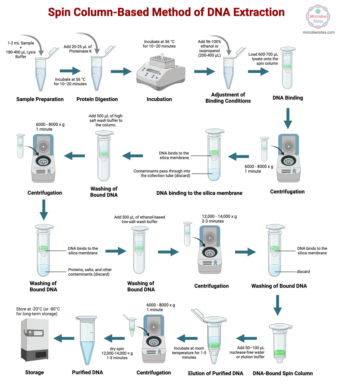
Sample Preparation
- Collect the biological sample:
- Bacterial pellet: 1–2 mL liquid culture
- Animal tissue: ~25 mg
- Plant tissue: 50–100 mg
- Transfer the sample into a sterile microcentrifuge tube.
- Add 180–400 µL of lysis buffer containing:
- Chaotropic salts (e.g., guanidinium salts)
- Detergents (e.g., SDS)
- Mix gently to ensure uniform contact between the sample and the lysis buffer.
- Incubate the mixture at room temperature or 56 °C for 10–20 minutes. This allows efficient disruption of cell membranes and release of DNA.
- For Gram-positive bacteria:
- Add 20–40 µL of lysozyme (10–20 mg/mL) before incubation to weaken the thick peptidoglycan layer.
- For tough plant tissues:
- Perform bead beating for 30–60 seconds or
- Grind the sample in liquid nitrogen before lysis to improve DNA release.
Protein Digestion
- Add 20-25 µL of Proteinase K (~20 mg/mL) directly to the lysate. Mix gently by inversion to distribute the enzyme evenly.
- Incubate at 56 °C for 10–20 minutes.
- For difficult samples (e.g., keratin-rich nail clippings or archival tissues), extend incubation up to 60 minutes to improve DNA yield and purity.
Adjustment of Binding Conditions
- Add 200-400 µL of 96–100% ethanol or isopropanol to the digested lysate.
- Mix gently by inversion to avoid DNA shearing.
- This step reduces water activity and promotes strong binding of DNA to the silica membrane.
Binding of DNA to the Spin Column
- Transfer the prepared lysate to a silica spin column placed in a collection tube.
- Load up to 600-700 µL of lysate per spin.
- Centrifuge at 6,000–8,000g for 1 minute.
- During centrifugation:
- DNA binds to the silica membrane.
- Contaminants pass through into the collection tube.
- If the lysate volume exceeds column capacity, repeat loading and centrifugation until all lysate has been processed.
Washing of Bound DNA
- Add 500 µL of high-salt wash buffer to the column.
- Centrifuge at 6,000–8,000g for 1 minute to remove proteins and polysaccharides.
- Discard the flow-through.
- Add 500 µL of ethanol-based low-salt wash buffer.
- Centrifuge at 12,000–14,000g for 2–3 minutes.
- Perform an additional dry spin for 1 minute to remove residual ethanol.
Elution of Purified DNA
- Transfer the spin column to a clean microcentrifuge tube.
- Add 50–100 µL of either nuclease-free water or Tris-EDTA (TE) buffer.
- Apply the elution buffer directly to the center of the silica membrane.
- Incubate at room temperature for 1–5 minutes.
- Centrifuge at 6,000–8,000g for 1 minute to collect purified DNA.
Modifications of the Spin Column-Based Method of DNA Extraction
- Bead-beating assisted lysis: Mechanical disruption improves DNA recovery from rigid plant, fungal, and Gram-positive cell walls when combined with spin columns, though excessive treatment causes DNA shearing and requires optimization.
- Automated spin-column workflows: Robotic integration reduces handling errors and variability while maintaining high DNA purity, particularly beneficial for high-throughput and long-read sequencing applications.
- High-molecular-weight DNA adaptations: Reduced centrifugation force, gentle mixing, and wider-pore membranes preserve long DNA fragments, improving suitability for long-read sequencing at the cost of slightly reduced yield.
- Direct boiling pre-treatment: Boiling simplifies microbial lysis and, when followed by column purification, yields DNA suitable for LAMP diagnostics with improved cleanliness over boiling alone.
- Forensic sample optimization: Extended proteinase K digestion and modified lysis buffers enhance DNA recovery from keratin-rich forensic samples such as nails and hair.
Troubleshooting of the Spin Column-Based Method of DNA Extraction
| Problem | Likely Cause | Solution |
| Low DNA yield | Incomplete lysis | Increase lysis time or mechanical disruption |
| DNA degradation | Nuclease contamination | Ensure proper Proteinase K digestion |
| Poor purity (low A260/280) | Protein carryover | Repeat the wash steps |
| PCR inhibition | Residual ethanol or salts | Air-dry membrane before elution |
| Column clogging | Excess tissue debris | Reduce sample input or pre-clarify lysate |
Quality Assessment of the Spin Column-Based Isolated DNA
- Spectrophotometric analysis (A260/A280 and A260/A230 ratios): DNA concentration and purity assessment using A260/A280 and A260/A230 absorbance ratios.
- Agarose gel electrophoresis: Evaluation of DNA integrity, fragmentation, and size distribution by gel visualization.
- Fluorometric quantification: Specific measurement of double-stranded DNA concentration using fluorescent dyes.
- PCR amplification test: Functional assessment of DNA quality and inhibitor absence through target gene amplification.
Safety Tips and Precautions of the Spin Column-Based Method of DNA Extraction
- Use of personal protective equipment (PPE): Wearing gloves, lab coats, and eye protection to prevent exposure to hazardous reagents and biological samples.
- Avoid mixing bleach with chaotropic salts: Preventing toxic gas formation by segregating guanidinium-containing waste from bleach.
- Proper waste disposal: Disposal of used columns, buffers, and consumables according to biosafety and chemical safety guidelines.
- Use nuclease-free consumables: Employing DNase-free tubes, tips, and reagents to prevent DNA degradation.
Storage and Long‑Term Stability of Spin Column-Based Isolated DNA
- Short-term storage: DNA can be stored at 4 °C for a few days without significant degradation, provided it is free of nucleases. This is suitable for samples awaiting immediate downstream processing.
- Long-term storage: For extended storage, freezing DNA prevents enzymatic degradation. −80 °C storage is recommended for archival samples or forensic material where long-term integrity is critical.
- Avoid repeated freeze–thaw cycles: Repeated temperature fluctuations can cause DNA shearing and degradation. Aliquoting DNA into smaller volumes minimizes this risk.
- Use of TE buffer for enhanced stability: Tris maintains pH stability, while EDTA chelates divalent cations required for nuclease activity. Storage in the TE buffer therefore enhances long-term DNA stability compared to water alone.
Applications of the Spin Column-Based Method of DNA Extraction
- PCR and qPCR analysis: Spin-column-purified DNA is routinely used for amplification-based assays due to its high purity and low inhibitor content, ensuring reliable and reproducible amplification.
- LAMP-based diagnostics: DNA extracted using spin columns supports isothermal amplification techniques such as LAMP, which are increasingly used in rapid diagnostics. Lim et al. (2022) demonstrated effective use of spin-column-purified DNA in Candida pan-LAMP assays.
- Next-generation sequencing: High-quality DNA obtained through optimized spin-column protocols is compatible with both short-read and long-read sequencing platforms, provided fragmentation is minimized.
- Forensic analysis: The method is widely applied in forensic genetics due to its reproducibility and ability to process degraded or low-quantity samples when appropriately modified.
- Food and environmental microbiology: Spin-column extraction enables the detection of bacterial DNA from complex matrices such as food products and environmental samples, supporting food safety and public health monitoring.
Advantages of the Spin Column-Based Method of DNA Extraction
- Rapid and user-friendly workflow: Spin-column protocols can be completed within 30–60 minutes (given there are many user-friendly commercial kits available) and require minimal technical expertise, making them suitable for teaching and diagnostic laboratories.
- High reproducibility: Standardized reagents and protocols ensure consistent results across samples and operators.
- Minimal use of hazardous chemicals: Unlike phenol-chloroform extraction, spin-column methods largely avoid toxic organic solvents, improving laboratory safety.
- Broad sample compatibility: The method can be adapted for blood, tissues, plants, microbes, and forensic samples.
- Scalability and automation potential: Spin-column systems are compatible with high-throughput and automated platforms, increasing laboratory efficiency.
Limitations of the Spin Column-Based Method of DNA Extraction
- Higher cost compared to crude methods: Commercial spin-column kits are more expensive than boiling or salting-out methods, which may limit use in low-resource settings.
- Potential DNA shearing: Centrifugation and vigorous mixing can fragment DNA, particularly affecting applications requiring long DNA molecules.
- Limited binding capacity of columns: Overloading the column can reduce DNA recovery and purity. Sample input must therefore be carefully optimized.
- Reduced efficiency for highly degraded samples: Extremely fragmented or chemically modified DNA may bind inefficiently to silica membranes, reducing yield.
Conclusion
The spin column–based method of DNA extraction is a fundamental technique in modern molecular biology because it offers a practical balance between speed, safety, and the production of high-quality DNA. This method takes advantage of the ability of DNA to bind selectively to silica membranes under chaotropic conditions, allowing contaminants to be efficiently removed during washing steps. As a result, the extracted DNA is suitable for a wide range of downstream applications, including routine PCR assays as well as more advanced sequencing technologies.
Although the method has certain limitations, such as higher cost and limited binding capacity compared to some traditional approaches, ongoing improvements and protocol modifications have greatly broadened its use. Today, spin column–based DNA extraction is widely applied in clinical diagnostics, forensic analysis, environmental studies, and research laboratories, making it an essential technique in both undergraduate teaching laboratories and professional molecular biology settings.
References
- Thermo Fisher Scientific. (2024). How do spin columns purify DNA? Retrieved from https://www.thermofisher.com/us/en/home/life-science/dna-rna-purification-analysis/spin-column-purification.html
- Hanselle, T., Otte, M., Schnibbe, T., Smythe, E., & Krieg-Schneider, F. (2003). Isolation of genomic DNA from buccal swabs for forensic analysis, using fully automated silica-membrane purification technology. Legal Medicine. https://doi.org/10.1016/s1344-6223(02)00099-8
- Le Clercq, L. S., Dalton, D. L., & Grobler, J. P. (2023). DNA extraction protocol for animal blood samples using the EZNA blood mini kit. https://doi.org/10.17504/protocols.io.ewov141xpvr2/v1
- Khan, A., Mansour, S. M., & El Samak, M. (2025). Narrative review of bacterial DNA extraction: Key points and challenges. SINAI International Scientific Journal, 1(3), 64‑72. https://sisj.journals.ekb.eg/article_407750_b0a7cc635f64b3fa089e524381ee2ac6.pdf
- Liu, A. W., Villar‑Briones, A., & Luscombe, N. M. (2022). Automated phenol‑chloroform extraction of high‑molecular‑weight genomic DNA for long‑read sequencing. bioRxiv. https://doi.org/10.1101/2022.01.26.477939
- De Gannes, M. K. (2014). Rapid Method of Processing Sperm for Nucleic Acid Extraction in Clinical Research. https://doi.org/10.7275/5457102
- Qiagen. (2024). DNeasy Blood & Tissue Kit – Reagents, Materials, and Protocol. Retrieved from https://www.qiagen.com/us/products/discovery-and-translational-research/dna-rna-purification/dna-purification/genomic-dna/dneasy-blood-and-tissue-kit
- Sepp, R., Szabó, I., Uda H., & Sakamoto, H. (1994). Rapid techniques for DNA extraction from routinely processed archival tissue for use in PCR. Journal of Clinical Pathology, 47, 318‑323. https://doi.org/10.1136/jcp.47.4.318
- Zhang, C., & Wang, L. (2024). Salting‑out DNA extraction: principles and recent advances. In Advances in DNA Extraction Techniques (pp. 215‑237). Springer. https://doi.org/10.1007/978-3-030-94230-4_12
- Dimitrakopoulou, M., Stavrou, V., Kotsalou, C., & Vantarakis, A. (2020). Boiling extraction method vs. commercial kits for bacterial DNA isolation from food samples. Journal of Food Science and Nutrition Research, 3, 311‑319. https://cdn.fortunejournals.com/articles/boiling-extraction-method-vs-commercial-kits-for-bacterial-dna-isolation-from-food-samples.pdf
- Gautam, A. (2022). Spin Column-Based Isolation of Nucleic Acid. In: DNA and RNA Isolation Techniques for Non-Experts. Techniques in Life Science and Biomedicine for the Non-Expert. Springer, Cham. https://doi.org/10.1007/978-3-030-94230-4_5
- Lim, D. H., Jee, H., Moon, K. C., & Jang, W. S. (2022). Development of a simple DNA extraction method and Candida pan LAMP assay for diagnosis of candidemia. Pathogens, 11, 111. https://doi.org/10.3390/pathogens11020111
- Gand, C. M., et al. (2023). Comparison of six DNA extraction methods for high‑molecular‑weight DNA for Nanopore sequencing. BMC Genomics, 24, 438. https://doi.org/10.1186/s12864-023-09537-5
- Cold Spring Harbor Laboratory Press. (2017). Phenol‑chloroform method of DNA extraction. CSH Protocols. https://cshprotocols.cshlp.org/content/2017/3/pdb.prot093484
- Chauhan, T. (2018, October 21). 10 different types of DNA extraction methods (updated). Genetic Education. https://geneticeducation.co.in/10-different-types-of-dna-extraction-methods-updated/
- Brown, T. A. (2020). Gene cloning and DNA analysis: An introduction (8th ed.). Wiley-Blackwell.
- Kathmandu University. (2025). Phenol–chloroform extraction manual for human urine samples. Unpublished laboratory protocol.
