
The salting-out method of DNA Extraction is a simple and widely used technique for extracting genomic DNA from various cell types, including blood cells and tissue samples.
Instead of relying on harsh chemicals, this method uses a high concentration of salt to precipitate proteins. Alcohol (usually ethanol or isopropanol) is then added to collect the DNA. The method was first clearly described for human nucleated cells in 1988 by Miller, Dykes, and Polesky, and since then, it has become a standard approach in many labs.
This method was originally developed as a safer alternative to traditional DNA extraction techniques that used hazardous organic solvents like phenol and chloroform (Chacon-Cortes & Griffiths, 2014). Because salting-out avoids these toxic chemicals, it quickly became popular in both research and clinical laboratories. It being straightforward, inexpensive, less hazardous, and easily scalable, whether working with a few samples or handling large batches, makes it a popular DNA extraction method (Gautam, 2022).
Due to its simplicity and non-toxic workflow, the salting-out method is an excellent choice for routine genomic DNA extraction. The DNA obtained through this method is typically clean enough for downstream applications, including PCR, restriction digestion, sequencing, and even high-throughput studies where large numbers of samples need to be processed efficiently (El-Ashram & Al-Shaikh, 2016).
Key Reagents of the Salting-Out Method of DNA Extraction
| Reagent | Typical Concentration / Amount | Purpose |
| Lysis Buffer | About 0.4 M NaCl, 10 mM Tris-HCl (pH ~8.0), and 2 mM EDTA | Helps break open the cells and keeps the DNA stable. Tris maintains pH, EDTA protects DNA from nucleases, and salt helps with later steps. |
| Detergent (SDS) | Usually 1–2% SDS | Breaks open cell and nuclear membranes and denatures proteins so the DNA can be released. |
| Protease (Proteinase K) | Varies, but commonly 20–100 µg/mL | Digests proteins—including nucleases and histones—to make sure DNA is free and protected. |
| High-Salt Solution (NaCl) | Often saturated NaCl (~6 M) | “Salts out” or precipitates proteins, leaving DNA in the liquid phase (Miller, Dykes, & Polesky, 1988). |
| Alcohol (Ethanol or Isopropanol) | Cold 100% ethanol or isopropanol, sometimes followed by 70% ethanol wash | Precipitates the DNA so it forms a visible pellet and removes leftover salts. |
| Elution Buffer or Water | Sterile water or TE buffer (10 mM Tris, 1 mM EDTA) | Used to dissolve the final DNA pellet. TE helps keep DNA stable over time. |
Principle of the Salting-Out Method of DNA Extraction
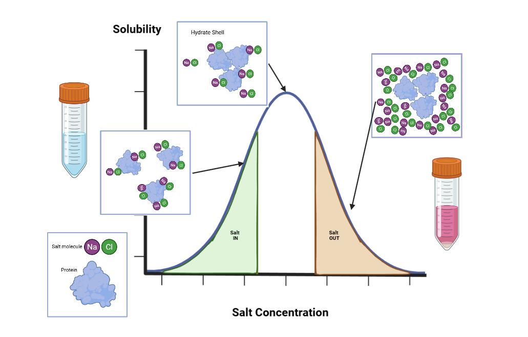
The salting-out method is based on the distinct solubility behaviors of proteins and nucleic acids in high–ionic strength environments (Miller, Dykes, & Polesky, 1988). In the initial lysis step, a detergent such as SDS disrupts cellular and nuclear membranes, while Proteinase K degrades structural and DNA-associated proteins. This combined action releases genomic DNA and other intracellular components into solution (Promega Corporation, n.d.).
Following lysis, the addition of a high-concentration salt solution (commonly saturated NaCl) markedly increases the ionic strength of the mixture. Under these conditions, denatured proteins lose solubility and aggregate, resulting in their precipitation- a process known as “salting out.” In contrast, DNA remains soluble in this high-salt environment. Centrifugation then separates the precipitated proteins as a pellet, leaving the DNA in the supernatant. To recover the DNA, alcohol (typically ethanol or isopropanol) is added to the supernatant. Alcohol reduces the dielectric constant of the solution, allowing nucleic acids to precipitate more readily. Salt ions present in the mixture further assist this process by neutralizing the negatively charged phosphate backbone of DNA. After centrifugation, the DNA forms a stable pellet (Chacon-Cortes & Griffiths, 2014; Promega Corporation, n.d.).
The pellet is subsequently washed with 70% ethanol to remove residual salts and contaminants, then dried and re-dissolved in sterile water or a buffer such as TE. Overall, this method leverages fundamental physicochemical properties, including solubility, ionic interactions, and molecular charge, to isolate high-quality DNA without the need for organic solvents (Miller, Dykes, & Polesky, 1988).
Steps / Protocol of the Salting-Out Method of DNA Extraction
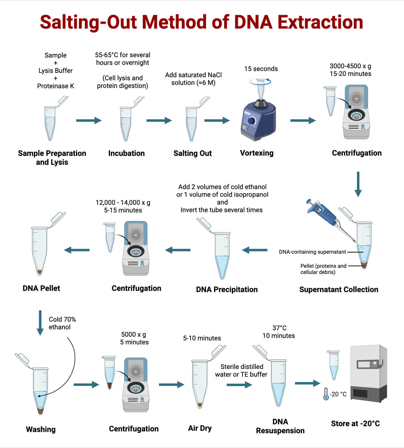
Sample Preparation and Lysis:
- Collect the cellular material you want to extract DNA from. This may include a buffy coat from blood, a tissue homogenate, or a pellet of cultured cells.
- Resuspend the cell pellet in about 3 mL of lysis buffer containing 0.4 M NaCl, 10 mM Tris–HCl (pH ~8.0), 2 mM EDTA, and SDS.
- Add Proteinase K to help break down proteins, including nucleases that could degrade the DNA.
Note: This mixture is then incubated at 55–65°C, often overnight. The warm temperature allows SDS and Proteinase K to fully disrupt cell membranes and digest proteins, ensuring the DNA is completely released into solution (Miller, Dykes, & Polesky, 1988).
Protein Precipitation (Salting-Out):
- After lysis, add an equal volume of saturated NaCl (≈6 M) to the mixture.
- Vortex the tube for about 15 seconds.
- Centrifuge the sample at ~2,500 rpm for 15 minutes to pellet the precipitated proteins and cellular debris.
Note: At this point, the proteins “salt out” and fall to the bottom of the tube, while the DNA stays dissolved in the clear supernatant (Chacon-Cortes & Griffiths, 2014).
DNA Precipitation:
- Carefully transfer the clear supernatant to a fresh tube. Avoid disturbing the protein pellet.
- Add two volumes of cold 100% ethanol (or isopropanol, depending on the variation of the method).
- Some protocols also include sodium acetate to help DNA clump together more efficiently.
- Mix gently by inverting the tube several times. The DNA may form visible threads or a cloudy mass.
- Chill the mixture on ice or at –20°C for a few minutes to improve precipitation.
- Centrifuge at 12,000-14,000g for 5–15 minutes to pellet the DNA.
Note: The DNA pellet will appear as a small, white or translucent mass at the bottom of the tube (Miller, Dykes, & Polesky, 1988).
Washing and Resuspension:
- Discard the supernatant.
- Wash the DNA pellet with cold 70% ethanol to remove leftover salts and impurities.
- Briefly centrifuge again, remove the ethanol, and allow the pellet to air-dry for a short time.
- Avoid over-drying, as this makes DNA difficult to dissolve later.
- Resuspend the purified DNA in sterile distilled water or TE buffer (10 mM Tris, 1 mM EDTA, pH ~8.0) for storage.
Note: TE buffer is often preferred for long-term storage because the EDTA helps protect DNA from nuclease activity (Promega Corporation, n.d.).
Modifications of the Salting-Out Method of DNA Extraction
- Single-Lysis Salting-Out (SLSO): Designed for dried blood spots (DBS), this version reduces the number of steps and minimizes reagent use. It is especially useful in large population studies or when laboratory resources are limited.
- Use of Alternative Detergents or Salts: Some laboratories modify the lysis buffer by substituting different detergents or salts. For example, ammonium acetate can be used instead of NaCl, which has proven useful for extracting DNA from formalin-fixed paraffin-embedded (FFPE) tissue samples.
- Adaptations for Non-Blood or Non-Tissue Samples: The method has been successfully applied to other biological materials, such as milk somatic cells in livestock genetics research, showing how adaptable the technique can be.
- Integration with Additional Clean-Up or Binding-Based Methods: For applications that require higher purity (such as sequencing), salting-out may be followed by a clean-up step using silica- or cellulose-based purification. Many modern nucleic acid purification systems use solid-phase binding after lysis and clearing instead of relying solely on alcohol precipitation.
- Optimization for High-Molecular-Weight (HMW) DNA: Recent studies have shown that the method can produce HMW DNA suitable for next-generation sequencing, even from frozen tissue stored for several years, when the protocol is carefully optimized.
Troubleshooting of the Salting-Out Method of DNA Extraction
| Problem | Likely Cause | Solution |
| Low DNA yield | Incomplete cell lysis or insufficient protein digestion (e.g., too little SDS or Proteinase K, or too short incubation) | Ensure enough lysis buffer is used, check SDS and Proteinase K concentrations, and incubate long enough (often overnight at 55–65°C) |
| Poor DNA purity (protein/salt contamination) | Incomplete removal of precipitated proteins, co-precipitation of proteins with DNA, or insufficient washing | Centrifuge properly to pellet proteins, carefully transfer supernatant, wash DNA thoroughly with 70% ethanol, and repeat salting-out if needed |
| Failure of DNA precipitation (no or tiny pellet) | Old or contaminated alcohol, wrong alcohol volume, or low salt concentration | Use fresh, cold ethanol or isopropanol, ensure correct volume and salt concentration, incubate on ice or at –20°C, and centrifuge at high speed |
| DNA resuspension issues | Over-dried pellet or water with improper pH | Resuspend in TE buffer (pH ~8) or low-salt buffer, and avoid over-drying the pellet |
| Residual salts or inhibitors affecting downstream use | Incomplete ethanol washes or insufficient clean-up | Perform proper ethanol washes; consider additional clean-up steps such as repeated ethanol washes, silica-based purification, or a small phenol/chloroform extraction for challenging samples |
| Problematic samples (tissues with proteins, lipids, polysaccharides) | High levels of interfering compounds | Optimize salt concentration, detergent type, incubation time, and washing steps empirically for best results |
Quality Assessment of the Salting-Out Isolated DNA
- Spectrophotometry (A260/A280 and A260/A230 ratios): Evaluates DNA purity by indicating protein and salt contamination.
- Gel electrophoresis: DNA integrity and fragmentation by visualizing band patterns.
- Downstream functional validation: Confirms DNA suitability for applications such as PCR, digestion, or sequencing.
- Yield measurement: DNA concentration can be determined using spectrophotometry or fluorometric methods.
In modern laboratories, it is standard practice to combine spectrophotometric analysis with agarose gel electrophoresis for a thorough assessment of DNA quality. For high-demand applications, such as long-read sequencing, confirming the presence of HMW DNA is often necessary.
Safety Tips and Precautions of the Salting-Out Method of DNA Extraction
- Handling concentrated salts: Use gloves and eye protection when working with high-molarity salt solutions, such as saturated NaCl, as they can irritate the skin or mucous membranes.
- Use of clean, nuclease-free reagents and equipment: To prevent DNA degradation, use sterile tubes, pipette tips, and reagents that are certified DNase- and RNase-free.
- Ethanol and isopropanol safety: Both alcohols are flammable. Handle them away from open flames and perform precipitation and drying steps in a well-ventilated area or under a fume hood.
- Proper disposal of waste: Dispose of protein pellets and lysates (biological wastes) according to biosafety guidelines.
- Avoid over-drying the DNA pellet: Over-drying can make DNA difficult to dissolve, increasing the risk of shearing or reducing yield.
Storage and Long‑Term Stability of Salting-Out Isolated DNA
- Buffer choice for storage: DNA is best eluted in a buffer such as TE rather than plain water. The EDTA component chelates divalent cations like Mg²⁺, which are required by nucleases, helping protect DNA from enzymatic degradation.
- Aliquoting samples: To avoid repeated freeze–thaw cycles, which can shear DNA and reduce yield, it is more sensible to divide the DNA into smaller aliquots if it will be used multiple times.
- Storage temperature: For long-term preservation, DNA should be stored at –20°C or –80°C, depending on the intended downstream applications. Many laboratories store genomic DNA at –20°C in TE buffer for routine use.
- Handling HMW DNA: When HMW DNA is needed (e.g., for long-read sequencing), minimize handling and freeze–thaw cycles. Resuspend DNA gently to avoid shearing.
Applications of the Salting-Out Method of DNA Extraction
- Genotyping, PCR, and restriction fragment analyses: Suitable for PCR, RFLP, Southern blotting, and other routine molecular assays.
- Plasmid and bacterial genomic DNA extraction: Adaptable for isolating bacterial genomic DNA and plasmids with modified protocols.
- Large-scale population, epidemiological, and forensic studies: Ideal for high-sample-volume studies due to low cost, safety, and scalability.
- Environmental and non-model organism genomics: Effective for extracting high–molecular-weight DNA for NGS-based studies.
- Non-invasive or alternative sample types: Applicable to samples such as milk somatic cells beyond conventional tissues.
- Diagnostics from bodily fluids: Salting-out has been used to extract parasite DNA, for example, Schistosoma mansoni from urine, enabling PCR-based diagnostic applications.
Advantages of the Salting-Out Method of DNA Extraction
- Non-toxic: Does not require hazardous organic solvents, improving laboratory safety and waste handling.
- Cost-effective: Uses inexpensive and easily available reagents, making it suitable for low-resource and large-scale studies.
- Scalable and suitable for high-throughput work: Easily adapted for processing multiple samples simultaneously with standard equipment.
- Versatile: Applicable to blood, tissues, cultured cells, and samples from non-model organisms.
- Yields high-quality DNA: Produces high–molecular-weight genomic DNA suitable for PCR, cloning, and sequencing applications.
Limitations of the Salting-Out Method of DNA Extraction
- Potential co-precipitation of proteins or contaminants: Incomplete removal of proteins, lipids, or salts can interfere with downstream enzymatic reactions.
- Variability in yield and purity across sample types: Complex samples may require additional clean-up steps to obtain high-quality DNA.
- Time-consuming for some sample types: Traditional protocols may involve long incubation steps, slowing overall processing time.
- Less effective for degraded or fixed samples: DNA from FFPE or highly degraded samples may show poor quality and integrity.
- Lower purity compared to column- or bead-based methods: High-sensitivity applications often require further purification.
- Manual handling and potential for variability: Multiple manual steps increase the risk of inconsistency between samples.
Conclusion
The salting-out method continues to be a widely used and reliable technique for genomic DNA extraction since its introduction by Miller, Dykes, and Polesky in 1988. Its main strengths are simplicity, safety, low cost, scalability, and versatility across different sample types. When performed correctly, it produces high-molecular-weight, high-purity DNA suitable for PCR, restriction digestion, sequencing, genotyping, and even next-generation sequencing of non-model organisms.
However, the method has limitations, including potential contamination, variable yield or purity depending on sample type, and sensitivity to procedural details. For challenging samples or applications requiring ultra-pure DNA, additional clean-up or alternative methods may be needed.
Overall, salting-out remains highly relevant in modern molecular biology. With careful optimization, attention to technique, and proper quality control and storage, it provides a safe, adaptable, and cost-effective approach for genomic DNA extraction across a wide range of research contexts.
References
- Miller, S. A., Dykes, D. D., & Polesky, H. F. (1988). A simple salting-out procedure for extracting DNA from human nucleated cells. Nucleic Acids Research, 16(3), 1215. https://doi.org/10.1093/nar/16.3.1215
- Gautam, A. (2022). Isolation of DNA from blood samples by salt-out method. In [DNA and RNA Isolation Techniques for Non-Experts]. Springer. https://link.springer.com/
- El-Ashram, S., & Al-Shaikh, S. (2016). Nucleic acid protocols: Extraction and optimization. https://www.ncbi.nlm.nih.gov/pmc/articles/PMCXXXXXXX
- Daniels, B. N., Nurge, J., Sleeper, O., et al. (2023). Genomic DNA extraction optimization and validation for genome sequencing using the marine gastropod Kellet’s whelk. PeerJ. https://doi.org/10.7717/peerj.16510
- Chacon-Cortes, D., & Griffiths, L. R. (2014). Methods for extracting genomic DNA from whole blood samples: Current perspectives. Dove Medical Press. https://www.dovepress.com/
- Promega Corporation. (n.d.). DNA purification. In DNA Purification Protocols and Applications Guide. https://worldwide.promega.com/resources/guides/nucleic-acid-analysis/dna-purification/
- Rivero, E. R. C., Neves, A. C., Silva‑Valenzuela, M. G., Sousa, S. O. M., & Nunes, F. D. (2006). Simple salting-out method for DNA extraction from formalin-fixed, paraffin-embedded tissues. Pathology – Research and Practice, 202(7), 523–529. https://doi.org/10.1016/j.prp.2006.02.007
- Chauhan, T. (2018, October 21). 10 different types of DNA extraction methods (updated). Genetic Education. https://geneticeducation.co.in/10-different-types-of-dna-extraction-methods-updated/
