Interesting Science Videos
Structure of Rotavirus
- The name rotavirus is derived from the Latin word rota, meaning “wheel,” which refers to the virion’s appearance in negative-stained electron micrographs.
- Rotavirus falls under Reoviridae family and subfamily Sedoreovirinae, which appear more smooth, lacking the large surface projections.
- The virion has icosahedral symmetry and is triple-layered capsid lacking an envelope and measures 70–75 nm in diameter.
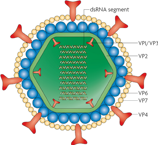
- The capsid consists of 132 capsomers for both inner and outer capsids.
- The outer capsid has two layers, an intermediate layer consisting of the major capsid protein (VP6) and an outer layer that contains the viral attachment protein (VP4) and glycoprotein (VP7).
- The RNA genome consists of 11 segments of double-stranded RNA.
- The genome size is 18,550 bp.
- The virion core contains several enzymes needed for transcription and capping of viral RNAs.
- Rotaviruses are stable to heat at 50°C, to a 3.0–9.0 range of pH, and to lipid solvents, such as ether and chloroform, but they are inactivated by 95% ethanol, phenol, and chlorine.
Genome of Rotavirus
- The RNA genome consists of 11 segments of double-stranded RNA coding for 12 proteins.
- RNA segments codes for 6 structural proteins VP1, VP2, VP3, VP4, VP6, and VP7.
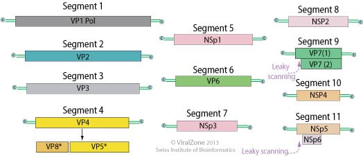
- Outer capsid proteins VP4 and VP7 carry epitopes important in neutralizing activity, with VP7 glycoprotein being the predominant antigen.
- The minor core proteins comprise of VP1 (which functions as the viral polymerase), VP2 (the main scaffolding protein), VP3 (the capping enzyme), VP6 which is the single most abundant rotavirus protein and which interacts with the core protein VP2 and the outer capsid proteins VP7 and VP4.
- The virus genome also encodes for six nonstructural proteins NSP1, NSP2, NSP3, NSP4, NSP5, NSP6.
- The viral mRNAs contain 5′-methylated cap structures but lack polyA tail.
- Instead, rotavirus mRNAs have at their 3′ end a consensus sequence (UGACC) that is conserved in all 11 viral genes.
Epidemiology of Rotavirus
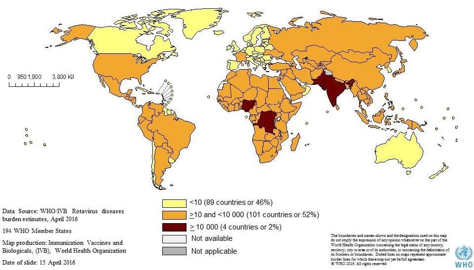
- Rotaviruses are ubiquitous worldwide, with 95% of children infected by 3 to 5 years of age.
- Rotaviruses are one of the most common causes of serious diarrhea in young children worldwide, affecting more than 18 million infants and children and accounting for close to 1600 deaths per day resulting from dehydration.
- In North America, outbreaks occur during the autumn, winter, and spring.
- More severe disease occurs in severely malnourished children.
Replication of Rotavirus
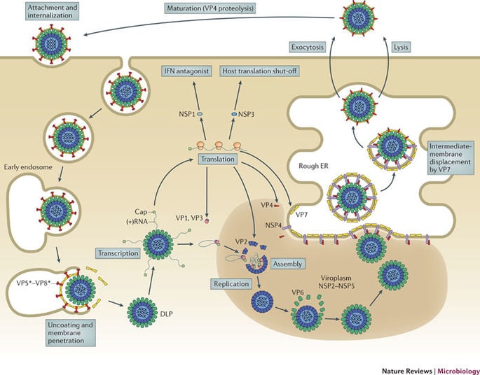
- Attachment of the viral VP4 protein to host receptors mediates clathrin-mediated endocytosis of the virus into the host cell.
- Particles are partially uncoated in endolysosomes (loss of the VP4-VP7 outermost layer) and penetrate into the cytoplasm via permeabilization of host endosomal membrane.
- When crossing the cell membrane, the outer capsid is removed, but the viral particle is never completely uncoated, leaving the viral genomic RNA in the core of the double-layered particle.
- RNA is transcribed by the virion-associated, RNA-dependent RNA polymerase complex to produce messenger RNA (mRNA) molecules from each genomic RNA segment.
- The mRNA molecules, which are capped but not polyadenylated, leave the subviral particle through channels penetrating the outer and inner capsids, and are then translated to generate the various viral proteins.
- The whole replication events occur within electron-dense structures (viroplasms) in the cytoplasm, which are localized adjacent to the nucleus and the endoplasmic reticulum (ER).
- As the particles move towards the interior of the ER cisternae, the transient lipid membrane and the nonstructural protein NSP4 are lost, while the virus surface proteins VP4 and VP7 rearrange to form the outermost virus protein layer, yielding mature infectious triple-layered particles.
- Mature virions are released by lysis of the cell.
Pathogenesis of Rotavirus
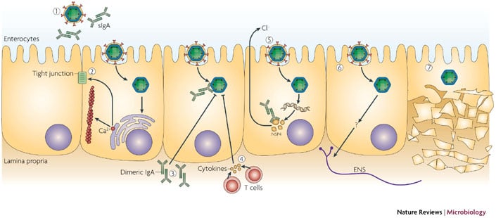
- Rotaviruses infect cells in the villi of the small intestine.
- They multiply in the cytoplasm of enterocytes and damage their transport mechanisms.
- Rotavirus infection prevents the absorption of water, causing a net secretion of water and loss of ions, which together result in watery diarrhea.
- The NSP4 protein of rotavirus acts in a toxin-like manner to promote calcium ion influx into enterocytes, the release of neuronal activators, and a neuronal alteration in water absorption.
- The loss of fluids and electrolytes can lead to severe dehydration and even death if therapy does not include electrolyte replacement.
- Diarrhea caused by rotaviruses may also be due to impaired sodium and glucose absorption as damaged cells on villi are replaced by nonabsorbing immature crypt cells.
- Damaged cells may slough into the lumen of the intestine and release large quantities of viruses, which appear in the stool.
- Viral excretion usually lasts from 2 to 12 days in otherwise healthy patients but may be prolonged in those with poor nutrition and immunocompromised patients.
- Immunity to infection requires the presence of antibody, primarily immunoglobulin A (IgA), in the lumen of the gut.
- Antibodies to the VP7 and VP4 neutralize the virus.
Clinical manifestations of Rotavirus
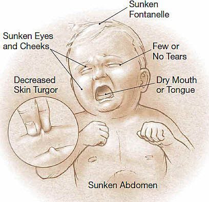
- There is an incubation period of 1–3 days.
- Typical symptoms include watery diarrhea, fever, abdominal pain, and vomiting, leading to dehydration.
- Vomiting is usually of short duration and may occur before or after the onset of diarrhea.
- Diarrhea appears as watery, green or yellow but does not contain mucus.
- Rotavirus diarrhea is self-limiting and patients recover completely within 5-10 days.
- It causes gastroenteritis in adults.
Lab Diagnosis of Rotavirus
- Rotavirus can be demonstrated in stool by Electron microscopy (EM) or Immunoelectron microscopy (IEM).
- EM detects typical 70 nm cartwheel shaped viruses in the stool, however, sensitivity is low.
- IEM utilizes the specific antibody to examine the viral specimens.
- Enzyme immunoassay and latex agglutination tests are used to detect viral antigen directly in the stool.
- Antibody detection by ELISA.
- Genotyping of rotavirus nucleic acid from stool specimens by the polymerase chain reaction (PCR) is the most sensitive detection method.
Treatment of Rotavirus
- Treatment of gastroenteritis is supportive to correct the loss of water and electrolytes that may lead to dehydration, acidosis, shock, and death.
- In selected clinical situations, anti-rotaviral immunoglobulin therapy has been used as prophylaxis against, and as treatment of, rotavirus gastroenteritis.
- The mainstay of therapy consists of oral rehydration with fluids of specified electrolyte and glucose composition.
- Intravenous rehydration therapy is reserved for patients with severe dehydration, shock, or reduced level of consciousness.
Vaccination of Rotavirus
- The rotavirus vaccines that have been developed are live-attenuated, orally administrable strains.
- The first licensed rotavirus vaccine was a Rhesus monkey rotavirus-based tetravalent human reassortant vaccine (RotaShield) which was withdrawn after this live oral vaccine was associated with the development of intestinal intussusceptions.
- Two further live-attenuated oral rotavirus vaccines were developed and extensively evaluated, and have now been recommended by the World Health Organization(WHO).
- Two rotavirus vaccines that are currently licensed for use in infants:
- RotaTeq (RV5) is given in 3 doses at ages 2 months, 4 months, and 6 months
- Rotarix(RV1) is given in 2 doses at ages 2 months and 4 months
Prevention and control of Rotavirus
- The best way to protect a child from rotavirus is with a rotavirus vaccine.
- Hospitalized patients with the disease must be isolated to limit the spread of the infection to other susceptible patients.
- Good hygiene like handwashing and cleanliness are important to control the spread of the disease.

One of the best I’ve read so far. It concise and to the point. I love every bit of the words. Keep it up.
Very brilliant note in fact so helpful to we microbiology students….. Thanks so much for the well done job.
perfect work..am also a masters medical microbiologist researching and specializing in Virology..invite me on medical fora..
Very nice artical
Thanks 🙂
Very beautiful artical. Write another
On other viral infections.
Thank you, 🙂
Good job Mr Sagar Aryal keep it up
Thanks,
Good job, keep it up
Excellent, keep it up