Interesting Science Videos
Reproductive system of Ascaris lumbricoides
- Only sexual reproduction occurs in Ascaris.
- Sexes are separate i.e., dioecious in Ascaris and these roundworms also show distinct sexual dimorphism.
- Males are smaller than the females and they also have a curved tail with pre and post anal papillae, cloaca, and a pair of spicules or penial setae.
- But the female possesses an anus and no spicules and no pre-and post- anal papillae.
- The male system is reduced to a single tube, but the female system is double.
- The gonads are typically long, tubular, and coiled.
- Gonads are attached at the genital pore in females and at the cloaca in the male worms.
Male reproductive organs of Ascaris lumbricoides
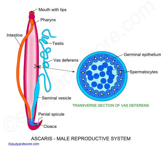
The male reproductive organs are enclosed to the posterior half of the body and consist of the testis, vasa deferens, seminal vesicles, ejaculatory duct, and penial setae.

Image Source: Study and Score.
1. Testis
- It is single. i.e. monorchic but it may be diorchic i.e. 2 testes in some nematodes.
- It is a long, thread-like, coiled tube.
- It continues into a vas deferens.
- Its wall is composed of a single layer of cuboidal cells covered by a basement membrane. It acts as a “growth zone”.
- The central axis of the testis is in the form of cytoplasmic rachis around which clusters of amoeboid sperms are present in various stages of development. In fact, these are developing sperms.
2. Vas deferens
- Testis continues into a short and thick coiled tube of the same diameter, the vas deferens.
- It has a central lumen instead of solid rachis, which differentiate it from the testis.
3. Seminal vesicles
- This is a long, straight, and relatively much thicker tube into which the vas deferens opens.
- It lies in the posterior third of the pseudocoel below the intestine.
- It serves to store the mature sperms.
4. Ejaculatory ducts
- The seminal vesicle narrows at its posterior end to form a short, narrow, and highly muscular and glandular ejaculatory duct.
- This duct opens behind the last part of the rectum from the ventral side to form the cloaca which receives both feces and sperms. It opens out by the cloacal aperture.
- This duct bears a number of prostatic glands whose secretion helps in copulation.
5. Penial setae
- Penial spicules are located in the spicular pouch. Two spicular pouches are situated on the dorsal side of the cloaca. These are basically evaginations of the cloaca.
- Each spicular pouch secretes and houses a club-shaped spicule or penial seta enclosed in a spicular sheath and it consists of a cytoplasmic core surrounded by the thick cuticle.
- Spicules which are 2 to 3.5mm long, can be protruded and retracted through the cloacal aperture by special protractor and retractor muscles.
- The spicules help in opening female gonopore during copulation. Sperms of Ascaris are the tail, asymmetrical and amoeboid.
Female reproductive organs of Ascaris lumbricoides
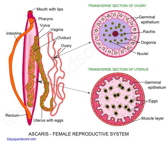
Female reproductive organs lie in the posterior two-thirds of the pseudocoel and consist of ovaries, oviducts, uteri, and vagina. There is an asset of 2 parallel tracts of female reproductive organs, i.e. an ovary, oviduct, and a uterus in one tract in Ascaris like other nematodes; this condition is called didelphic although monodelphic and polydelphic condition are also found.

Image Source: Study and Score.
1. Ovaries
- There are two ovaries.
- They are long, thread-like, and highly twisted tube-like and terminate blindly in the pseudocoel.
- Its wall is made of a single layer of cuboidal epithelial cells lined externally by a basement membrane.
- The central axis of the ovary is in the form of cytoplasmic rachis which is encircled by a group of developing ova.
- There is no lumen in the ovaries.
2. Oviducts
- From the distal end of each ovary, there arises a thick, wider, and twisted oviduct.
- The wall is similar to that of the ovary.
- The major difference is that the cytoplasmic rachis of the ovary is replaced by lumen in the oviducts.
3. Uteri
- Each oviduct leads into a much wider, thicker, and almost untwisted tube, the uterus.
- The uterine wall thick and formed of a layer of tufted secretory cells surrounded by a muscular layer with inner circular and outer oblique fibers.
- Uteri serve to store the eggs after fertilization.
4. Vagina
- At the anterior one-third of the body two uteri combine to form a short and highly muscular tube called a vagina, having an inner lining of the cuticle.
- The wall of the vagina is quite muscular and contractile.
- The vagina opens out by a slit-like female pore or vulva.
Gamete formation in Ascaris lumbricoides
- In Ascaris, the gametogonia are budded off from the proximal end of the gonad.
- As they move towards the distal end of the gonad these gametogonia undergo gametogenesis. This method of formation of a germ cell is called as telogonic and it occurs commonly in most nematodes.
- In a telogonic gonad, the following three zones can be identified:
Germinal zone
- This zone is also known as the proliferation zone.
- This zone lies at the proximal blind end of the gonad.
- Usually, a single large terminal cell constitutes this zone.
- A large apical cell which is an epithelial cell of the gonad wall lies in front of the terminal cell is present.
- From this apical cell, gametogonia are budded.
Growth zone
- It follows the germinal zone.
- In this growth zone, the gametogonia lie attached to the cytoplasmic rachis.
- Here it grows and gets differentiated into amoeboid gametocytes.
Maturation zone
- It is the distal-most zone, where the gametocytes separate from the cytoplasmic rachis to undergo maturation.
- After maturation, gametocytes transform into gametes. The maturation zone is followed by a gonoduct.
The sperms (spermatozoa) are amoeboid while the ova are elliptical in shape. The ova at this stage are actually secondary oocytes that have developed from a single maturation division. The second maturation division occurs after fertilization.
References and Sources
- Kotpal RL. 2017. Modern Text Book of Zoology- Invertebrates. 11th Edition. Rastogi Publications.
- Jordan EL and Verma PS. 2018. Invertebrate Zoology. 14th Edition. S Chand Publishing.
- 11% – https://www.studyandscore.com/studymaterial-detail/ascaris-male-reproductive-system-female-reproductive-system
- 5% – https://www.biologydiscussion.com/invertebrate-zoology/phylum-aschelminthe/ascaris-lumbricoides-habitat-sense-organs-and-life-history/29004
- 3% – https://www.studyandscore.com/studymaterial-detail.php?Id=ascaris-male-reproductive-system-female-reproductive-system
- 2% – https://edurev.in/course/quiz/attempt/-1_Test-Ascaris-Lower-Animal/6d6f162b-7c05-43b1-b808-3dfd2df4968b
- 1% – https://www.sciencedirect.com/topics/medicine-and-dentistry/reproductive-organ
- 1% – https://clinicalgate.com/anatomy-of-the-vagina/
- <1% – https://www.webmd.com/digestive-disorders/digestive-system
