- Pulsed Field Gel Electrophoresis (PFGE) is a technique used for the separation of large deoxyribonucleic acid (DNA) molecules by applying to a gel matrix an electric field that periodically changes direction.
- As DNA larger than 15-20kb migrating through a gel essentially moves together in a size-independent manner, the standard gel electrophoresis technique was unable to separate very large molecules of DNA effectively which led to the practice of pulsed field gel electrophoresis.
- In 1982, Schwartz introduced the concept that DNA molecules larger than 50 kb can be separated by using two alternating electric fields.

Interesting Science Videos
Principle of Pulsed Field Gel Electrophoresis (PFGE)
While in general small fragments can find their way through the gel matrix more easily than large DNA fragments, a threshold length exists above 30–50 kb where all large fragments will run at the same rate, and appear in a gel as a single large diffuse band.
However, with periodic changing of field direction, the various lengths of DNA react to the change at differing rates. That is, larger pieces of DNA will be slower to realign their charge when field direction is changed, while smaller pieces will be quicker. Over the course of time with the consistent changing of directions, each band will begin to separate more and more even at very large lengths. Thus separation of very large DNA pieces using PFGE is made possible.
The Method of Pulsed Field Gel Electrophoresis (PFGE)

- The procedure for this technique is relatively similar to performing a standard gel electrophoresis except that instead of constantly running the voltage in one direction, the voltage is periodically switched among three directions; one that runs through the central axis of the gel and two that run at an angle of 60 degrees either side.
- The pulse times are equal for each direction resulting in a net forward migration of the DNA.
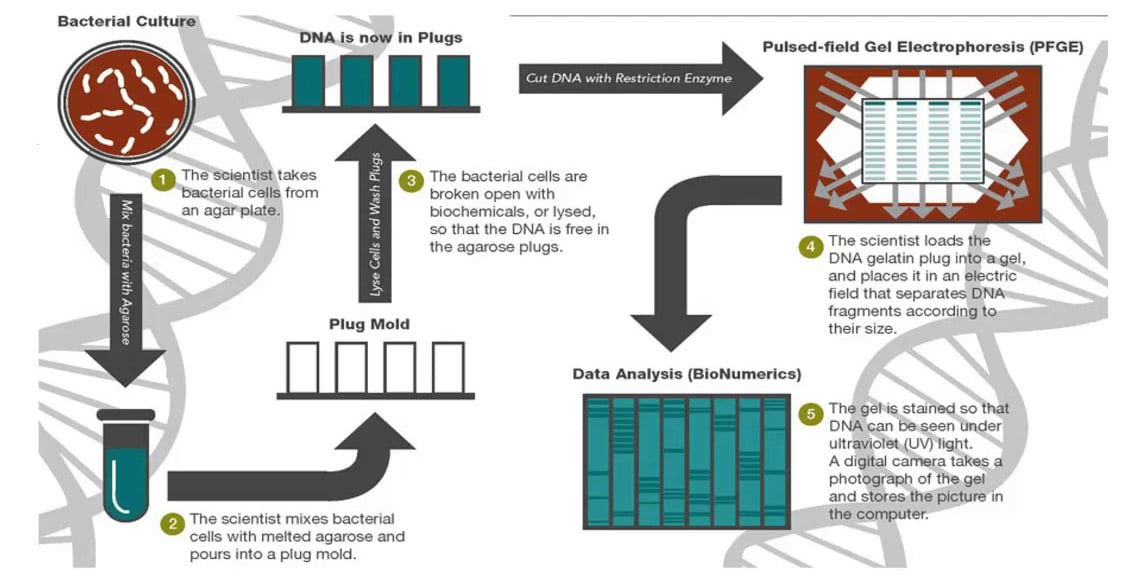
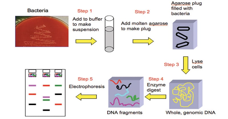
The major steps involved in Pulsed-field gel electrophoresis are:
- Lysis: First, the bacterial suspension is loaded into an agarose suspension. This is done to protect the chromosomal DNA from mechanical damage by immobilizing it into agarose blocks. Then the bacterial cells are lysed to release the DNA. The agarose-DNA suspension is also known as plug mold.
- Digestion of DNA: The bacterial DNA is treated with unusual cutting restriction enzymes so that it yields less number of larger size DNA fragments (in contrast to frequently used restriction enzymes used in RFLP which produces large number of smaller fragments).
- Electrophoresis: The larger pieces of DNA are subjected to pulse field gel electrophoresis by applying electric current and altering its direction at regular intervals (in contrast to the conventional agarose gel electrophoresis done to separate the smaller fragments where the current is applied in a single direction).
- Analysis: The fragments of different organisms generated by PFGE are compared to standards manually or by computer software like BioNumerics.
Applications of Pulsed Field Gel Electrophoresis (PFGE)
- Since, field gel electrophoresis allows the separation of DNA fragments containing up to 100,000 bp (100 kilobase pairs, or kbp), characterization of such large fragments has allowed construction of a physical map for the chromosomes from several bacterial species.
- PFGE may be used for genotyping or genetic fingerprinting.
- It is commonly considered a gold standard in epidemiological studies of pathogenic organisms.
- Subtyping has made it easier to discriminate among strains of Listeria monocytogenes and thus to link environmental or food isolates with clinical infections.
Advantages of Pulsed Field Gel Electrophoresis (PFGE)
- PFGE separates DNAs from a few kb to over 10 Mb pairs.
- PFGE subtyping has been successfully applied to the subtyping of many pathogenic bacteria and has high concordance with epidemiological relatedness.
- PFGE has been repeatedly shown to be more discriminating than methods such as ribotyping or multi- locus sequence typing for many bacteria.
- PFGE in the same basic format can be applied as a universal generic method for subtyping of bacteria. (Only the choice of the restriction enzyme and conditions for electrophoresis need to be optimized for each species.)
- DNA restriction patterns generated by PFGE are stable and reproducible.
Limitations of Pulsed Field Gel Electrophoresis (PFGE)
- Time-consuming.
- Requires trained and skilled technicians.
- Does not discriminate between all unrelated isolates.
- Pattern results vary slightly between technicians.
- Can’t optimize separation in every part of the gel at the same time.
- Don’t really know if bands of the same size are the same pieces of DNA.
- Bands are not independent.
- Change in one restriction site can mean more than one band change.
- “Relatedness” should be used as a guide, not true phylogenetic measure.
- Some strains cannot be typed by PFGE.
References
- Bailey, W. R., Scott, E. G., Finegold, S. M., & Baron, E. J. (1986). Bailey and Scott’s Diagnostic microbiology. St. Louis: Mosby.
- Sastry A.S. & Bhat S.K. (2016). Essentials of Medical Microbiology. New Delhi : Jaypee Brothers Medical Publishers.
- Parija S.C. (2012). Textbook of Microbiology & Immunology.(2 ed.). India: Elsevier India.
- https://www.slideshare.net/pankajgaonkar31/pulse-field-gel-electrophoresis
- https://en.wikipedia.org/wiki/Pulsed-field_gel_electrophoresis
- https://www.slideshare.net/jitenderanduat/pulsed-field-gel-electrophoresis-pfge
- https://bitesizebio.com/29971/pulsed-field-gel-electrophoresis/

Good Compilation, Great work. Keep it up