Interesting Science Videos
What is Pineal Gland?
Definition of Pineal Gland
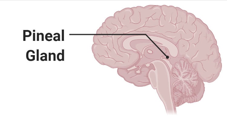
The pineal gland is an endocrine gland present at the geometric center of the brain that is essential in the circadian cycle of sleep and wakefulness of the body.
- Since the gland is present at the posterior area of the cranial fossa in the brain, it is also known as the epiphysis cerebri.
- The pineal gland occurs in all vertebrates, and it has been studied that pineal-like organs can also be found in non-vertebrate animals like insects.
- It is a photo-neuro-endocrine gland that secretes hormones and other compounds like serotonin, melatonin, and N, N-dimethyltryptamine.
- The most important and notable function of the pineal gland is the production of melatonin which is produced in a rhythmic pattern.
- The rhythmic pattern of melatonin production by the pineal gland is often used as a marker of the phase of the internal circadian clock.
- The activity of the gland is influenced by the light received by the retina, which is then converted from the neural input into endocrine output by the pineal gland.
- The pineal gland has also been called ‘The Third Eye’ due to the histological similarities between the pineal gland and the lateral eyes of amniotic vertebrates.

Structure of Pineal Gland
- The pineal gland is a tiny pine cone-shaped gland that hangs from the roof of the third ventricle of the brain.
- The pineal gland is a secretory neuroendocrine organ that is highly vascularized and weighs about 100-150 mg.
- The size of the pineal gland in vertebrates is associated with the environment, and geographical locations as the glands tend to be larger in the vertebrates living in harsh environments.
- The secretory cells of the pineal gland are called pinealocytes that are arranged in the form of compact cords and clusters.
- In between the cells are calcerous bodies that are prone to calcification, the risk of which increases with age.
- The calcareous deposits are also called acervuli and are used as the most distinguishable radiographic characteristics of the pineal gland.
- The deposits are assumed to be formed by the combination of the polypeptide secreted by the pinealocytes and calcium accumulated interstitially.
- The pinealocytes in humans have a prominent nucleus and a granular cytoplasm. The cytoplasm also contains cytoplasmic processes that terminate into fenestrated capillaries.
- Besides, the extracellular space of the gland is occupied by neuroglia that surrounds pinealocytes and peripheral patches.
Hormones of Pineal Gland
- The exact function and secretions of the gland are still unknown; however, the most important secretory product produced by the gland is melatonin.
- Melatonin is an amine-derived hormone formed from serotonin. The release of melatonin by the pineal gland occurs in a rhythmic pattern where the levels rise and fall depending on the diurnal cycle.
- The synthesis of melatonin is stimulated by darkness by utilizing the post-ganglion beta-adrenergic sympathetic fibers of the cervical sympathetic ganglia.
- The precursor of melatonin is tryptophan which is hydroxylated by the pinealocytes into 5-hydroxytryptophan in the presence of hydroxylase.
- The most important effect of melatonin is to coordinate with the diurnal rhythms in the body by stimulating the hypothalamus.
- Besides, several studies have indicated that melatonin can have antigonadotrophic effects in children.
- Other effects of the hormone include downregulation of thyroid secretion, hypothermic, sleep induction, and hypotension by stimulating norepinephrine levels.
Functions of Pineal Gland
The following are some of the functions of the pineal gland;
- The hormone produce by the gland is essential for the regulation of the circadian or diurnal cycle of the body.
- The gland functions as a mediator between the nervous system and the endocrine system as the ganglion on the gland help to convert the photo input from the eyes into neural output.
- In the case of other mammals like rodents, the pineal gland influences the action of different drugs like antidepressants and cocaine.
Diseases and Disorders of Pineal Gland
The following are some of the diseases and disorders associated with the pineal gland;
Calcification
- Calcification of the pineal gland is a common condition, resulting from the deposition of calcium and phosphate in the cytoplasm of the gland.
- The concentration and degree of calcification depend on the age of the individual and increases with the increase in age.
- A correlation has also been established between the function of the pineal gland and conditions like migraine and headaches.
Tumors
- Tumors can form on the pineal gland, which, if it reaches the hypothalamus, can cause weakness and loss of sensation in most parts of the body.
- The diagnosis of pineal tumors is crucial for the treatment of the condition. An MRI can be performed in order to detect the location and size of the tumor.
- Surgical removal of the tumor is the most effective means of treatment of pineal tumors.
References
- Hall JE and Guyton AC. (2011) Textbook of Medical Physiology. Twelfth Edition. Elsevier Saunders.
- Waugh A and Grant A. (2004) Anatomy and Physiology. Ninth Edition. Churchill Livingstone.
- Marieb EN and Hoehn K. (2013) Human Anatomy and Physiology. Ninth Edition. Pearson Education, Inc.
- Rastogi SC. (2007) Essentials of Human Physiology. Fourth Edition. New Age International Limited.
- Ilahi S, Beriwal N, Ilahi TB. Physiology, Pineal Gland. [Updated 2020 May 4]. In: StatPearls [Internet]. Treasure Island (FL): StatPearls Publishing; 2021 Jan-. Available from: https://www.ncbi.nlm.nih.gov/books/NBK525955/
- Aulinas A. Physiology of the Pineal Gland and Melatonin. [Updated 2019 Dec 10]. In: Feingold KR, Anawalt B, Boyce A, et al., editors. Endotext [Internet]. South Dartmouth (MA): MDText.com, Inc.; 2000-. Available from: https://www.ncbi.nlm.nih.gov/books/NBK550972/
- Gheban, Bogdan Alexandru et al. “The morphological and functional characteristics of the pineal gland.” Medicine and pharmacy reports vol. 92,3 (2019): 226-234. doi:10.15386/mpr-1235
- Tan, Dun Xian et al. “Pineal Calcification, Melatonin Production, Aging, Associated Health Consequences and Rejuvenation of the Pineal Gland.” Molecules (Basel, Switzerland) vol. 23,2 301. 31 Jan. 2018, doi:10.3390/molecules23020301
- Macchi MM, Bruce JN. Human pineal physiology and functional significance of melatonin. Front Neuroendocrinol. 2004 Sep-Dec;25(3-4):177-95. doi: 10.1016/j.yfrne.2004.08.001. PMID: 15589268.
- Booth FM. The human pineal gland: a review of the “third eye” and the effect of light. Aust N Z J Ophthalmol. 1987 Nov;15(4):329-36. doi: 10.1111/j.1442-9071.1987.tb00092.x. PMID: 3435677.
