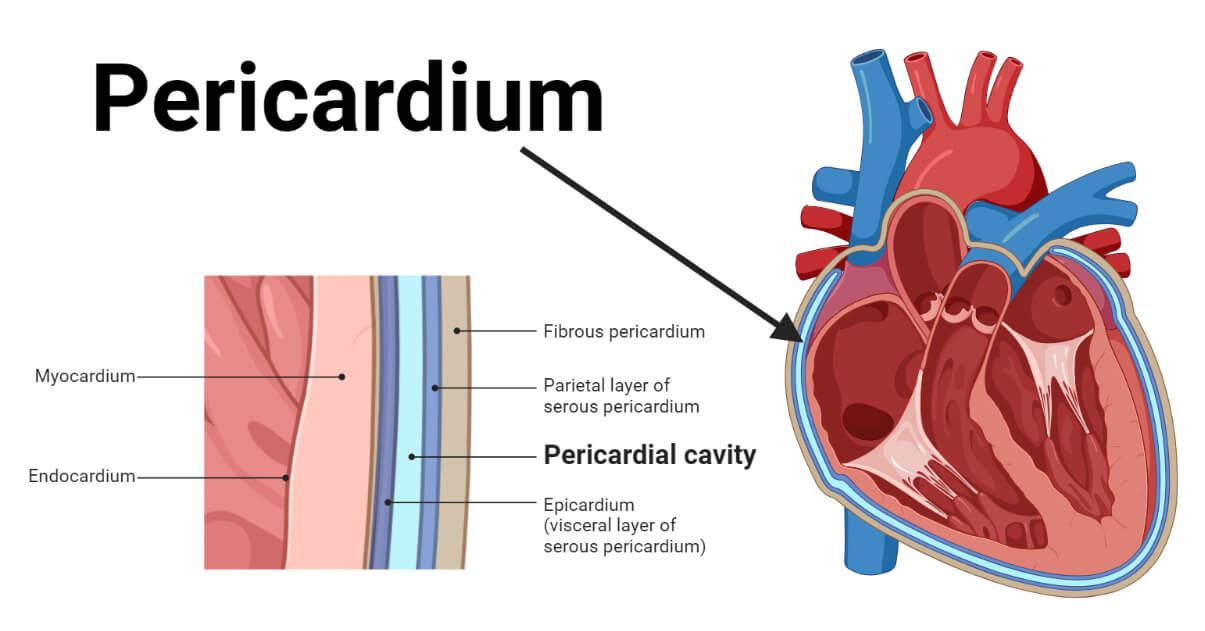
The pericardium is a double-layered sac that surrounds the heart externally. It is a double-walled sac-like structure; hence, it is also called the pericardial sac.
- The pericardium has two layers separated by a space called the pericardial space containing pericardial fluid. The outer layer is called the fibrous pericardium, and the inner layer is called the serous pericardium. It defines the middle mediastinum – a space where the heart is located and provides protection and lubrication to the heart.
- Pericardium Location: The pericardium is located in the thoracic cavity, behind the sternum, and just ahead of the spine. It externally encloses the heart and the roots of the aorta, the main pulmonary vein, the main pulmonary artery, and the two vena cava.
Interesting Science Videos
Pericardium Structure
Histologically, the pericardium is mainly made of outer fibrous and elastic connective tissue and inner mesothelial cells.
Anatomically, the pericardium is divided into two layers, the fibrous pericardium and the serous pericardium. Its thickness varies from region to region; according to cardiac magnetic resonance imaging, it is usually about 1.5 to 2.0 mm thick.

a. Fibrous Pericardium
It is the relatively inelastic outer tough layer made of connective tissues with collagen and elastin fibers. This layer provides toughness and structural support to the pericardium. It is connected to the central tendon of the diaphragm, the posterior part of the sternum, cervical fascia, and is blended with the tunica externa of the great vessels. Several pericardial ligaments affix it to the thorax. This connection secures its location and provides strong anchorage to the heart in the mediastinum.
b. Serous Pericardium
It is the inner thin double-layered membrane directly in contact with the heart. It is primarily composed of mesothelial cells with numerous microvilli and cilia.
- Parietal Serous Pericardium
It is the outer layer of the serous pericardium that lines the fibrous pericardium and marks the outer boundary of the pericardial cavity.
- Visceral Serous Pericardium
It is the inner thin mesothelial cell monolayer that adheres firmly to the epicardium.
- Pericardial Cavity
In between the parietal and the visceral layers of the serous pericardium, there is a fluid-filled space called the pericardial cavity. It contains up to 50 mL of serous fluid, called the pericardial fluid.
Pericardium Functions
- It provides toughness and mechanical protection to the heart.
- It provides constraint to ventricular expansion i.e. expansion of the heart.
- It also acts as a barrier against infection of the heart.
- It equalizes gravitational, hydrostatic, and inertial forces.
- The pericardial fluid reduces friction over the heart’s surface.
- The mesothelial cells of the serous pericardium generate prostacyclins and other chemicals that regulate sympathetic neurotransmission, fibrinolysis, and epicardial coronary arterial tone.
Pericardial Diseases
The pericardium is affected by various medical conditions known as pericardial diseases. Some common types of pericardial diseases are tabulated below:
| Disease | Introduction |
| Pericarditis | Inflammation of the pericardium. |
| Pericardial Effusion | Accumulation of excessive fluid in the pericardial sac. |
| Cardiac tamponade | A condition arises due to excessive pericardial effusion forcing the heart to compress resulting in decreased cardiac output. |
| Constrictive pericarditis | A condition arises due to excessive pericardial effusion forcing the heart to compress, resulting in decreased cardiac output. |
References
- Hoit, Brian D. (2017). Anatomy and Physiology of the Pericardium. Cardiology Clinics, 35(4), 481–490. doi:10.1016/j.ccl.2017.07.002
- Pericardium—Anatomy and Function (thoughtco.com)
- The Pericardium – TeachMeAnatomy
- Pericardium: Anatomy, Function, and Treatment (verywellhealth.com)
- What is the function of the pericardium? – eNotes.com
- Pericardium: Function and Anatomy (clevelandclinic.org)
- Pericardium: Function, Role in the Body, and Associated Conditions (healthline.com)
- Pericardium. (2023, May 21). In Wikipedia. https://en.wikipedia.org/wiki/Pericardium
- Willner DA, Goyal A, Grigorova Y, et al. Pericardial Effusion. [Updated 2023 Feb 20]. In: StatPearls [Internet]. Treasure Island (FL): StatPearls Publishing; 2023 Jan-. Available from: https://www.ncbi.nlm.nih.gov/books/NBK431089/
- Stashko E, Meer JM. Cardiac Tamponade. [Updated 2022 Aug 8]. In: StatPearls [Internet]. Treasure Island (FL): StatPearls Publishing; 2023 Jan-. Available from: https://www.ncbi.nlm.nih.gov/books/NBK431090/
- Yadav NK, Siddique MS. Constrictive Pericarditis. [Updated 2023 Feb 13]. In: StatPearls [Internet]. Treasure Island (FL): StatPearls Publishing; 2023 Jan-. Available from: https://www.ncbi.nlm.nih.gov/books/NBK459314/
