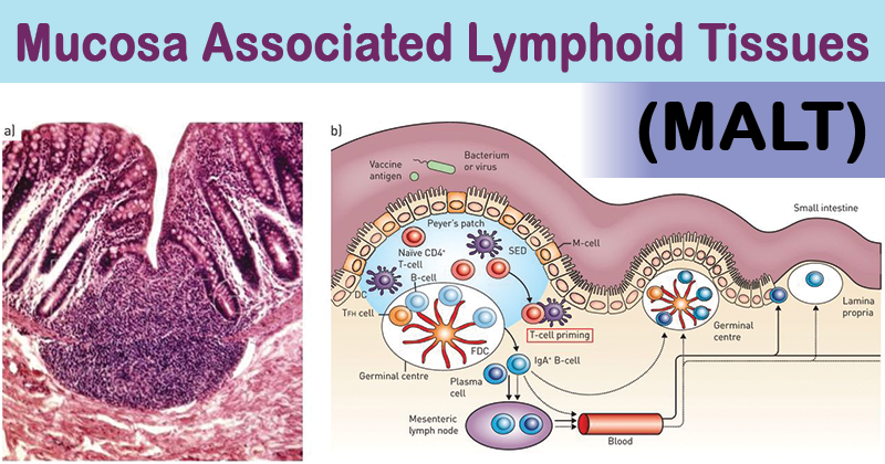
- The main sites of entry for microbes into the body are through mucosal surfaces.
- Hence, the majority (>50%) of lymphoid tissue in the human body is located within the lining of the respiratory, digestive and genitourinary tracts.
- Small concentrations of lymphoid tissue are also found in thyroid, breast, lung, salivary glands, eye, and skin.

Image Source: DOI: 10.1183/13993003.01701-2015
- These lymphoid tissues collectively are thus referred to as mucosa associated lymphoid tissues (MALT). Examples include tonsils, the Peyer patches within the small intestine, and the vermiform appendix.
- Depending upon their location, they can be sub-divided into Nasal-associated lymphoid tissue (NALT), Gut-associated lymphoid tissue (GALT), Bronchus- associated Lymphoid Tissue (BALT) and lymphoid tissue associated with the genitourinary system.
Interesting Science Videos
Structure of MALT
All mucosal lymphoid tissues although present at various sites, contain the same basic compartments-follicles, interfollicular regions, subepithelial dome regions, and follicle-associated epithelium.
- Nasal-associated lymphoid tissue (NALT)
- It includes immune cells underlying the throat and nasal passages and especially the tonsils.
- Their structure is similar to that of lymph nodes but they are not encapsulated and are without lymphatics.
- It consists of follicles composed mainly of B cells surrounded by T cells and the germinal center.
- Within the follicle is the site of antigen-dependent B cell proliferation.
- Antigens and foreign particles are trapped within the deep crypts of their lympho-epithelium from where they are transported to the lymphoid follicles.
- Gut-associated lymphoid tissue (GALT)
- It is composed of lymphoid complexes that consist of specialized epithelium, antigen-presenting cells and intraepithelial lymphocytes.
- These structures occur strategically at specific areas in the digestive tract for example Peyer’s patches in the terminal ileum.
- The specialized epithelial cells are called as M cells which are intimately associated with antigen presenting cells (APCs) which together carry out antigen sampling in the gut. M cells sample antigen from the lumen and deliver it to the lymphoid tissue through APCs.
- Bronchus- associated lymphoid tissue (BALT)
- The lymphoid tissue associated with the bronchus (BALT) is structurally similar to Peyer’s patches and other lymphoid tissues of the gut.
- It is composed mainly of aggregates of lymphocytes organized into follicles that are found in all lobes of the lung and along the main bronchi.
- The majority of lymphocytes in the follicles are B cells.
- Antigen sampling is carried out by epithelial cells lining the surface of the mucosa and by way of M cells which transport antigens to underlying APCs and lymphocytes.
Functions of MALT
- The mucosa-associated lymphoid tissue initiates immune responses to specific antigens encountered along all mucosal surfaces.
- Diffuse lymphoid tissue along all mucosal surfaces are the sites of IgA transport across the mucosal epithelium.
- The primary role of GALT is to protect the body against microbes entering the body via the intestinal tract.
- M cells take up foreign molecules and pass them to underlying APCs, which present them in the context of class I and class II MHC molecules to T cells. The helper T cells help to activate B cells and both T and B cells can migrate to other parts of the GI tract (including salivary glands) and other MALT sites, e.g. lactating mammary glands and respiratory and genitourinary tracts, and protect these surfaces from invasion by the same microbes.
- The location of nasal and bronchus associated lymphoid tissues in the airway suggest that they are directly involved in handling airborne microbes.
- MALT tissues comprise the mucosal immune system which can function independently of the systemic immune system and are, therefore, an important aspect of immunity.
References
- Cesta, M. (2006). Normal structure, function, and histology of mucosa associated lymphoid tissue. Toxicol Pathol , 34 (5), 599-608.
- Lydyard, P.M., Whelan,A.,& Fanger,M.W. (2005).Immunology (2 ed.).London: BIOS Scientific Publishers.
- Tuitui, R., & Suwal, D. S. (2010). Human Anatomy and Physiology. Kathmandu: Vidyarthi Prakashan.

Thank you so much! it’s super helpful
How can we get PDF form of these valuable topics..? Or download process please.
You need to copy paste the content, Thanks !!!