Interesting Science Videos
Definition of Monocot Leaves
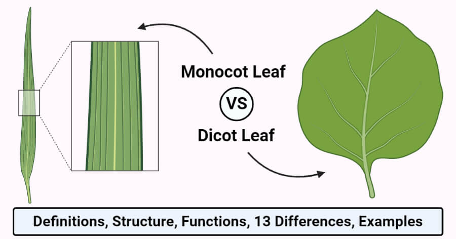
Monocotyledonous leaves are narrow and elongated with parallel venation, which is often used to distinguish monocotyledonous plants from dicots. Monocot leaves are isobilateral as both the surfaces of the leaves are similar to the same coloration.
- The primordial monocot leaves consist of a proximal leaf base or hypophyll and a distal hyperphyll. The hyperphyll is the dominant part of the leaf in dicots, but in monocots, the hypophyll acts as the dominant structure.
- The leaves are narrow and linear with a sheath covering around the stem at the base, but there are many exceptions within monocots that might not have similar structures.
- The venation, as mentioned, is of the striate type which is mostly longitudinally striate and sometimes, palmate-striate or pinnate-striate.
- The veins on the leaf surface emerge at the base of the leaf and move together to the apex.
- Most monocotyledonous plants consist of a single leaf per node because the leaf base occupies more than half of the circumference of the plant stem.
- The presence of a larger leaf base has been attributed to the differences in the development of the stem during zonal differentiation.
Definition of Dicot Leaves
Dicotyledonous leaves are usually rounded with reticulate venation that can be distinguished from monocotyledonous leaves in their structure and anatomy. A typical dicot leaf consists of a lead blade which is also called the lamina. The lamina is the widest part of a leaf.
- Dicot leaves are dorsoventral as the dorsal and ventral part of the leaves can be differentiated based on the coloration on the leaves. The dorsal side of the leaf is usually more pigmented than the ventral side.
- Dicot leaves are attached to the stem via a petiole which distinguished them from monocot leaves that are directly attached to the stem.
- Small green appendages called stipules might be present at the base of the petiole in some dicot leaves.
- Dicot leaves have a midrib that runs through the leaf blade and travels the length of the leaves. Many branches develop on either side of the midrib giving rise to the reticulate venation.
- The number of leaves on a node in the stem depends on the species, but dicots usually have two or more leaves arising from a single node.
- Dicot leaves can be further divided into different groups on the basis of the leaf form as some are simple leaves while others are compound.
Structure of Monocot and Dicot Leaves
The internal structure or anatomy of both monocot and dicot leaves can be expressed on the basis of the following structures;
1. Epidermis
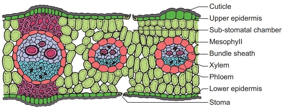
- The epidermis is the outermost tissue in leaves that is composed of a compact layer of thin-walled barrel-shaped cells. The epidermis is present both on the upper and lower part of the leaf.
- The epidermis is surrounded by an outer waxy layer of the cuticle which functions to protect the leaf and prevent water loss.
- The cuticle on the upper layer of the leaf is thicker than that on the lower surface in dicot leaves as the leaves are dorso-ventral. In monocots, however, the layer of the epidermis is about the same thickness on both surfaces.
- The epidermis is essential as it prevents the loss of excess water as it is a waxy waterproof layer. Besides, it also plays an important role in gas exchange as it contains tiny pores called stomata.
- The number of stomata is equal on both surfaces in the case of monocot leaves. The stomata are more on the lower epidermis than on the upper epidermis in dicot leaves.
- Stomata is a small opening present between the bean-shaped cells called the guard cells that regulate the size of the stomata. The guard cells are dumb-bell shaped in dicots.
- The epidermal cells do not have chloroplasts, but the guard cells contain chloroplasts that give the characteristic green color to the leaves.
- Besides the epidermal cells and the guard cells, the epidermis also contains other cells called subsidiary cells that are usually present around the guard cells.
- In the case of dicot cells, the upper epidermis contains large thin-walled cells called bulliform cells or motor cells. These cells assist in the rolling of the leaves in response to weather change.
- In monocot leaves, some epidermal cells are filled with silica, which is called silica cells.

Figure: T.S. of a monocot leaf (grass). Image Source: BrainKart.
2. Mesophyll
- The mesophyll is the ground tissue of leaves present between the upper and lower epidermis of the leaves.
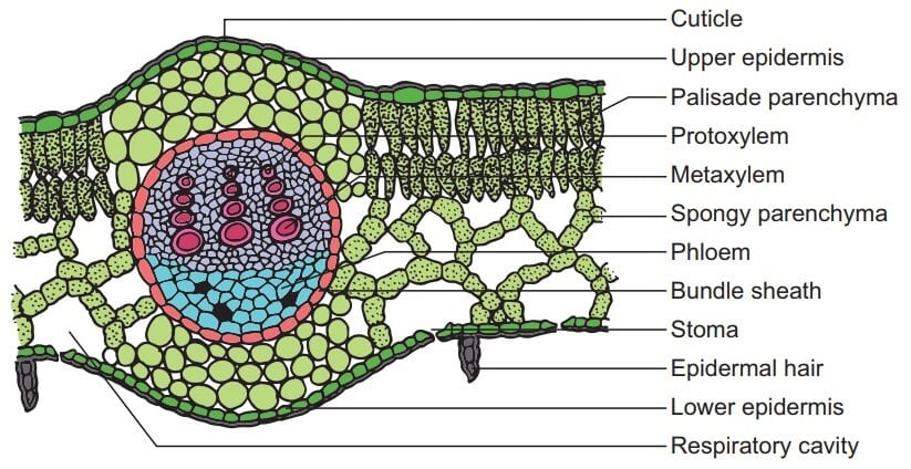
- The mesophyll is differentiated into palisade parenchyma, and spongy parenchyma in dicot leaves both no such differentiation can be found in monocot leaves.
- The palisade parenchyma is present directly below the upper epidermis and is composed of vertically elongated cylindrical cells in one or more layers. The cells are compactly placed with no intercellular spaces.
- The cells of the palisade parenchyma contain more chloroplasts than the cells of the spongy parenchyma.
- The spongy parenchyma is present below the palisade parenchyma where the cells are irregularly shaped. These cells have fewer chloroplasts than the palisade parenchymal cells.
- The cells of the spongy parenchyma are loosely arranged with numerous spaces, thus the name. These air spaces facilitate the exchange of gases between the cells. The spaces present next to the stomata is called respiratory cavities or sub-stomatal cavities.
- The mesophyll of the dicot leaves is not differentiated into palisade and spongy parenchyma and is composed of isodiametric and thin-walled cells. The cells are compactly arranged with some intercellular air spaces.

Figure: T.S. of a dicot leaf (sun-flower). Image Source: BrainKart.
3. Vascular Bundles
- The innermost layer of tissues in plant leaves is the vascular bundle which is present underneath the mesophyll and along the veins of the leaves.
- All the vascular bundles are not the same size, and the larger bundles occur at regular intervals along the veins. The large vascular bundles have two patches of sclerenchyma cells above and below.
- The vascular bundles of the leaves are a part of the vascular system of the plant that extends through different organs, encompassing the entire plant.
- The vascular bundles in leaves display highly divergent patterns in terms of size and position.
- In dicot leaves, the veins are of different size orders forming a highly branched network. The vascular bundles present underneath such veins are thus, diverse.
- In monocot leaves, the longitudinal veins lie parallel along the leaf blade and are connected transversally by tiny commissural veins, which results in less diverse vascular bundles.
- The vascular bundles are conjoint, collateral, and closed and are surrounded by a bundle sheath. The bundle sheath in dicot leaves is parenchymatous, but the sheath of dicot leaves in sclerenchymatous.
- The xylem tissue in the vascular bundle is present towards the upper epidermis, whereas the phloem is present towards the lower epidermis.
- The xylem consists of vessels and xylem parenchyma. The xylem in monocot leaves is differentiated into metaxylem and protoxylem. The phloem consists of sieve cells, companion cells, and phloem parenchyma.
Functions of Monocot and Dicot Leaves
The functions of leaves are mostly similar in both dicot and monocot plants. The functions of leaves depend on the plant species, their environment, and their age. The following are some of the functions of monocot and dicot plants;
- The most important function of leaves in green plants is the preparation of food by the process of photosynthesis. The cells in the leaves contain chlorophyll as one-fifth of the cells in the mesophyll contain chlorophyll-containing chloroplasts. The large, broad surface of the leaves allows absorption of a larger amount of sunlight which is essential for photosynthesis.
- The epidermis and cuticle of the leaves prevent excess loss during transpiration which protects the plant from drying up.
- The leaves consist of stomata which are an essential structure for the movement of water from the plant into the atmosphere via the transpiration process. This is important in order to draw water with minerals from the soil by the roots.
- In addition to facilitating transpiration, stomata are also involved in the exchange of gases. The stomata take in carbon dioxide from the air and give out oxygen during photosynthesis.
- In some plants like cabbage and lettuce, the food prepared by the cells in the leaves is stored in the leaves in different forms.

Monocot vs. Dicot Leaves (13 Key Differences)
| Characteristics | Monocot leaves | Dicot leaves |
| Definition | Monocotyledonous leaves are narrow and elongated with parallel venation, which is often used to distinguish monocotyledonous plants from dicots. | Dicotyledonous leaves are usually rounded with reticulate venation that can be distinguished from monocotyledonous leaves in their structure and anatomy. |
| Shape | Monocot leaves are narrow, slender, and longer than dicot leaves. | Dicot leaves are broad and relatively smaller than monocot leaves. |
| Symmetry | Monocot leaves are isobilateral in symmetry. | Dicot leaves are dorsoventral as the upper and lower surfaces of the leaves are distinguished. |
| Venation | Monocot leaves have parallel venations as the longitudinal veins run along the length of the leaf that is connected by tiny commissural veins. | Dicot leaves have reticulate venation consisting of veins of different sizes connected to form a complex network. |
| Stomata | The number of stomata on the upper and lower surfaces of the leaves is equal, and thus monocot leaves are also termed amphistomatous. | Dicot leaves contain more stomata on the lower surface than the upper surface. Some dicot leaves do not have any stomata on the upper surface, and such plants are termed hypostomatous. |
| Guard cells | The guard cells in the monocot leaves are dumb-bell shaped. | The guard cells in the dicot leaves are kidney-shaped. |
| Intercellular spaces | Monocot leaves have smaller intercellular spaces as the cells are compactly arranged. | Dicot leaves have larger intercellular spaces as the cells are loosely packed. |
| Vascular bundles | Both large and small vascular bundles occur in the monocot leaves. | Dicot leaves contain larger vascular bundles. |
| The xylem of monocot leaves is differentiated into metaxylem and protoxylem. | The xylem in dicot leaves is not differentiated into metaxylem and protoxylem. | |
| The bundle sheath of the monocot leaves is sclerenchymatous. | The bundle sheath of the dicot leaves is parenchymatous. | |
| Epidermis | Epidermal cells of monocot leaf have heavy deposition of silica. | Epidermal cells of dicot leaf do not have silica deposition. |
| The epidermis of monocot leaves has bulliform or motor cells. | The epidermis of dicot leaves doesn’t have bulliform or motor cells. | |
| Mesophyll | The mesophyll of monocot leaves is not differentiated. | The mesophyll of dicot leaves is differentiated into spongy mesophyll and palisade mesophyll. |
Examples of Monocot Leaves
1. Maize leaves
- Maize leaves are considered the most typical monocot leaves with simple and orderly structures.
- A typical maize plant contains about 20 leaves that might be present in different stages of development. A mature maize leaf is about 70 cm long and 8 cm wide.
- A maize leaf is divided into three regions; an upper blade, lower sheath, and auricle. The leaf cells are highly oriented and aligned to form parallel lines and elongated shape.
- The leaf blade of maize leaf contains adaxial and abaxial epidermal tissue that encloses the mesophyll and vascular tissue.
- The cells of the epidermis of maize leaf are divided into two types; specialized cells and nonspecialized intercostals cells.
- Specialized cells consist of stomatal complexes and three types of hair cells; large macrohairs, microbars, and bicellular hairs. These cells serve a protective function.
2. Grass
- A grass leaf is a monocot leaf with an elongated structure that arises from the node consisting of a basal cylindrical sheath encircling the stem and other younger leaves.
- The sheaths on the outer surface are hollow cylinders that split down on one side. These sheaths usually form overlapping structures.
- The leaf contains an auricle that might exist either as an ear-like projection or may be reduced to a hairy edge present at the base of the leaf blade.
- The epidermis of a grass leaf consists of specialized hair cells that protect the plant against various harmful agents.
- The shape, texture, and hairiness of the leaves are often used as diagnostic features for the differentiation of grass as they are variable within species or even within the same plant.
Examples of Dicot Leaves
1. Mustard leaves
- Mustard is a typical dicot plant commonly used for studies regarding dicotyledonous plants.
- Mustard leaves begin growing within 4 weeks when the leaves reach 6-8 inches in length. A mature mustard leaf measures about 15-18 inches in length in about 6 weeks.
- The leaves are green and broad with reticulate venation. The leaves are dorsoventrally flattened, and the ventral surface is lighter than the dorsal surface.
- The epidermis of mustard leaves contains few specialized hair cells that serve as protection against water loss and harmful agents.
- Mustard leaves are used as vegetables as they contain a nutritional value for human health.
2. Mint leaves
- Mint is a fast-growing plant consisting of round lanceolate leaves that are arranged in opposing pairs on the stem.
- The leaves are small in size ranging between 4-5 inches in length. The size of the leaves, however, depends on the age of the leaves and their maturity.
- The leaves have reticulate venation with a central midrib from which branched veins arise.
- The surface of the leaves is covered with tiny hairs that are present both on the upper and lower surface of the leaves.
- Mint leaves are aromatic with a characteristic mint odor. These leaves are used as species in different food in different cultures.
References and Sources
- Lee Yuen Lew (2000) Storage Organs and Plant Growth, Science Activities, 37:2, 39-46, DOI: 10.1080/00368120009603566
- Nelissen H, Gonzalez N, Inzé D. Leaf growth in dicots and monocots: so different yet so alike. Curr Opin Plant Biol. 2016 Oct;33:72-76. doi: 10.1016/j.pbi.2016.06.009. Epub 2016 Jun 23. PMID: 27344391.
- Sylvester A.W., Smith L.G. (2009) Cell Biology of Maize Leaf Development. In: Bennetzen J.L., Hake S.C. (eds) Handbook of Maize: Its Biology. Springer, New York, NY. https://doi.org/10.1007/978-0-387-79418-1_10
- 2% – https://courses.lumenlearning.com/boundless-biology/chapter/leaves/
- 1% – https://vivadifferences.com/monocot-leaf-vs-dicot-leaf/
- 1% – https://nph.onlinelibrary.wiley.com/doi/full/10.1111/j.1469-8137.2004.01191.x
- 1% – https://en.mimi.hu/biology/lower_epidermis.html
- 1% – https://de.answers.yahoo.com/question/index?qid=20060909022759AAG460M
- 1% – https://botit.botany.wisc.edu/botany_130/anatomy/Leaf/Leaf.html
- 1% – http://mahatmaacademy.com/uncategorized/ncert-botany-anatomy-flowering-plant-online-model-paper/
- <1% – https://www.visiblebody.com/learn/biology/monocot-dicot/leaves
- <1% – https://www.vedantu.com/biology/difference-between-monocot-and-dicot-leaf
- <1% – https://www.thoughtco.com/plant-stomata-function-4126012
- <1% – https://www.sciencedirect.com/science/article/pii/S167420521500204X
- <1% – https://www.sciencedirect.com/science/article/pii/0020740381900539
- <1% – https://www.quora.com/What-is-the-arrangement-of-leaves-on-a-stem
- <1% – https://www.medicinenet.com/epidermis/definition.htm
- <1% – https://www.differencebetween.com/difference-between-upper-and-vs-lower-epidermis/
- <1% – https://www.diffen.com/difference/Dicot_vs_Monocot
- <1% – https://www.britannica.com/science/mesophyll
- <1% – https://www.bioexplorer.net/epidermis-cells.html/
- <1% – https://www.answers.com/Q/Why_do_palisade_mesophyll_cell_contains_more_chloroplast_than_the_spongy_mesophyll_cell
- <1% – https://www.answers.com/Q/What_is_the_function_of_the_leaf_sheath_in_the_monocotyledon_plant
- <1% – https://wikimili.com/en/Monocotyledon
- <1% – https://vivadifferences.com/difference-between-protoxylem-and-metaxylem/
- <1% – https://study.com/academy/lesson/structure-of-leaves-the-epidermis-palisade-and-spongy-layers.html
- <1% – https://socratic.org/questions/59a2a6de11ef6b48d6fdf17b
- <1% – https://samacheerkalvi.guide/samacheer-kalvi-10th-science-guide-chapter-12/
- <1% – https://quizlet.com/220346710/leaf-characteristics-flash-cards/
- <1% – https://quizlet.com/169169427/epidermis-flash-cards/
- <1% – https://plantvillage.psu.edu/topics/mint/infos/diseases_and_pests_description_uses_propagation
- <1% – https://ph.answers.yahoo.com/question/index?qid=20070930201800AAAmKBH
- <1% – https://pediaa.com/difference-between-stoma-and-stomata/
- <1% – https://newikis.com/en/Monocotyledon
- <1% – https://medium.com/@biologynotes/anatomy-of-dicotyledonous-and-monocotyledonous-plants-fb6cc6b34fcf
- <1% – https://en.wikipedia.org/wiki/Epidermis_(botany)
- <1% – https://en.wikipedia.org/wiki/Endogen
- <1% – https://coredifferences.com/difference-between-monocot-leaf-and-dicot-leaf/
- <1% – https://biologyreader.com/difference-between-dicot-and-monocot-leaf.html
- <1% – https://biologydictionary.net/leaf/
- <1% – https://biology4alevel.blogspot.com/2015/03/52-summary-of-gas-exchange.html
- <1% – https://biology.stackexchange.com/questions/56660/tissue-identification-in-vascular-bundle-sections
- <1% – https://biology.homeomagnet.com/vascular-bundle/
- <1% – https://au.answers.yahoo.com/question/index?qid=20070210075215AAn5fHB
- <1% – https://answers.yahoo.com/question/index?qid=20110206071916AA6wNrk
- <1% – https://americangardener.net/difference-between-monocot-and-dicot-leaf-with-similarities/

Thanks for the great round up of differences between monocots and dicots. In the final table, for mesophyll, I think you have dicot and monocot leaves reversed. The monocot leaves are the ones that have undifferentiated mesophyll, while the dicot leaves have both palisade and spongy mesophyll.
Hi Harley,
Thank you so much for the correction. We have updated the page with correction in the table.
Thanks,
Sagar