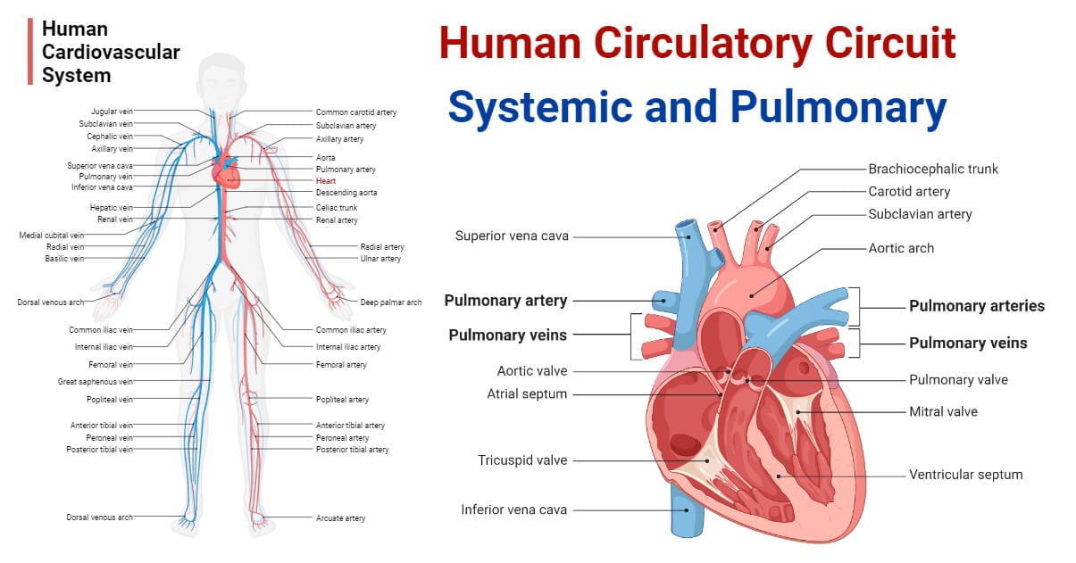
Our cardiovascular system comprises the heart and a closed circuit of blood vessels for the closed continuous blood circulation throughout the body. This closed, interconnected network of blood vessels defines the circulatory system circuit of our body.

Functionally, the human circulatory circuit can be divided into two types; the systemic circulation circuit and the pulmonary circulation circuit. The heart functions as the central organ or junction point, interconnecting these two distinct circuits.
Interesting Science Videos
Systemic Human Circulation Circuit
- It is composed of the systemic arteries, capillaries, and the systemic veins. It serves to deliver oxygenated blood to every tissue and cell of our body and in return collects metabolic wastes from each cell.
- The systemic circuit starts from the heart as a single main and largest artery called the aorta. The aorta emerges from the left ventricle of the heart and soon divides into four sections – ascending aorta, aortic arch, thoracic aorta, and abdominal aorta. From each section, different arteries, called the major arteries arise. The major arteries supply blood to specific organs or parts of our body.
- The major arteries gradually divide and become smaller vessels called the small arteries which supply blood to various parts of an organ or region. The small arteries also divide into finer forms called the arterioles which are connected to the capillaries in order to supply blood to each tissue and cell within the organ or region of our body.
- The arterioles convey the oxygenated blood to the incredibly small and finer blood vessels of just about 5 to 10 μm, called the capillaries. These capillaries are very thin-walled (composed of a very thin permeable layer of simple squamous endothelial cells) and allow for the diffusion of oxygen and nutrients out of the blood in the interstitial fluid. In return, the carbon dioxide and other metabolic wastes diffuse inside the capillaries from the interstitial fluid. The difference in concentration of materials permits this diffusion process.
- The capillaries are connected to the finer tubules of about 8 to 100 μm in diameter, called the venules. Several venules merge forming a smaller vein. Different small veins unite forming larger veins that finally connect to form two major veins called the vena cava; the superior vena cava and the inferior vena cava. The vena cava drains the deoxygenated blood back into the heart at the right atrium. The connection of the major veins into the right atrium completes the systemic circulation circuit.
- In summary, the systemic circulation circuit begins from the left ventricle, travels consecutively as the aorta, arteries, capillaries, veins, and vena cava, and finally terminates into the right atrium.
Pulmonary Human Circulation Circuit
- It is composed of the pulmonary arteries and the pulmonary veins. It serves to deliver deoxygenated blood from the heart to the lungs for re-oxygenation and carry the re-oxygenated blood to the heart for recirculation in the systemic circuit.
- The pulmonary circuit starts from the right atrium as the major pulmonary artery, called the pulmonary trunk. The pulmonary trunk grows about 5 cm and soon branches into the two pulmonary arteries; the left and the right pulmonary artery.
- The left pulmonary artery delivers blood to the left lung. It branches into two lobar arteries, the left upper lobar artery, and the left lower lobe artery, each supplying one lobe of the left lung. The right pulmonary artery also divides into two lober arteries, the right upper lobar artery supplying the upper right lobe and the interlobar artery supplying the middle and the lower right lobes.
- Each lobar artery in the right and the left lung further divides into the segmental arteries. These segmental arteries divide into finer vessels called the sub-segmental arteries that eventually divide and become finer vessels called the pulmonary capillaries. These pulmonary capillaries form a dense web in the alveolar wall called the pulmonarycapillary bed.
- The pulmonary capillaries allow for gaseous exchange at the alveoli of the lungs making the blood oxygenated.
- The pulmonary capillaries are connected to the pulmonary venules. These pulmonary venules surround alveoli and merge to form smaller pulmonary veins. These small pulmonary veins unite and form four main pulmonary veins, two emerging from each lung. The left superior and the left inferior pulmonary vein arise from the left lung; whereas, the right superior and the right inferior pulmonary vein arise from the right lung. As these four pulmonary veins ascend towards the heart, they merge to form two larger pulmonary veins, namely the left pulmonary vein and the right pulmonary vein. These two veins supply the oxygenated blood into the left atrium.
- In summary, the pulmonary circuit begins from the right atrium, and travels consecutively as the pulmonary trunk, left and right pulmonary arteries, lobar pulmonary arteries, segmental and sub-segmental pulmonary arteries, pulmonary capillaries, pulmonary venules, smaller pulmonary veins, main pulmonary veins, and larger pulmonary veins, and terminates in the left atrium.
References
- Kandathil, A., & Chamarthy, M. (2018). Pulmonary vascular anatomy & anatomical variants. Cardiovascular Diagnosis and Therapy, 8(3), 201-207. https://doi.org/10.21037/cdt.2018.01.04
- Tucker WD, Weber C, Burns B. Anatomy, Thorax, Heart Pulmonary Arteries. [Updated 2022 Jul 25]. In: StatPearls [Internet]. Treasure Island (FL): StatPearls Publishing; 2023 Jan-. Available from: https://www.ncbi.nlm.nih.gov/books/NBK534812/
- https://my.clevelandclinic.org/health/body/23242-pulmonary-veins
- Pulmonary artery. (2023, May 1). In Wikipedia. https://en.wikipedia.org/wiki/Pulmonary_artery
- https://www.kenhub.com/en/library/anatomy/pulmonary-arteries-and-veins
- https://my.clevelandclinic.org/health/articles/21486-pulmonary-arteries
- https://radiopaedia.org/articles/pulmonary-veins
- https://training.seer.cancer.gov/anatomy/cardiovascular/blood/pathways.html
- Pulmonary vein. (2023, January 4). In Wikipedia. https://en.wikipedia.org/wiki/Pulmonary_vein
- https://www.uc.edu/content/dam/uc/ce/docs/OLLI/Page%20Content/OLLI%20Circulatory_System.pdf
- https://study.com/academy/lesson/special-circuits-in-the-circulatory-system.html
- https://my.clevelandclinic.org/health/body/21775-circulatory-system
- https://www.visiblebody.com/learn/circulatory/circulatory-pulmonary-systemic-circulation
- https://www.kenhub.com/en/library/anatomy/circulatory-system
- https://www.khanacademy.org/science/high-school-biology/hs-human-body-systems/hs-the-circulatory-and-respiratory-systems/a/hs-the-circulatory-system-review
- https://www.theexpertta.com/book-files/OpenStaxBio2e/Chapter%2040%20-%20The%20Circulatory%20System.pdf

Typo: You state that the pulmonary system starts with the right atrium in one paragraph and the right ventricle in the summary paragraph. Otherwise, excellent work.
Thanks for the correction. The page has been updated. 🙂