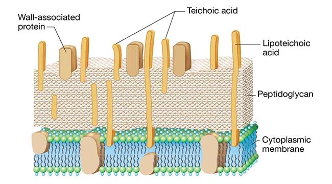
Prokaryotic cells almost always are bounded by a fairly rigid and chemically complex structure present between the cell membrane and capsule/slime layer called the cell wall. Peptidoglycan is the main component of the cell wall and is responsible for the shape and strength of the cell. It is a disaccharide and contains two sugar derivatives—N-acetylglucosamine and N-acetylmuramic acid—joined together by short peptide chains. N-acetylmuramic acid carries a tetrapeptide side chain consisting of D- and L-amino acids (D-glutamic acid and L-alanine) with mesodiaminopimelic acid (Gram-negative bacteria) or L-lysine (Gram-positive bacteria). Tetrapeptide side chains are interconnected by pentaglycine bridges. Most Gram-negative cell walls lack interpeptide bridge. Cell wall provides shape to the cell and protects bacteria from changes inosmotic pressure, which within the bacteria cell measures 5–20 atmospheres.

Bacterial cells can be classified into Gram-positive or Gram-negative based on the structural differences between Gram-positive and Gram-negative cell walls. The cell walls of the Gram-positive bacteria have simpler chemical structures compared to Gram-negative bacteria.
Interesting Science Videos
Gram-positive cell wall
The Gram-positive cell wall is thick (15–80 nm) and more homogenous than that of the thin (2 nm) Gram-negative cell wall. The Gram-positive cell wall contains large amount of peptidoglycan present in several layers that constitutes about 40–80% of dry weight of the cell wall. The Gram-positive cell wall consists primarily of teichoic and teichuronic acids. These two components account for up to 50% of the dry weight of the wall and 10% of the dry weight of the total cell.
1. Teichoic acids
Teichoic acids are polymers of polyribitol phosphate or polyglycerol phosphate containing ribitol and glycerol. These polymers may have sugar or amino acid substitutes, either as side chain or within the chain of polymer. Teichoic acids are of two types—wall teichoic acid (WTA) and lipoteichoic acids (LTA). They are connected to the peptidoglycan by a covalent bond with the six hydroxyl of N-acetylmuramic acid in the WTA and to plasma membrane lipids in LTA.
Teichoic acids have many functions:
- They constitute major surface antigens of those Gram-positive species that possess them. In Streptococcus pneumoniae, the teichoic acids bear the antigenic determinants called Forssman antigen. In Streptococcus pyogenes, LTA is associated with the M protein that protrudes from the cell membrane through the peptidoglycan layer. The long M protein molecules together with the LTA form microfibrils that facilitate the attachment of S. pyogenes to animal cells;
- They are also used as antigen for serological classification of bacteria;
- They serve as substrates for many autolytic enzymes;
- They bind magnesium ion and may play a role in supply of this ion to the cell;
- They play a role in normal functioning of the cell wall and provide an external permeability barrier to Gram-positive bacteria; and
- Membrane teichoic acid serves to anchor the underlying cell membrane.
2. Teichuronic acid
Teichuronic acid consists of repeat units of sugar acids (such as N-acetylmannuronic or D-glucuronic acid). They are synthesized in place of teichoic acids when phosphate supply to the cell is limited. Gram-positive cell wall also contains neutral sugars (such as mannose, arabinose, rhamnose, and glucosamine) and acidic sugars (such as glucuronic acid and mannuronic acid), which occur as subunits of polysaccharides in the cell wall.

Anyone knows how to quantify Gram positive bacteria cell wall by a chemical or immunological assay?
I will be grateful for any help~
I was wondering that how to quantify the Gram positive bacteria cell wall by a chemical or immunological assay? Anyone have a solution?
Superb and precisely described bacterial wall structure.
Super liked.