- Vitamins refer to any of a group of organic compounds which are essential for normal growth and nutrition and are required in small quantities in the diet.
- Vitamins are biologically important. Although a micronutrient, it enhances the metabolism of macronutrients like proteins, carbohydrates and fats.
- Vitamins are also required for growth in children, formation of hormones, blood cells, tissues and bones.
- Vitamins cannot be synthesized or produced by the human body, thus, our diet must contain vitamins.
- Vitamins crucial for the body include:
- Vitamin A, or Retinol
- Vitamin B1, or Thiamin
- Vitamin B2, or Riboflavin
- Vitamin B3, or Niacin
- Vitamin B5, or Pantothenic Acid
- Vitamin B6, or Pyridoxine
- Vitamin B7, or Biotin
- Vitamin B9, or Folic Acid (Folate)
- Vitamin B12, or Cobalamin
- Vitamin C
- Vitamin D
- Vitamin E
- Vitamin K
They are divided into two groups-
(1) Fat soluble vitamins, and
(2) Water soluble vitamins.
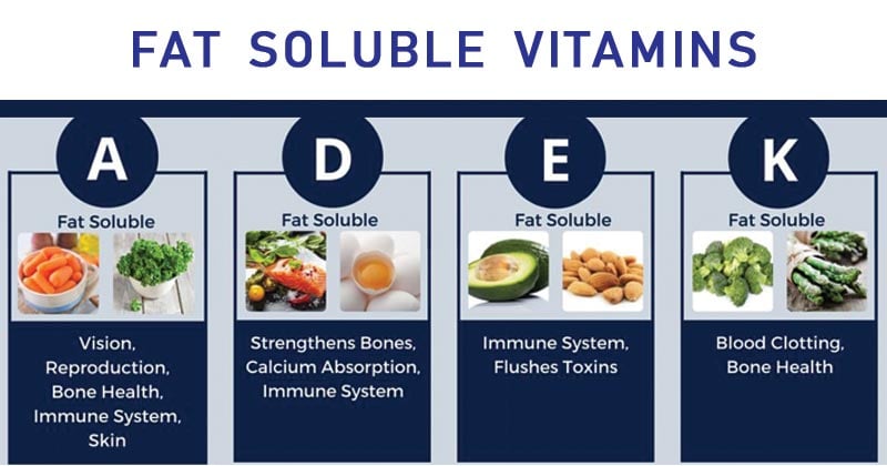
- Fat soluble vitamins– A, D, E and K.
- Water soluble vitamins – Vitamin B1, B2, B3, B5, B6, B12, C, Folic acid and Biotin.
Interesting Science Videos
Solubility, Source, Functions and Deficiencies of Fat Soluble Vitamins

Vitamin A
- Solubility
Fat
- Source
Carrots; leafy greens (eg, spinach); sweet potato
- Function
- Synthesis of rhodopsin; cell differentiation and growth; antioxidant.
- There are several derivatives of vitamin A, which are involved in multiple different metabolic processes.
- 11-cis-retinol is involved in the synthesis of rhodopsin, the visual pigment in retinal cells. Retinoic acid acts to regulate cell growth and differentiation.
- β-Carotene, a precursor for vitamin A, has been known to act as an antioxidant.
- Vitamin A (Retinol) Deficiency
Cause
Causes include fat malabsorption syndromes (pancreatic insufficiency, cholestatic liver disease, celiac sprue, inflammatory bowel disease, gastrectomy), mineral oil laxative abuse, and malnutrition
Clinical Manifestation
Early symptoms include night blindness, poor wound healing, increased susceptibility to infection, and Bitot spots (white patches on the conjunctiva). Later symptoms include hyperkeratinization and resulting skin dryness, ulceration and keratinization of the cornea, and complete blindness.
Treatment
Treat with vitamin A supplementation (30 000 IU daily for 1 week for early deficiency; 20 000 IU/kg for 5 days for late deficiency). Early signs of deficiency can often be reversed with supplementation.
Toxicity
Toxicity occurs with ingestion of more than 50 000 IU per day of vitamin A for longer than 3 months. Symptoms include scaly skin, nausea, diarrhea, headache, papilledema, and hepatosplenomegaly, which may lead to eventual cirrhosis.
Vitamin D
- Solubility
Fat
- Source
Fish; milk; cereals. Skin production via sunlight.
- Supply
Vitamin D can be either absorbed intestinally or synthesized in the skin by ultraviolet
radiation and then hydroxylated by either the liver or the kidney to form potent metabolites.
- Function
- Increases Ca2+ through increased absorption in kidney and GI tract
- Vitamin D is involved in stimulating the synthesis of a calcium-binding protein found in the intestine and thus is involved in increasing intestinal calcium absorption.
- Vitamin D also acts in conjunction with PTH to stimulate osteoblast activity, leading to bone demineralization and calcium release into the blood, and calcium reabsorption by the distal tubules of the kidney.
- Vitamin D Deficiency
Cause
Causes include malnutrition, fat malabsorption syndromes, decreased exposure to the sun, liver disease, and chronic renal failure.
Clinical Manifestation
- Rickets: Seen in young children; skeletal deformities (resulting from disruption of mineralization at epiphyseal plates); shortened stature; pigeon breast (resulting from sternum protrusion); rachitic rosary (costochondral junction thickening); late closing of fontanelles; craniotabes (thinning of occipital and parietal bones).
- Osteomalacia: Seen in adults; diffuse bone pain; muscle weakness; pathologic fractures; hypocalcemia; radiographs demonstrate diffuse radiolucency with thinning of cortical bone.
Treatment
Vitamin D supplementation; adequate sunlight exposure.
Toxicity
- Clinical manifestations of vitamin D toxicity include hypercalcemia, calcification of soft tissues, kidney stones, and bone demineralization.
- Vitamin D toxicity can also be seen in sarcoidosis, in which abnormal cells convert vitamin D into active metabolites.
Vitamin E
- Solubility
Fat
- Source
Cereals; almonds; vegetable oils
- Function
- Antioxidant
- Vitamin E is believed to act as an antioxidant, reacting with free radicals and thereby protecting cellular membranes from damage.
Deficiency of Vitamin E
Deficiency occurs in fat malabsorption syndromes and abetalipoproteinemia. Vitamin E deficiency manifests clinically with vision disturbances, hemolytic anemia (because of the increased fragility of RBC membranes), neurologic dysfunction (ataxic gait, areflexia, decreased proprioception), and myopathies.
Vitamin K
- Solubility
Fat
- Source
Leafy green vegetables (eg, spinach);cabbage
- Supply
Vitamin K is supplied to the body through diet and endogenous synthesis by intestinal bacteria.
- Function
- Facilitates γ-carboxylation of clotting factors II, VII, IX, and X
- Vitamin K acts as a cofactor for glutamate carboxylase, which catalyzes the posttranslational f-carboxylation of glutamic acid residues on clotting factors II, VII, IX, and X and thereby results in superior activation of the coagulation cascade.
- Vitamin K Deficiency
Causes
Caused by fat malabsorption syndromes and use of broad-spectrum antibiotics, which serve to suppress bowel bacterial flora, thereby decreasing the synthesis of vitamin K.
Clinical Manifestations
May be asymptomatic in mild cases or may present with bleeding from mucous membranes, impaired blood clotting, and increased bruising.
Treatment
- Vitamin K supplementation.
- Infants are always given a dose of vitamin K at birth because they are born with a mild vitamin K deficiency, resulting from poor diffusion of vitamin K across the placenta as well as decreased intestinal flora and subsequent decreased vitamin K synthesis.
References
- Suzanne J. Baron and Christoph I. Lee (2013).Biochemistry & Genetics. Second Edition. Mc Graw Hill: New York.
- David Hames and Nigel Hooper (2005). Biochemistry. Third ed. Taylor & Francis Group: New York.
- https://www.sscadda.com/2015/12/short-notes-on-vitamins.html
- https://medlineplus.gov/vitamins.html
