Endocytosis is a cellular mechanism by which, a cell internalizes substances including proteins, fluids, electrolytes, microorganisms, and some macromolecules, from its external environment.
These substances undergo certain processes of breaking down to smaller elements either for use by the cell or for elimination purposes.
White blood cells, of the immune system, are the most common cells that use endocytosis mechanisms to eliminate microbial pathogens from the body. They entrap the pathogens, break them down and destroy them, for elimination from the body.
Endocytosis was first described by Christian de Devu, A Belgium Cytologist and Biochemist who won several Nobel prizes for his role in discovering cellular elements such as lysosomes, peroxisomes, endosomes, and even exocytosis cellular mechanisms including the endocytosis mechanism.

Figure: Types of Endocytosis mechanisms. Image Source: Wikipedia.
Interesting Science Videos
Process of Endocytosis: Summary
- The cell membrane folds in forming a cavity filled with extracellular fluid, dissolved molecules, food particles, foreign matter, pathogens, and/or other substances, a process known as invagination.
- After invagination, the cell membrane folds back to itself until it forms a uniformly enclosed membrane around the trapped molecules and this enclosed membrane or cavity is known as a vesicle. Some cells form extension channels into the cell cytoplasm.
- The formed vesicle detaches off from the cell membrane which then undergoes processing by the cell.
Types of Endocytosis mechanisms
There are three types of endocytosis mechanisms:
- Phagocytosis
- Pinocytosis
- Receptor-Mediated Endocytosis (Clathrin-Mediated Endocytosis)
1. Phagocytosis
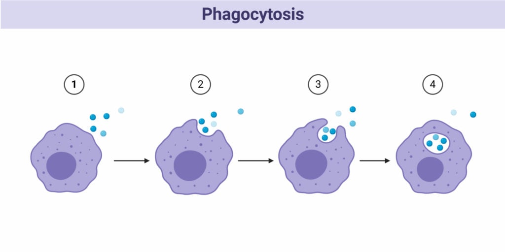
- Also known as cell eating; This is the process whereby the cell membrane of a cell extends toward a particle, engulfing it and encloses it within this folded membrane forming a phagosome. The ingested material in the phagosome is later processed by cellular enzymes. Phagocytosis is a common mechanism in multicellular organisms by the White blood cells (macrophages, monocytes, neutrophils, Eosinophils, dendritic cells), in the elimination of pathogens from the system. Some protozoans such as Entamoeba spp use phagocytosis to acquire nutrients;
- Phagocytosis mechanism was first noted by Canadian physician William Osler (1876).
- Phagocytosis takes place in 5 steps:
- The phagocytic cells detect the molecule of interest or an antigen and moves towards it.
- The phagocyte then attaches itself to the target molecule or antigen. Phagocytes have the ability to extend their membrane (pseudopodia) to the target particle and surround the particle of the pathogen. The pseudopodia extend toward each other while enclosing the particles.
- The particle is then enclosed within the vesicle formed from the extended pseudopodia that have fused. The vesicle with the enclosed particles is known as a phagosome. This is the vesicle that is digested by the phagocyte.
- The phagosome fuses with the lysosomes of the phagocyte forming a phagolysosome. The lysosomes have digestive enzymes that degrade or digest the materials contained in the vesicle.
- The degraded particles are then expelled from the phagocytic cell by exocytosis.

Example of phagocytosis
- A typical example of phagocytosis is the mechanism of the immune cells such as the macrophages, dendritic cells, and neutrophils. Macrophages are the largest phagocytic cells in the immune system. They function by detecting, attaching, ingesting, digesting, and releasing digested particles from its cytoplasm by exocytosis. The antigens vary and they include bacteria, fungi, dust particles, dead cells, etc. Macrophages are the major phagocytic cells in the immune system. They have a pseudopodial membrane. On detection of an antigen, they move toward the antigen and extend their pseudopodia toward the antigen, and engulf it. On engulfment, the cell ingests the antigen forming a cell vesicle otherwise known as the phagosome. Within the macrophage, the vesicle encounters the lysosomes, forming a phagolysosome, which is digested my the lysosomic enzymes breaking the particle down which are then released from the cell by exocytosis.
- Adherence of the particle with the phagocyte highly depends on the chemical nature of the particle. Some bacterial antigens bind directly and some need a protein component from blood, known as an opsonin (such as complements of antibodies), to form a film on the bacterial surface for it to adhere to the phagocyte, a process known as opsonization. So the phagocytes first will bind to the opsonin for phagocytosis to take place.
- Some bacteria with encapsulated cell walls are rather difficult to digest even with an opsonin. Therefore they must be bound to specific antibodies after the body responds to their presence. The antibody-bound encapsulated bacteria can then be acted upon by the phagocytes.
2. Pinocytosis
- Also known as cell drinking or fluid endocytosis; it is a form of endocytosis where small particles in extracellular fluids enter the cell through the cell membrane by invagination forming a small vesicle with suspended small molecules or particles within a cell. The pinocytic vesicle fuses with the cell endosome for the digestion of the particles.
- Its mechanisms are similar to the other endocytic processes, the major difference between pinocytosis and phagocytosis is that in pinocytosis particles ingested are contained within the cells extracellular fluids. The cell membrane invaginated together the vesicle containing the fluid particles, transporting them into the cell lysosomes. The vesicle and the lysosomes fuse, releasing digestive enzymes from the lysosomes. The enzymes degrade the vesicle, releasing its content into the cell cytoplasm, for utilization by the cell.
- Sometimes the vesicles do not interact with the lysosomes, instead, they move across the cell, fusing with the cell membrane causing a recycling effect of the membrane proteins and lipids.
- Pinocytosis takes place by two mechanisms:
- Micropinocytosis – This is the formation of small vesicles of about 0.1 um diameter; it takes place in the body cells forming tiny budding vesicles on the cell membrane known as caveolae. They are found in blood vessel endothelium.
- Macropinocytosis – This is the formation of larger vesicles of about 0.5-5um in diameter; they are found on the white blood cells; the large vesicles are formed by the cell membrane ruffles (villi), which are projections that extend to the extracellular fluids and have the ability to fold back by themselves. While folding, they shovel in some of the extracellular fluid forming a vesicle that pulls into the cell.
Example of pinocytosis:
The intake or absorption of nutrients in the small intestines.
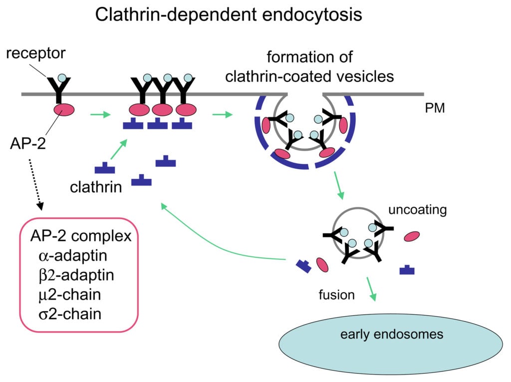
3. Receptor-Mediated Endocytosis (Clathrin Mediated Endocytosis)
- This is a type of endocytosis also known as clathrin-mediated endocytosis; It involves the internalization and recycling of receptors that are used in processes such as signal transduction (G-protein and tyrosine kinase receptors), nutrient uptake and synaptic vesicle reformation.
- This process is initiated by the accumulation of phosphatidylinositol-4,5-bisphosphate (PIP2) within the cell membrane. PIP2 accumulation is because of the catalyzation process of phosphoinositide within the plasma membrane. PIP2 accumulates as a result of phosphoinositide by the lipid kinase and the hydrolyzation of phosphatases. The combination of adapter proteins (AP proteins) with the Phosphatidylinositol-4,5-bisphosphate (PIP2), leads to the attachment of a cytosol protein known as clathrin to the vesicle. This forms Clathrin-coated vesicles (CCV).
- The Clathrin-coated vesicles (CCV)must invaginate and mature to form the clathrin-coated pits. The Clathrin-coated vesicles bind to the cell membrane recruiting several proteins including Actin-binding proteins, Adapter proteins (AP) all of which play a major role in the maturation of the vesicle.
- The CCV, which contains several receptors bound with ligands and adapter proteins, then invaginated into the membrane and by the assistance of dynamin protein, it matures and scissions from the cell membrane, forming a clathrin-coated pit.
- How are the Clathrin-coated pits formed? Clathrin-coated vesicles are found in most if not all cells, and therefore, after the detection of a signal, these vesicles recruit the adaptor proteins in the plasma membrane which accumulate on the lipid layer of the plasma membrane.
- The adapter proteins incorporate the Clathrin from the clathrin-coated vesicles into the cell membrane lipids along with the Actin-binding proteins. Due to the negative charge of the lipids layer, it gets attracted to the positive charge of the clathrin, forming a concave shape that is raised up from the membrane, thus forming pits all over the plasma membrane.
- The clathrin on the pits acts as a sensor for signals that activate endocytosis while the vesicle from the Clathrin Coated vesicles gets recycled to the cell membrane. The cycle between the clathrin-coated pits and clathrin-coated vesicles formation is continuous as long as there are signaling receptors and ligands that activate them.
- The process of Receptor-mediated endocytosis has the following steps:
- The particles (ligands) that need to be synthesized are bound to the receptors on the cell membrane, The receptors with the ligands cluster forming the coated pits. The pits then undergo invagination with the help of the dynamin proteins forming a vesicle and the vesicle pinches-off within the cell membrane. The vesicles then lose the clathrin and the adaptor proteins.
- The uncoated vesicle then fuses with an early endosome to form the late endosome or the sorting vesicle. The late endosome segregates the particles within the vesicle i.e the receptors from the ligands recycling them into the cell membrane.
- The released particles interact with the lysosomes which contain digestive enzymes that hydrolyze the content in the vesicles. The digested particles are then released for utilization by the cell.
- This mechanism of receptor-mediated endocytosis (clathrin-coated Endocytosis) can best be used to bring macromolecules into the cell.

Figure: Clathrin Mediated Endocytosis. Image Source: Barth D. Grant and Miyuki Sato
Examples of Clathrin-Mediated Endocytosis
There are two classic examples of the Clathrin-Mediated Endocytosis which include
- iron-bound transferrin recycling
- receptor-mediated endocytosis is the uptake of cholesterol bound to low-density lipoprotein (LDL), a complex of phospholipid, protein, and cholesterol.
References and Sources
- Color Atlas of Cytology, Histology and Microscopic Anatomy by Kuehnel
- http://www.brooklyn.cuny.edu/bc/ahp/LAD/C5/C5_Endocytosis.html
- https://www.thoughtco.com/what-is-endocytosis-4163670
- https://bio.libretexts.org/Bookshelves/Cell_and_Molecular_Biology/Book%3A_Basic_Cell_and_Molecular_Biology_(Bergtrom)/17%3A_Membrane_Function/17.4%3A_Endocytosis_and_Exocytosis
- https://www.pathwayz.org/Tree/Plain/ENDOCYTOSIS+%26+EXOCYTOSIS
- https://www.britannica.com/science/pinocytosis
- MBINFO Defining Mechabiology:www.mechabio.info
- https://www.biologyonline.com/dictionary/opsonization
- https://www.britannica.com/science/phagocytosis
- https://www.britannica.com/science/pinocytosis

Shòw more diagram for it