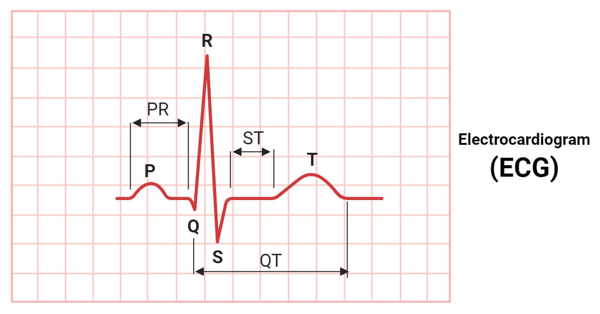
Electrocardiogram, commonly known as ECG or EKG is a medical test report indicating the heart’s electrical activity and rhythm during repeated cardiac cycles.

It is printed on grid paper called the ECG strip or ECG tracing. The process of producing an electrocardiogram is termed electrocardiography. It is a non-invasive medical procedure performed by recording the cardiac impulses on the surface of our body using specialized electrodes over specific regions of our body.
The main purpose of ECG is to detect and diagnose any underlying cardiovascular issues or structural abnormalities in the heart. Electrocardiography is one of the most commonly prescribed tests by cardiologists to obtain information about the patient’s cardiac electrical functioning.
Interesting Science Videos
Electrocardiogram Instrumentation
ECG test is done using an ECG machine containing a set of electrodes connected to a central signal processor through lead wires and a monitor and a printer to display and print the ECG.
Typically 10 electrodes, that receive, collect and transmit the electric potentials from our body (biopotentials), are present in a standard 12-lead ECG-producing machine. The biopotentials collected by the electrodes are carried by lead wires to the central impulse processing machine. The central ECG machine amplifies the received biopotential, filters the signal, and processes it to produce ECG waves in specific intervals and segments. The processed signals are displayed on the monitor and are printed on grid papers to produce an ECG.
Principle of Electrocardiogram
The ECG is based on the cardiac action potential and cardiac conduction. The SA node generates the cardiac action potential which is relayed down the cardiac conduction pathway resulting change in the membrane voltage (potential) across the membrane of cardiomyocytes. This change in cardiac action potential results in the continuous running of the cardiac cycle.
The cardiac impulse transmits through the heart, spreads around the surrounding tissue, and finally reaches the skin of our body. This electric impulse in the skin is received by the electrodes of the ECG machine and processed to produce an ECG.
Procedure of Obtaining an Electrocardiogram
- Guide the patient to lie flat on a bed in a comfortable and relaxed position. Ensure that the patient is not wearing any metallic objects or electrical devices.
- Attach the electrodes in their respective location. In 12-lead ECG, 10 electrodes are attached at specific sites of the patient’s body.
There are four arm electrodes, the RA, the LA, the RL, and the LL electrodes. The RA electrode is placed on the right arm, the LA electrode is placed on the left arm, the RL electrode is placed on the right leg, and the LL electrode is placed on the left leg.
There are 6 chest electrodes named V1 to V6. The V1 and V2 are attached in the fourth intercostal space just right and left of the sternum respectively. The V4 is attached in the fifth intercostal space in the mid-clavicular line. The V3 is attached in between the V2 and V4. The V5 is attached horizontally left of the V4 in the left anterior axillary line. The V6 is attached horizontally left of the V5 in the mid-axillary line.
- Lead wires are connected to the electrodes and the ECG machine is turned on.
- The ECG machine is an automated machine that auto-records the cardiac electric impulse activity and develops an ECG.
- Once the adequate ECG is recorded, the leads are disconnected from the electrodes and the electrodes are detached.
Types of Electrocardiogram
There are three main types of ECG, that are:
- Resting ECG
It is the standard ECG type routinely used for diagnostic purposes in hospital settings. It is performed while staying lying still in a bed.
- Exercise ECG
It is the ECG obtained while performing physical activities, generally walking on a treadmill or peddling a stationary bicycle. It is also called the stress test and is performed to monitor the heart and its electrical activity during physical stress.
- Ambulatory ECG
It is a type of ECG that represents the continuous electric activity of 24 hours or more of a heart. In this type, a small portable ECG machine called the Holter monitor is attached to the waist, and the person is allowed to work normally for at least a day. After the desired time, the ECG is studied from the Holter machine.
Waves in a Normal Electrocardiogram
In a normal ECG, we can see three distinct waves; the P-wave, the QRS-complex, and the T-wave.
- P-wave
It is the first small upward wave in an ECG that represents atrial depolarization. It represents the electric activity that triggers the atrial systole i.e. impulse generated and transmitted by the SA node just before and starting of atrial systole.
- QRS-complex
The QRS complex is the second sharply steeper larger upright triangular-shaped wave representing rapid ventricular depolarization. The QRS complex comprises three individual waves; the Q wave represents the initial downward deflection, the R wave represents the sharp ascending deflection, and the S wave represents the sharp descending deflection in the larger upright triangular wave of an ECG. The QRS complex coincides with the ventricular depolarization stage or ventricular systole stage of a cardiac cycle.
- T-wave
It is the final dome-shaped upward deflection representing ventricular repolarization. In a few ECGs, a low amplitude barely noticeable small upward deflection is seen just after the T-wave, known as the U-wave. It is believed that the interventricular septum repolarization is responsible for this wave.
Intervals in a Normal Electrocardiogram
In an ECG, the time period between the beginnings of two waves is measured and analyzed. These time periods represent the time required by impulses to transmit in the cardiac conduction pathway causing a coordinated cycle of depolarization and repolarization of the heart’s wall. These time periods are termed ‘intervals’ and there are two main intervals, viz. PR interval and the QT interval.
- PR Interval
It is represented in an ECG by the gap between the beginnings of the P-wave to the beginning of the QRS-complex. The PR interval represents the time required by the cardiac impulse to transmit from the SA node, through the atria, and AV bundle, and finally reach the ventricles. It is usually about 120 to 200 milliseconds.
- QT Interval
It is represented in an ECG by the gap between the start of the QRS complex to the end of the T wave. It represents the duration between consecutive ventricular depolarization and repolarization. It is usually shorter than 440 milliseconds.
Segments in a Normal Electrocardiogram
A flat horizontal line between two successive waves i.e. from the end of one wave to the beginning of another in an ECG is termed a ‘segment’. There are two main segments in an ECG, viz. the PR segment and the ST segment.
- PR Segment
The flat line between the P-wave’s conclusion and the beginning of the QRS complex is known as the PR segment. It represents the time delay between the atrial systole and the ventricular systole. It is slightly longer than 440 milliseconds.
- ST Segment
It is the flat line between the QRS complex and T wave representing the electrically neutral area between the end of ventricular depolarization and the beginning of ventricular repolarization. It is usually around 80 milliseconds long.
Interpretation of ECG
An ECG is analyzed and interpreted by trained medical personnel only. While reading an ECG the P wave, QRS complex, T wave, PR interval, QT interval, PR segment, and ST segment are mainly focused. The elevation and depression of each wave, the duration of the waves, and the duration of the segments and intervals are studied. Additionally, the heart rate, rhythm, and axis on an ECG are also studied. Based on a collective result of all these various components, an ECG report is interpreted.
A normal ECG will show the following results:
- Heart Rate: Normal heart rate of 60 to 100 beats per minute
- Heart Rhythm: Heart rhythm will be consistent and even
- PR Interval: 0.12 to 0.20 seconds
- QRS Duration: 0.06 to 0.10 seconds
- QT Interval: 0.40 seconds
- ST Segment: 0.08 seconds
- P-wave
- Upright, uniform, and consistent before each QRS complex.
- P duration < 0.12 seconds
- P amplitude < 2.5 mm
- T-wave: Upright in lead I, II, V3 to V6, and inverted in aVR.
Application of ECG
- Diagnosing arrhythmias and related cardiovascular diseases.
- Studying the heart’s electrical activity and the cardiac cycle of the heart.
- Assessing heart diseases and heart attacks.
- It is used to evaluate the progress after medication or cardiac surgery.
- It is coupled with ECHO to access the overall structural and functional health of the heart and valves.
- Used to check how the heart performs under stress.
Limitations of ECG
- It provides information about the heart’s electrical activity only but doesn’t provide information about the heart’s anatomy.
- ECG reports have a high chance to show false results due to problems in electrodes, the body’s electric potential, and due to metallic or electric devices nearby.
- Interpretation of the ECG report is subjective; hence, the reporting might be slightly variable among different healthcare professionals.
- ECG reports the electrical activity for a brief period of time only.
- It is unable to predict future cardiac events, the formation of abnormal tissues and clot, and some cardiac events that are unrelated to cardiac electric potential.
References
- InformedHealth.org [Internet]. Cologne, Germany: Institute for Quality and Efficiency in Health Care (IQWiG); 2006-. What is an electrocardiogram (ECG)? 2019 Jan 31. Available from: https://www.ncbi.nlm.nih.gov/books/NBK536878/
- Kashou AH, Basit H, Malik A. ST Segment. [Updated 2022 Aug 8]. In: StatPearls [Internet]. Treasure Island (FL): StatPearls Publishing; 2023 Jan-. Available from: https://www.ncbi.nlm.nih.gov/books/NBK459364/
- Ashley EA, Niebauer J. Cardiology Explained. London: Remedica; 2004. Chapter 3, Conquering the ECG. Available from: https://www.ncbi.nlm.nih.gov/books/NBK2214/
- https://johnsonfrancis.org/professional/ecg-basics-a-brief-review/basic-principles-of-electrocardiography/
- https://www.mayoclinic.org/tests-procedures/ekg/about/pac-20384983
- https://www.nhs.uk/conditions/electrocardiogram/
- https://www.hopkinsmedicine.org/health/treatment-tests-and-therapies/electrocardiogram
- https://my.clevelandclinic.org/health/diagnostics/16953-electrocardiogram-ekg
- https://www.betterhealth.vic.gov.au/health/conditionsandtreatments/ecg-test
- https://www.heart.org/en/health-topics/heart-attack/diagnosing-a-heart-attack/electrocardiogram-ecg-or-ekg
- https://www.robots.ox.ac.uk/~neil/teaching/lectures/med_elec/notes2.pdf
- https://www.egr.msu.edu/classes/ece480/capstone/spring13/group03/documents/ElectrocardiographyCircuitDesign.pdf
- https://www.onlinebiologynotes.com/electrocardiogram-ecg-working-principle-normal-ecg-wave-application-of-ecg/
- https://litfl.com/pr-segment-ecg-library/
- https://www.healio.com/cardiology/learn-the-heart/ecg-review/ecg-interpretation-tutorial/pr-segment
- https://ecgwaves.com/topic/the-pr-interval-pr-segment/
- https://ayu.health/blog/three-main-types-of-ecg/
- https://byjus.com/biology/electrocardiograph/
- Kenny BJ, Brown KN. ECG T Wave. [Updated 2022 Dec 22]. In: StatPearls [Internet]. Treasure Island (FL): StatPearls Publishing; 2023 Jan-. Available from: https://www.ncbi.nlm.nih.gov/books/NBK538264/
- https://elentra.healthsci.queensu.ca/assets/modules/ECG/normal_ecg.html
- https://ecg.utah.edu/lesson/3
- https://www.mountsinai.org/health-library/tests/electrocardiogram
- Gonçalves, M. A., Pedro, J. M., Silva, C., Magalhães, P., & Brito, M. (2020). Normal limits of the electrocardiogram in Angolans. Journal of Electrocardiology, 63, 68-74. https://doi.org/10.1016/j.jelectrocard.2020.10.011
- https://ecglibrary.com/norm.php

You can help me
Great site for Educational porpus