Interesting Science Videos
Structure of Ebola Virus
- It falls under the Filoviridae family.
- The virion is filamentous, enveloped measuring 800 nm length and 80 nm in diameter.
- It comprises of negative sense, single stranded RNA genome.
- Structural proteins associated with the nucleocapsid are the nucleoprotein (NP), VP30, VP35, and the polymerase (L) protein.
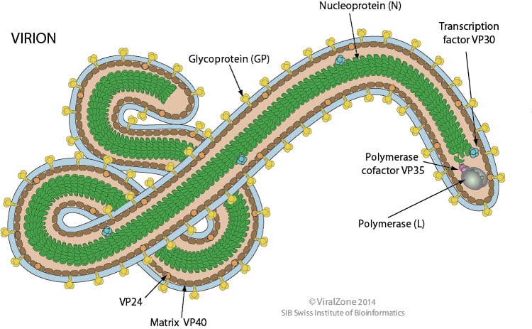
- Membrane-associated proteins are the matrix protein (VP40), VP24, and the GP (peplomer glycoprotein).
- The nucleocapsid is encapsulated by an outer viral envelope originating from the host cell membrane with characteristic 10 nm long viral glycoprotein (GP) spikes that initiates attachment.
Genome of Ebola Virus
- It comprises of linear, negative-stranded RNA genome, about 18-19 kb in size.

- The genome encodes for seven proteins which includes
- GP- transmembrane glycoprotein that helps in attachment
- NP- nucleoprotein necessary for capsid assembly and packaging
- VP24- antiviral inhibitor, suppresses interferon production in the host cell
- VP35- inhibits interferon production or antiviral response to ds RNA.
- VP30- transcription anti-terminator
- VP40- maintaining the structural integrity of the virion, necessary for capsid assembly and budding
- L protein- viral polymerase
Epidemiology of Ebola Virus
- Fruit bats of the Pteropodidaefamily, including the species Hypsignathus monstrosus, Epomops franquetiand Myonycteris torquata, are believed to be the natural hosts of Ebola viruses, with humans and other mammals serving as accidental hosts.
- Ebola virus was discovered in 1976 when two severe epidemics of hemorrhagic fever occurred in Sudan and Zaire.
- The outbreaks involved more than 500 cases and at least 400 deaths caused by clinical hemorrhagic fever.
- Subsequent outbreaks of Ebola hemorrhagic fever have occurred in Uganda Congo, Gabon, Sudan, Ivory Coast.
- Ebola viruses are mainly found in primates in Africa and possibly the Philippines; there are only occasional Ebola outbreaks of infection in humans.
- The West Africa Ebola outbreak was the largest in history, affecting multiple countries in, and beyond, West Africa.
- According to the WHO, 28,616 suspected, probable, and confirmed cases with a total of 11,310 deaths were recorded in Guinea, Liberia, and Sierra Leone.
Transmission of Ebola Virus
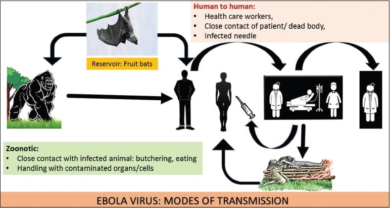
- It spreads to people by contact with the skinor bodily fluids of an infected animal, like a monkey, chimp, or fruit bat.
- Human to human transmission via direct contact with blood and secretions, by contact with blood and secretions that remain on clothing, and by needles and/or syringes or other medical supplies used to treat Ebola-infected patients.
Replication of Ebola Virus
- Entry is mediated by attachment of virus to host receptorslike DC-SIGN and DC-SIGNR through GP glycoprotein.
- The virion enters the cell by Macropinocytosis or Clathrin-mediated endocytosis.
- GP1 interacts with host NPC1, in late macropinosome and promotes fusionof virus membrane with the vesicle membrane.
- The ribonucleocapsid is then released into the cytoplasm.
- Sequential transcription, viral mRNAs are capped and polyadenylated by polymerase stutteringin the cytoplasm.
- VP30 is an important transcription activation factor for viral genome transcription, while VP24 is an inhibitor to this process.
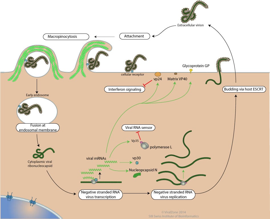
- Replication presumably starts by binding of RNA dependent RNA polymerase complex to the leader sequence on the encapsidated (-)RNA genome.
- The antigenome is concomitantly encapsidated during replication.
- The ribonucleocapsid interacts with the matrix protein VP40, and buds via the host ESCRT (endosomal sorting complex required for transport) complexes from the plasma membrane, releasing the virion.
Pathogenesis of Ebola Virus
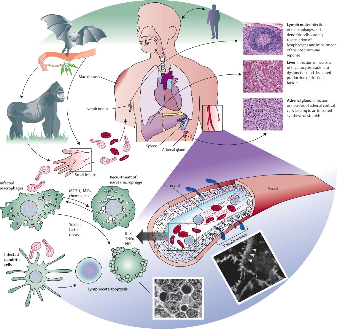
- The Ebola virusenters the host through basic entry paths. It can enter through mucous membranes, broken skin, or by parental transmission. It is capable of binding to these membranes of essentially every human cell.
- The entry of the cell is controlled by a glycoprotein that is responsible for binding the virus to the cell receptors.
- The specific cytokines the virus provokes are the proteins that prompt the natural inflammatory response.
- The inflammatory response induced by the infection of the virus is so drastic that it begins to cause damage to the host cells.
- By deregulating the cytokines, the virus is able to spread rapidly throughout the host.
- Ebola virus capability to inhibit the synthesis of proteins that activate the response when the cell has been infected.
- A protein produced by the Ebola virus, VP35 is capable of inhibiting the synthesis of a specific INFs protein.
- Another way that the virus disrupts the INF response is through the production of protein VP24.
- A third way the virus is capable of defending itself from the antiviral response of the human body is through the glycoprotein inhibition of tetherin expression.
- When the virus spreads throughout the body, one of its targets are hepatic cells, or liver cells.
- The Ebolavirusis capable of inducing necrosis of the hepatic cells, which can result in multiple outcomes.
- When the virus is killing the hepatic cells, the liver can’t synthesize enough coagulant proteins, allowing the hemorrhaging to happen.
- The necrosis doesn’t only shut down the hemorrhaging proteins but also shuts down the liver functions, rendering the liver useless to the infected human.
- Other organs that undergo a similar attack from the virus are the spleen, thymus, and lymph nodes.
- Similar to the liver, these organs see lymphatic depletion and necrosis due to the infection.
- The overall pathogenesis and pathology by Ebola virus include:
- Cell entry and tissue damage
- Gastrointestinal dysfunction
- Systemic inflammatory response
- Coagulation defects
- Impairment of adaptive immunity
Clinical manifestations of Ebola Virus
- In early stages patients feel like the fluor other illnesses.
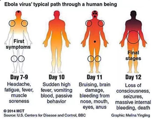
- The incubation period is 2 to 21 days and symptoms usually include:
- High fever
- Headache
- Joint and muscle aches
- Sore throat
- Weakness
- Stomach pain
- Lack of appetite
- Progression of Ebola symptoms includes diarrhea, vomiting, stomach pain, hiccups, rash, and internal and external bleeding in many patients.
- Laboratory finding include low white blood count, low platelet count and elevated liver enzymes.
- Other complications includes sever bleeding, failure of multiple organs, shock.
Lab Diagnosis of Ebola Virus
- Specimen: Blood or body fluids, semen, Oral swabs,
- Antibodies appear later in disease course or after recovery (IgM and IgG) that are detected by ELISA.
- Viral antigens in serum can be detected by ELISA, providing a rapid screening test of human samples.
- RT-PCR is also used on clinical specimens.
- Virus isolates can be cultured in cell lines such as Vero and MA-104 monkey cell lines.
Treatment of Ebola Virus
- No proven treatment in use and no approved vaccine.
- Secondary treatment during disease progression include:
- Providing intravenous fluids (IV) and balancing electrolytes (body salts).
- Maintaining oxygen status and blood pressure.
- Treating other infections if they occur.
Prevention and control of Ebola Virus
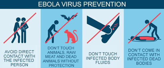
- Avoidance of funeral or burial rituals that require handling the body of person who has died from Ebola.
- Avoidance of contact with bats and non human primates or blood, fluids, and raw meat prepared from these animals.
- Healthcare workers who may be exposed to people with Ebola should wear appropriate personal protective equipment (PPE) and practice proper infection control and sterilization measures.
- Early testing and isolation of the patient plus barrier protection for caregivers (mask, gown, goggles, and gloves) is very important to prevent other people from getting infected.

Thanks for your well presented Dr