
Studies involving the analysis of materials including food, water, milk, and, in some cases, air require quantitative enumeration of microorganisms in the substances.
Many methods have been devised to accomplish this, including direct microscopic counts, the use of an electronic cell counter such as the Coulter Counter, chemical methods for estimating cell mass or cellular constituents, and turbidometric measurements for increases in cell mass, and the serial dilution–agar plate method.
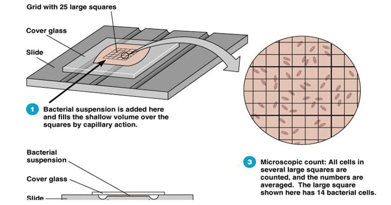
What is a Direct Microscopic Count?
Direct microscopic counts require the use of a specialized slide called the Petroff-Hausser counting chamber, in which an aliquot of a eukaryotic cell suspension is counted and the total number of cells is determined mathematically.
The Petroff-Hausser counting chamber is a thick glass microscope slide with a chamber 0.02 mm (1/50 mm) deep in the center.
The chamber contains an etched grid and has improved Neubauer rulings (1/400 square mm).
The rulings cover 9 mm2. The boundary lines (Neubauer rulings) are the center lines of the groups of three.
The center square millimeter is ruled into groups of 16 small squares, and each group is separated by triple lines, the middle one of which is the boundary.
The ruled surface is 0.02 mm below the cover glass, which makes the volume over a square millimeter 0.02 mm3 (cubic mm). All cells are counted in this square millimeter.
The number of cells counted is calculated as follows:
Number of cells per mm = number of cells counted * dilution * 50,000
[The factor of 50,000 is used in order to determine the cell count for 1 ml: 1 ml = 1000 mm3 = (50 times the chamber depth of 0.02 mm) * 1000.]
A variation of the direct microscopic count has been used to observe and measure the growth of bacteria in natural environments. In order to detect and prove that thermophilic bacteria were growing in boiling hot springs, T.D. Brock immersed microscope slides in the springs and withdrew them periodically for microscopic observation. The bacteria in the boiling water attached to the glass slide naturally and grew as microcolonies on the surface.
Advantages of Direct Microscopic Count
- Rapid, Simple and easy method requiring minimum equipment.
- Morphology of the bacteria can be observed as they counted.
- Very dense suspensions can be counted if they are diluted appropriately.
Limitations of Direct Microscopic Count
- Although rapid, a direct count has the disadvantages that both living and dead cells are counted.
- Only dense suspensions can be counted (>107 cells per ml), but samples can be concentrated by centrifugation or filtration to increase sensitivity.
- It is not sensitive to populations of fewer than 1 million cells.
- Small cells are difficult to see under the microscope, and some cells are probably missed.
- Precision is difficult to achieve
- A phase contrast microscope is required when the sample is not stained.
References
- Cappuccino, J. and Welsh, C. (2014). Microbiology: A Laboratory Manual, Global Edition. 1st ed. Pearson Education
- Sastry A.S. & Bhat S.K. (2016). Essentials of Medical Microbiology. New Delhi : Jaypee Brothers Medical Publishers.
- http://textbookofbacteriology.net/growth_2.html
- http://repository.uobabylon.edu.iq/mirror/resources/paper_2_13669_749.pdf
- https://courses.lumenlearning.com/boundless-microbiology/chapter/counting-bacteria/
- http://ecoursesonline.iasri.res.in/mod/resource/view.php?id=101513
