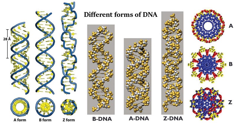
- The right-handed double-helical Watson – Crick Model for B-form DNA is the most commonly known DNA structure.
- In addition to this classic structure, several other forms of DNA have been observed.
- The helical structure of DNA is thus variable and depends on the sequence as well as the environment.

Interesting Science Videos
Why do different forms of DNA exist?
- There is simply not enough room for the DNA to be stretched out in a perfect, linear B-DNA conformation. In nearly all cells, from simple bacteria through complex eukaryotes, the DNA must be compacted by more than a thousand fold in order even to fit inside the cell or nucleus.
- Refined resolution of the structure of DNA, based on X-ray crystallography of short synthetic pieces of DNA, has shown that there is a considerable variance of the helical structure of DNA, based on the sequence. For example, a 200-bp piece of DNA can run as if it were more than 1000 bp on an acrylamide gel if it has the right sequence. The double helix is not the same uniform structure.
The Different Forms of DNA
B-form DNA
- B-DNA is the Watson–Crick form of the double helix that most people are familiar with.
- They proposed two strands of DNA — each in a right‑hand helix — wound around the same axis. The two strands are held together by H‑bonding between the bases (in anti-conformation).
- The two strands of the duplex are antiparallel and plectonemically coiled. The nucleotides arrayed in a 5′ to 3′ orientation on one strand align with complementary nucleotides in the 3′ to 5′ orientation of the opposite strand.
- Bases fit in the double helical model if pyrimidine on one strand is always paired with purine on the other. From Chargaff’s rules, the two strands will pair A with T and G with C. This pairs a keto base with an amino base, a purine with a pyrimidine. Two H‑bonds can form between A and T, and three can form between G and C.
- These are the complementary base pairs. The base‑pairing scheme immediately suggests a way to replicate and copy the genetic information.
- 34 nm between bp, 3.4 nm per turn, about 10 bp per turn
- 9 nm (about 2.0 nm or 20 Angstroms) in diameter.
- 34o helix pitch; -6o base-pair tilt; 36o twist angle
A-form DNA
- The major difference between A-form and B-form nucleic acid is in the confirmation of the deoxyribose sugar ring. It is in the C2′ endoconformation for B-form, whereas it is in the C3′ endoconformation in A-form.
- A second major difference between A-form and B-form nucleic acid is the placement of base-pairs within the duplex. In B-form, the base-pairs are almost centered over the helical axis but in A-form, they are displaced away from the central axis and closer to the major groove. The result is a ribbon-like helix with a more open cylindrical core in A-form.
- Right-handed helix
- 11 bp per turn; 0.26 nm axial rise; 28o helix pitch; 20o base-pair tilt
- 33o twist angle; 2.3nm helix diameter
Z-form DNA
- Z-DNA is a radically different duplex structure, with the two strands coiling in left-handed helices and a pronounced zig-zag (hence the name) pattern in the phosphodiester backbone.
- Z-DNA can form when the DNA is in an alternating purine-pyrimidine sequence such as GCGCGC, and indeed the G and C nucleotides are in different conformations, leading to the zig-zag pattern.
- The big difference is at the G nucleotide.
- It has the sugar in the C3′ endoconformation (like A-form nucleic acid, and in contrast to B-form DNA) and the guanine base is in the synconformation.
- This places the guanine back over the sugar ring, in contrast to the usual anticonformation seen in A- and B-form nucleic acid. Note that having the base in the anticonformation places it in the position where it can readily form H-bonds with the complementary base on the opposite strand.
- The duplex in Z-DNA has to accommodate the distortion of this G nucleotide in the synconformation. The cytosine in the adjacent nucleotide of Z-DNA is in the “normal” C2′ endo, anticonformation.
- Discovered by Rich, Nordheim &Wang in 1984.
- It has antiparallel strands as B-DNA.
- It is long and thin as compared to B-DNA.
- 12 bp per turn; 0.45 nm axial rise; 45o helix pitch; 7o base-pair tilt
- -30o twist angle; 1.8 nm helix diameter
Conditions Favoring A-form, B-form, and Z-form of DNA
- Whether a DNA sequence will be in the A-, B-or Z-DNA conformation depends on at least three conditions.
- The first is the ionic or hydration environment, which can facilitate conversion between different helical forms.
- A-DNA is favored by low hydration, whereas Z-DNA can be favored by high salt.
- The second condition is the DNA sequence: A-DNA is favored by certain stretches of purines (or pyrimidines), whereas Z-DNA can be most readily formed by alternating purine-pyrimidine steps.
- The third condition is the presence of proteins that can bind to DNA in one helical conformation and force the DNA to adopt a different conformation, such as proteins which bind to B-DNA and can drive it to either A-or Z forms.
- In living cells, most of the DNA is in a mixture of Aand B-DNA conformations, with a few small regions capable of forming Z-DNA.
Other rare forms of DNA
C-DNA
- Formed at 66% relative humidity and in presence of Li+ and Mg2+ ions.
- Right-handed with the axial rise of 3.32A° per base pair
- 9.33 base pairs per turn
- Helical pitch 3.32A°×9.33°A=30.97A°.
- Base pair rotation=38.58°.
- Has a diameter of 19 A°, smaller than that of A-&B- DNA.
- The tilt of base is 7.8°
D-DNA
- Rare variant with 8 base pairs per helical turn
- These forms of DNA found in some DNA molecules devoid of guanine.
- The axial rise of 3.03A°per base pairs
- The tilt of 16.7° from the axis of the helix.
E- DNA
- Extended or eccentric DNA.
- E-DNA has a long helical axis rise and base perpendicular to the helical axis.
- Deep major groove and the shallow minor groove.
- E-DNA allowed to crystallize for a period time longer, the methylated sequence forms standard A-DNA.
- E-DNA is the intermediate in the crystallographic pathway from B-DNA to A-DNA.
References
- https://www.researchgate.net/publication/10837288_A_glossary_of_DNA_structures_from_A_to_Z
- http://people.bu.edu/mfk/restricted566/dnastructure.pdf
- Alberts, B., Johnson, A., Lewis, J., Raff, M., Roberts, K., & Walter, P. (2002). Molecular biology of the cell. New York: Garland Science.
- http://www.newworldencyclopedia.org/entry/Deoxyribose
- https://www.nature.com/scitable/definition/phosphate-backbone-273
- http://www.newworldencyclopedia.org/entry/Deoxyribose
- https://www.slideshare.net/vinithaunnikrishnan16/forms-of-dna-49312507
- David Hames and Nigel Hooper (2005). Biochemistry. Third ed. Taylor & Francis Group: New York.
- Bailey, W. R., Scott, E. G., Finegold, S. M., & Baron, E. J. (1986). Bailey and Scott’s Diagnostic microbiology. St. Louis: Mosby.
- https://bio.libretexts.org/Bookshelves/Genetics/Book%3A_Working_with_Molecular_Genetics_(Hardison)/Unit_I%3A_Genes%2C_Nucleic_Acids%2C_Genomes_and_Chromosomes/2%3A_Structures_of_Nucleic_Acids/2.5%3A_B-Form%2C_A-Form%2C_and_Z-Form_of_DNA
- http://eagri.org/eagri50/GBPR111/lec15.pdf

very nice update
Simple, helpful and understandable; thanks.
Under C-DNA you say there are 33bp per turn. That should be 9.33bp per turn.
Thanks, it has been corrected. 🙂
I was in need of a lot of help but after I got it
reading Thanks a lot of
Do you have PPTs for your syllabus?
You are very helpful during the exams
wow…thank you so much ..this is very helpful …
Educative!
Very Educative