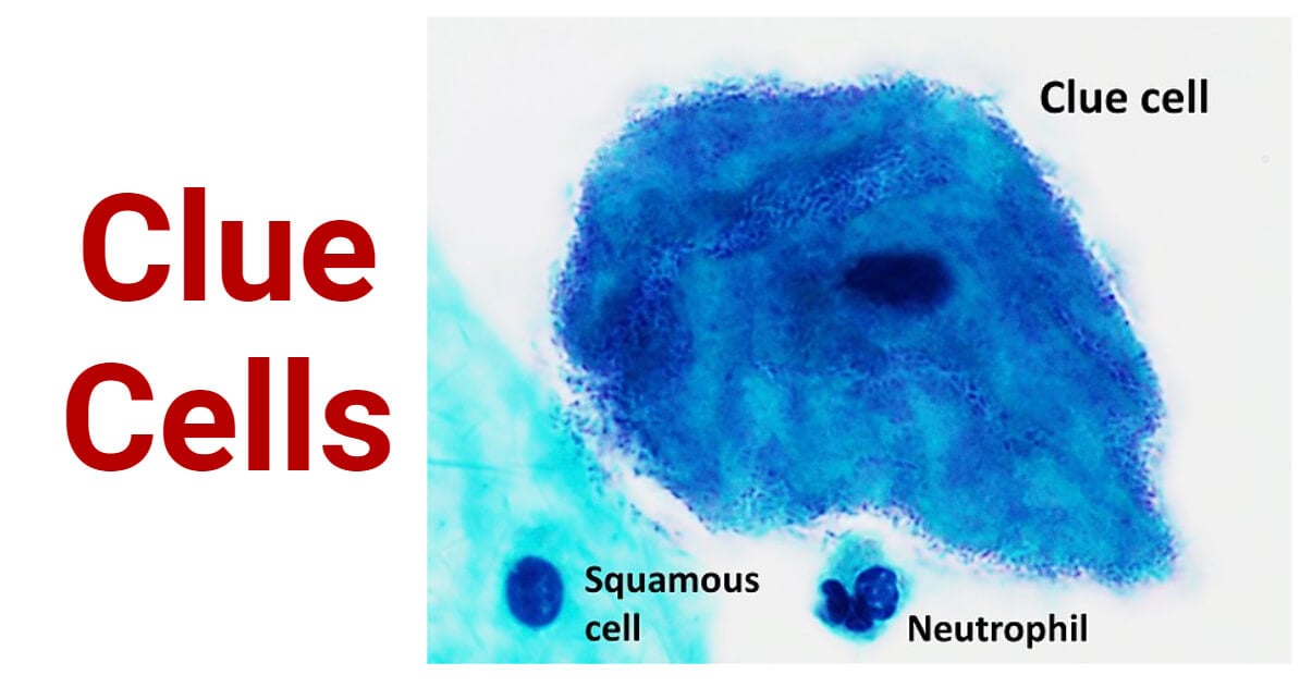
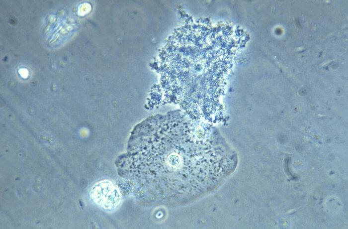
Clue cells are specialized vaginal epithelial cells that appear stippled or granulated due to being covered with numerous bacteria.
Meanwhile, these cells are a critical diagnostic feature in gynecology, particularly for diagnosing bacterial vaginosis (BV), a common vaginal infection.
The term “clue” in clue cells refers to the microscopic clues they provide regarding the presence of BV.

Interesting Science Videos
Overview of Bacterial Vaginosis
Bacterial vaginosis (BV) is a common vaginal infection resulting from an imbalance in the vaginal microbiota.
It occurs when there’s an overgrowth of harmful bacteria, disrupting the natural balance of the vaginal flora.
BV is often associated with symptoms like abnormal vaginal discharge, itching, and a characteristic “fishy” odor.

Appearance and Identification of Clue Cells
Clue cells, when observed under a microscope, display a distinct appearance. They are vaginal epithelial cells with a stippled or granulated surface due to the adherence of bacteria.
Additionally, the granular appearance distinguishes them from normal vaginal epithelial cells and is a key characteristic for identifying them.
Methods of Identification of Clue Cells
Wet Mount Examination: In this method, a sample of vaginal discharge is mixed with a solution and placed on a slide for microscopic examination. Clue cells can be observed directly under the microscope using this technique.
Gram Stain: Gram staining involves using a series of staining steps to differentiate bacteria based on their cell wall characteristics. Clue cells will retain the stain and can be observed under the microscope after this staining process.
Other Laboratory Techniques: Besides wet mount and Gram stain, other laboratory techniques may include the use of specific dyes or culture methods to identify clue cells. These techniques enhance the accuracy and reliability of clue cell identification.
Association with Bacterial Vaginosis (BV)
Bacterial vaginosis (BV) is a common vaginal infection resulting from an imbalance in the vaginal flora, characterized by a decrease in beneficial Lactobacillus species and an overgrowth of various anaerobic bacteria, such as Gardnerella vaginalis.
The exact cause of this imbalance is not fully understood, but various factors, including sexual activity, douching, hormonal changes, and certain behaviors or practices, can influence it.
Link Between Clue Cells and Bacterial Vaginosis (BV)
Clue cells are vaginal epithelial cells covered with bacteria, making them appear stippled or granulated when viewed under a microscope. In the context of BV, the presence of clue cells is a key diagnostic criterion.
The bacteria adhering to these cells are often indicative of an overgrowth of anaerobic bacteria associated with BV, especially Gardnerella vaginalis.
In contrast, the abundance of clue cells in a microscopic examination of vaginal discharge suggests the presence of bacterial vaginosis.
Importance of Clue Cells in Bacterial Vaginosis (BV) Diagnosis
Clue cells are crucial in diagnosing BV due to their strong association with the condition. Their presence in a wet mount or Gram stain examination of vaginal discharge provides a reliable indication of bacterial vaginosis.
Healthcare providers use clue cells as a significant diagnostic marker to initiate appropriate treatment for the patient. It ensures timely management of BV and minimizes potential complications.
Clinical Implications
Symptoms of Bacterial Vaginosis (BV)
- Often thin, grayish-white, and may have a “fishy” odor.
- Discomfort and itching in the vaginal area.
- Discomfort or burning during urination.
- Unpleasant or noticeable fish-like odor, particularly after intercourse.
- Pain or discomfort during sexual intercourse.
Diagnostic Criteria Involving Clue Cells
The diagnostic criteria for BV often include the presence of clue cells (>20% of the epithelial cells) in a high-power field on microscopic examination of vaginal discharge, along with clinical symptoms and other laboratory findings.
The presence of clue cells guides healthcare providers in choosing the appropriate treatment for BV. It helps in determining the type and duration of antibiotic therapy needed to eradicate the overgrowth of anaerobic bacteria and restore a balanced vaginal flora.
Diagnostic Procedures
A sample of vaginal discharge is collected using a swab. The swab is gently inserted into the vagina to obtain a specimen of the discharge, which is then used for microscopic examination to identify clue cells.
Laboratory Analysis Techniques
- Wet Mount Examination: A drop of vaginal discharge is placed on a slide, mixed with saline, and examined under a microscope to observe clue cells.
- Gram Stain: The discharge is spread on a slide, fixed, and stained using the Gram staining technique. Clue cells appear characteristic under this stain.
Interpretation of Results
If clue cells are observed in significant numbers during microscopic examination, this indicates a probable case of BV.
The abundance of clue cells is an important diagnostic parameter in identifying BV and determining appropriate treatment strategies.
Conclusion
Clue cells, vaginal epithelial cells covered with bacteria, serve as a critical diagnostic feature for bacterial vaginosis (BV). Their presence in vaginal discharge, indicative of an overgrowth of certain bacteria, is a key element in diagnosing BV accurately.
The presence of clue cells is a significant indicator for healthcare professionals in diagnosing BV and tailoring appropriate treatment.
Early detection through clue cells facilitates timely interventions, effectively addressing symptoms and reducing potential complications associated with bacterial vaginosis.
References
- https://myhealth.alberta.ca/Health/Pages/conditions.aspx?hwid=aa73146&lang=en-ca
- https://ijdvl.com/clue-cell/
- https://www.mayoclinic.org/diseases-conditions/bacterial-vaginosis/diagnosis-treatment/drc-20352285
- https://www.peacehealth.org/medical-topics/id/hw3367
- https://youngwomenshealth.org/guides/bacterial-vaginosis/
- https://screening.iarc.fr/atlasglossdef.php?key=Clue%20cells&lang=1
- http://www.bioline.org.br/request?dv06137
