Interesting Science Videos
Heart Conduction System
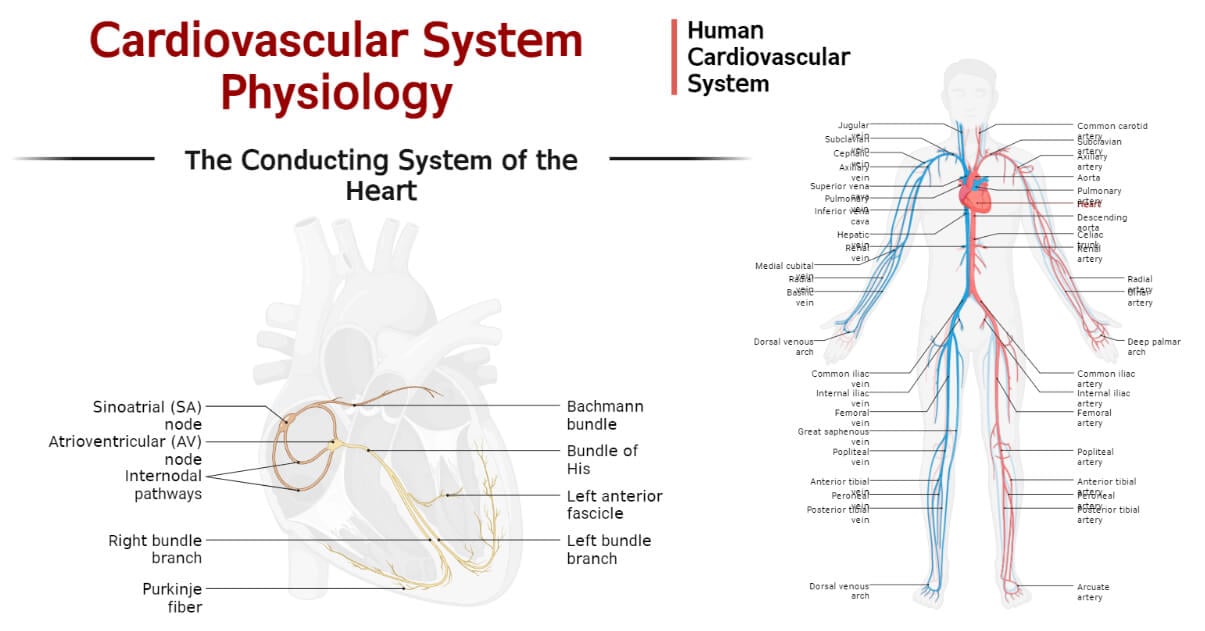
Also called the cardiac conduction system or the intrinsic conduction system of the heart or electrical conduction system of the heart, is a group of specialized cells and tissues that spontaneously generate and transmit the electrical impulse across the heart for regulation of the pumping action of the heart.

Each myocyte (cells of the heart) is capable of being excited i.e. conducting the electric impulse; however, only a few of them are specialized for the conduction of the cardiac action potential. And some specific myocytes are capable of generating the cardiac action potential (electric impulse). The network of these modified (specialized) myocytes collectively forms the conduction system of the heart. This conduction system of the human heart is intrinsic i.e. the myocytes produce the impulse themselves without the involvement of neurons. The major components of the human heart conduction system include the sinoatrial node, atrioventricular node, Bundle of His, and Purkinje fibers.
The Sinoatrial Node (SA node)
It is a small oval-shaped mass of specialized myocytes called the pacemaker cells that produces a cardiac action potential. It is also called the sinus node or sinoatrial node or simply the SA node. It is the natural pacemaker of the heart. It is 15 mm 3 mm 1 mm in dimension and is located in the upper back side of the epicardium of the right atrium just below and side of the superior vena cava. It is surrounded by the perinodal cells and the connective tissues insulating the action potential generated within the SA node.
Its main function is to spontaneously generate the cardiac action potential (electrical impulses), hence it is called the natural pacemaker. The impulse conducted by the pacemaker cells is transmitted to the perinodal cells from which it is transmitted over other structures of the conduction system. The activity of the SA node is regulated by the sympathetic and the parasympathetic nervous system, but the impulse is produced by the pacemaker cells.
The Atrioventricular Node (AV node)
It is a small oval-shaped node of specialized myocytes that relay the cardiac impulse from the atrium towards the ventricle for ventricular contraction. It is very small measuring only about 5 mm 3 mm 1 mm. It is located near the center of the heart at the lower right end of the interatrial septum in close proximity to the ventricles.
Its main function is to coordinate the atrial contraction and the ventricular contraction. It collects the cardiac impulses from the atrium, delays the impulse by about 0.09s, and sends the impulse down the other structures and fibers for contraction of the ventricles.
AV node can spontaneously produce an electric impulse at the rate of 40 to 60 times per minute and transmit it down to compensate for the loss of impulse during the atrial conduction and contraction and to run the cardiac cycle if there is a disturbance in the SA node. Hence, the AV node is considered the second pacemaker of the human heart.
Bundle of His
Bundle of His, also known as the atrioventricular (AV) bundle, is a collection of special myocytes that conducts the cardiac impulse from the AV node to the Purkinje Fiber for conduction across the ventricles. It descends the AV node and divides into two branches; the right bundle branch transmits the impulse to the Purkinje Fiber of the right ventricle, and the left bundle branch transmits the impulse to the Purkinje Fiber of the left ventricle.
Purkinje Fiber
Purkinje fiber is the network of specialized impulse-conducting myocytes that transmit the cardiac electric impulse to every part of the heart ventricles. They are originated from the Bundle of His and are branched and distributed across the ventricular wall below the endocardium within the space called the subendocardium.
They are made of electrically excitable cells that can conduct impulses at a higher rate and more effectively than any other myocytes. The cells are larger than other myocytes with many mitochondria and few myofibrils.
Besides conducting the electric impulse, the Purkinje Fibers are also capable of generating a cardiac action potential. They can spontaneously produce cardiac electric impulses at the rate of 20 to 40 times per minute and can compensate for the cardiac impulse and contraction if the pacemaker fails to function properly.
Pathway of Cardiac Conduction
The heart’s contraction and relaxation are regulated by the cardiac electric impulse (cardiac action potential) and its transmission. This rate of the generation and transmission of the cardiac impulse is called cardiac conduction.
The overall cardiac conduction can be summarized in the following four steps.
1. Impulse Generation by SA Node
The SA node continuously contracts and relaxes spontaneously generating the cardiac action potential 60 to 100 times per minute. The impulse is then transmitted via the internodal tract passing through the right atrial wall and through Bachmann’s bundle which transmits the impulse to the left atrial wall.
2. Impulse Conduction by AV Node
The AV node then collects all the impulses from the atrium, delay them for 0.09s, and relays the impulse to the AV bundle.
3. Relay of Impulse by AV Bundle
The AV bundle functions as the link between the AV node and the Purkinje fiber for the cardiac impulse to travel.
4. Conduction of Impulse by Purkinje Fibers
The cardiac impulse finally reaches the Purkinje Fibers which then branch and spread the impulse all over the ventricular wall.
Cardiac Action Potential
The cardiac action potential is the change in the membrane voltage (potential) across the membrane of cardiomyocytes due to the movement of ions in and out of the cardiomyocytes. Na+, K+, Ca+2, Cl– ions play a major role in generating the cardiac action potential. This cardiac action potential, also called cardiac impulses, is generated intrinsically by the Pacemaker cells. This potential causes the heart to regularly contract and relax facilitating the continuous running of the cardiac cycle. There is a continuous change in the cardiac action potential, which is recorded in an ECG (electrocardiogram) to study the cardiac conduction system and monitor the heartbeat rhythm.

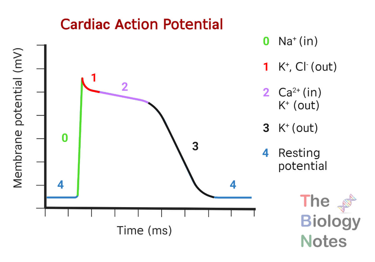
The cardiac action potential in cardiomyocytes occurs in a closed cycle of 5 phases, which is designated as phase 0 to phase 4.
Phase 4
Also called the resting membrane potential (RMP) phase, is an initial stage where the chambers are in the diastole stage and the non-pacemaker cardiomyocytes are at an RMP of -90 mV. In this stage there is the continuous efflux of the K+ ions via inward rectifier channels; additionally, the Na+ and Ca+2 channels are also in the closed stage.
In this stage, the action potential in the Pacemaker cells increases and reaches around -40 mV. This is believed either due to the HCN channels which allow Na+ and K+ ions to enter the cell at very low action potential or due to the calcium clock which allows the entrance of three Na+ ions in exchange for one Ca+2. Hence, for the Pacemaker cells, this stage is called Pacemaker Potential.
Phase 0
This phase is called the depolarization phase because the cardiac action potential increase around +50 mV from -90 mV. The opening of a voltage-gated sodium channel causes the rapid influx of Na+ ions in the non-Pacemaker cardiomyocytes increasing the potential to around +50 mV.
In the Pacemaker cells, the L-type (long opening type) calcium channel causes an inflow of Ca+2 increasing the action potential across the Pacemaker cells.
Phase 1
This phase is called the early repolarization phase where the Na+ channels are rapidly inactivated limiting the influx of the Na+ ions inside the cardiomyocytes. Concurrently, there is an opening of the K+ and Cl– ion gates, which further promotes repolarization. There is no significant Phase 1 in the Pacemaker cells.
Phase 2
This phase is also called the Plateau phase where the membrane potential is maintained just below 0 mV. In this phase, the L-type calcium channels and the delayed rectifier potassium channels are open causing a constant influx of Ca+2 ions and outflux of the K+ ions. The activated Ca+2 ion channels and flow of the Ca+2 result in the opening of Cl– ion channels that cause an influx of the Cl– ions. There is no significant Phase 2 in the Pacemaker cells.
Phase 3
This phase is also called the rapid repolarization phase and is characterized by the closing of the L-type calcium channels and opening of the K+ ion channels. The rapid outflux of the K+ ions exceeds the influx of the Ca+2 ions causing the membrane potential to come to -90 mV. This triggers the inactivation of the Ca+2 channels. Rapidly, the Ca+2 and Na+ ions efflux to extracellular space, and K+ ions enter the cell restoring the resting membrane potential of about -90 mV.
Closing of the L-type Ca+2 ion channel and opening of the K+ ion channel (rapid delayed rectifier type K+ channel) results in the rapid repolarization of the Pacemaker cells.
Cardiac Cycle
The cardiac cycle is a continuous closed sequence of events that results in the continuous and systematic contraction and relaxation of the chambers of the heart. Under the influence of the cardiac action potential, the heart continuously completes the cardiac cycle. The overall time required to complete one cardiac cycle is about 0.8 seconds.
The human cardiac cycle can be divided into the following five stages:
1. Atrial Systole
It is the contraction of the atrial myocardium to pump the blood out of the atria into the ventricles. The impulse generated by the SA node triggers the atrial systole. This stage is also known as presystole or the last rapid filling phase or atrial kick.
This stage is coupled with the late stage of ventricular diastole where about 20% of the remaining atrial blood is forced into the ventricle. This stage is completed in about 0.11 seconds.
2. Ventricular Systole
It is the stage where the ventricle contracts expelling the blood outside of the ventricles. It completes in about 0.3 seconds. The ventricular systole can be further subdivided into three stages:
Isovolumetric Ventricular Contraction
As the name suggests, in this stage volume of the ventricles does not change but the muscle tension is increased and the ventricles begin to contract. It is the first stage of ventricular systole. In this stage, the pressure inside the ventricle is increased making it higher than atrial pressure. This pressure difference causes the (atrioventricular) AV valves to close. The aortic and pulmonary valves are also closed in this stage, so the blood can’t leave the ventricle increasing the internal pressure of the ventricles. This stage lasts for about 0.05 seconds and the heart experiences another stage, ventricular ejection.
Rapid Ventricular Ejection
The ventricular pressure at the right ventricle will be slightly above 8 mm of Hg and the ventricular pressure at the left ventricle will be about 80 mm of Hg. This pressure forces both of the semilunar valves to open. When the semilunar valves open, immediately, about 70% of the ventricular blood in high pressure will be forced out of the ventricles through the aorta and the pulmonary arteries within about 0.13 seconds.
Reduced Ventricular Ejection
It is the second stage of ventricular ejection of blood when the remaining 30% of the blood is ejected from the ventricles. By this stage, the ventricular pressure will be dropped and the blood will have traveled up to smaller arteries. This stage usually lasts for about 0.09 seconds.
3. Protodiastole
It is the intermediary stage which indicates the end of systole and the beginning of the diastole stage. It is the first stage of ventricular diastole. After the ejection of blood, the pressure inside the ventricles drops, and the ventricular pressure becomes lower than the pressure in the pulmonary artery and aorta. This triggers the closing of the semilunar valves. This stage lasts for about 0.04 seconds.
4. Atrial Diastole
It can be marked as the beginning of a new cardiac cycle. This stage occurs immediately before the ventricular diastole stage. During this stage, the AV valves are closed and the blood is poured into the atria by systemic (vena cava) and pulmonary veins. This stage lasts about 0.7 seconds and overlaps the ventricular systole phase.
5. Ventricular Diastole
It is the stage when the blood enters the ventricles increasing the pressure inside the ventricles. This stage lasts for about 0.5 seconds and can be further subdivided into the following stages:
Isovolumetric Ventricular Relaxation
As the name suggests, the volume inside the ventricles does not change but the tension in the ventricular wall is reduced. This reduced pressure causes the semilunar valves to completely close. The AV valves are also closed in this stage so blood can’t flow in the ventricles and the volume of the ventricles remains the same. However, the rapid pressure drop will make the atrial pressure higher and the ventricular pressure lower which triggers the next stage. This stage usually lasts about 0.08 seconds.
Rapid Ventricular Filling
This pressure difference causes the AV valves to open rushing about 70% of the atrial blood into the ventricles. This stage lasts for about 0.11 seconds.
Reduced Ventricular Filling
This is the second stage of ventricular filling, also called diastasis, where about 20% of the atrial blood enters the ventricles at a slower rate. The time required for the completion of this stage varies according to the heart rate and completes in about 0.19 seconds.
Rapid Ventricular Filling
This is the last stage which overlaps the atrial systole stage and the remaining 10% of blood is pumped into the ventricles in this stage. This stage lasts for about 0.1 seconds.
Heart Beat and Heart Sound
The regular contraction and relaxation of the chambers of the heart in a rhythmic cycle are called heartbeat. The cardiac action potential generated by the SA node and conducted by the heart conduction system causes the heart to operate a continuous cardiac cycle. This cardiac cycle results in a heartbeat. The heartbeat is characterized by the two phages; the systole and diastole of the heart chambers.
The sound produced by the regular opening and closing of the heart valves during each cardiac cycle is called the heart sound. It is referred to as the “lub-dub” sound of the heart. There are four types of heart sounds; two primary heart sounds that are due to the closing of the heart valves, and the two secondary heart sounds that are due to blood rush.
The First Heart Sound
It is also called the S1 sound, which is produced as a result of the sudden closure of the two atrioventricular (AV) valves. It is produced during the ventricular systole stage of the cardiac cycle at the Isovolumetric ventricular contraction phage. The S1 sound is the low-pitched, soft, and long sound of about 25 to 45 Hz which is heard and referred to as the “Lub” sound. This is the longest sound and lasts for about 0.14 seconds.
The Second Heart Sound
It is also called the S2 sound and is produced as a result of the sudden closing of the two semilunar valves. It is produced during the protodiastole (end of the ventricular systole and beginning of the ventricular diastole) stage of the cardiac cycle. The S2 sound is a high-pitched, loud, and short sound of about 50 Hz which is heard and referred to as the “Dub” sound. This sound lasts for about 0.1 seconds.
The Third Heart Sound
It is also called the S3 sound or the Protodiastolic Gallop or the Kentucky Gallop or the Ventricular Gallop and is produced as a result of the back-and-forth oscillation of the blood in the ventricular wall during the inflow of the blood from the atria. The sound is usually low-pitched and weak and is rarely heard in normal conditions in adult persons. When this sound is heard loudly, the heart sound is referred to as the “Lub-Dub-Ta” sound and is an indication of congestive heart failure or increased blood volume.
The Fourth Heart Sound
It is also called the S4 sound or the Atrial Gallop or the Presystolic Gallop and is caused by blood flow during the atrial systole. It is a very weak, low-intensity sound and is rarely heard in normal conditions just before the “Lub” sound. When this sound is heard loudly, the heart sound is heard and referred to as the “Ta-Lub-Dub” sound. Loud hearing of this sound also indicates a high chance of heart disease and heart failure.
Cardiac Output
- The cardiac output is the total amount of blood pumped by the ventricles per minute. On average, for a heartbeat of 70 beats per minute, the cardiac output (CO) is about 4.9 (5) Liters per minute.
- Mathematically, CO is defined as the product of the heart rate (HR) and stroke volume (SV). It is calculated as: CO=HR × SV
- HR or heart rate is defined as the number of beats per minute and SV or stroke volume is defined as the volume of blood pumped by the left ventricle in one beat.
- The higher the CO, the faster and stronger the heart beats pumping a larger volume of blood quickly than in normal conditions. Hence, when the body needs rapid cellular respiration and release of energy, like during physical activities, the CO increases.
Blood Pressure
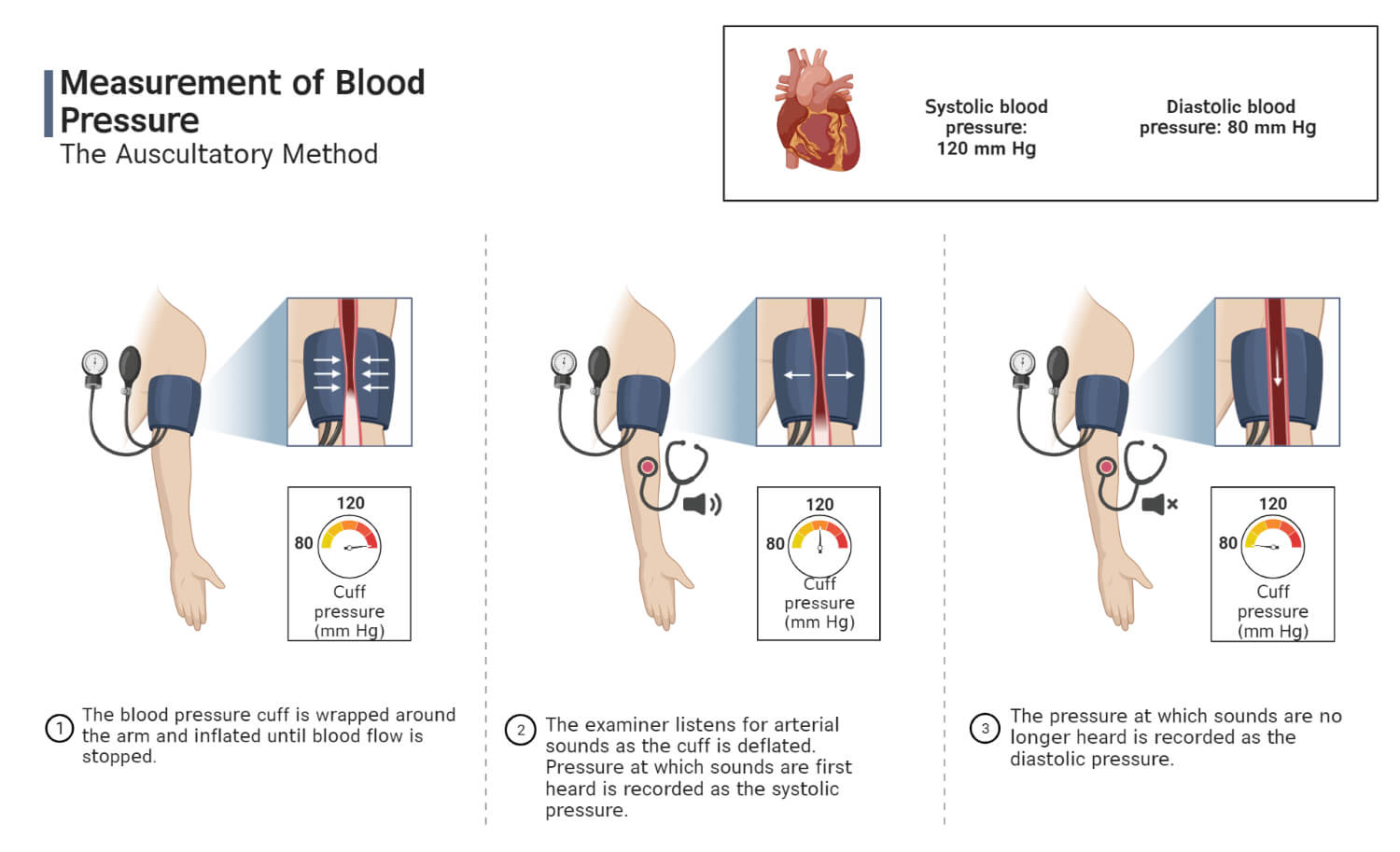
Blood pressure is the measure of the pressure of the blood in the major arteries (blood vessels) during the systole and diastole stages of the cardiac cycle. It is measured in mm of Hg and the reading is taken at the extreme point i.e. at the systole and diastole stage. The blood pressure is expressed as systolic pressure and diastolic pressure. The normal blood pressure of a human is 120/80 (systolic/diastolic) mm of Hg. The instrument used to measure human blood pressure is called a sphygmomanometer.

Systolic Pressure
The pressure build inside the blood vessels (arteries) due to ventricular systole is called systolic blood pressure. It is the maximum pressure generated during a heartbeat. In normal conditions, it is about 120 mm of Hg.
If the systolic pressure exceeds 129 mm of Hg, then the situation is called hypertension. The raise of systolic pressure up to 129 mm of Hg is termed elevated pressure, but if it increases about 130 to 139 mm of Hg it is called stage 1 hypertension, if it increases above 140 mm of Hg then it is called stage 2 hypertension, and if it crosses 180 mm of Hg it is referred as hypertensive crisis.
If the systolic pressure drops below 90 mm of Hg, it is called hypotension.
Diastolic Pressure
The pressure build inside the blood vessels (arteries) due to ventricular diastole is called diastolic blood pressure. It is the minimum blood pressure generated during a heartbeat. In normal conditions, it is 80 mm of Hg.
If the diastolic pressure exceeds 80 mm of Hg, then the situation is defined as hypertension. If it increases about 80 to 89 mm of Hg it is called stage 1 hypertension, if it increases above 90 mm of Hg then it is called stage 2 hypertension, and if it crosses 120 mm of Hg it is referred to as a hypertensive crisis.
If the diastolic pressure drops to and below 60 mm of Hg, it is referred to as hypotension.
Conduction System Related Disorders
- Sick Sinus Syndrome (Sinus Node Dysfunction): It is a condition characterized by the abnormality in cardiac impulse production due to either abnormality in pacemaker cells or their function or due to a defect in conduction through the perinodal cells.
- Sinus Bradycardia: It is a condition characterized by reduction in heartbeat below 60 beats per minute.
- Sinus Tachycardia: It is a condition characterized by elevation of heartbeat above 100 beats per minute.
- Decremental Conduction: It is a condition characterized by declination of the speed and amplitude of the cardiac action potential and impulse conduction.
- Bundle Branch Block: It is a condition characterized by delay in the conduction of the cardiac impulse across the Bundle of His.
References
- Conduction System Tutorial (umn.edu)
- Sinoatrial node. (2023, January 7). In Wikipedia. https://en.wikipedia.org/wiki/Sinoatrial_node
- Kashou AH, Basit H, Chhabra L. Physiology, Sinoatrial Node. [Updated 2022 Oct 3]. In: StatPearls [Internet]. Treasure Island (FL): StatPearls Publishing; 2022 Jan-. Available from: https://www.ncbi.nlm.nih.gov/books/NBK459238/
- Boyett MR, Honjo H, Kodama I. The sinoatrial node, a heterogeneous pacemaker structure. Cardiovasc Res. 2000 Sep;47(4):658-87. doi: 10.1016/s0008-6363(00)00135-8. PMID: 10974216.
- Atrioventricular Node (AV Node): Function and Purpose (verywellhealth.com)
- Atrioventricular node | Kenhub
- Patra C, Zhang X, Brady MF. Physiology, Bundle of His. [Updated 2022 May 8]. In: StatPearls [Internet]. Treasure Island (FL): StatPearls Publishing; 2022 Jan-. Available from: https://www.ncbi.nlm.nih.gov/books/NBK531498/
- Chaudhry R, Miao JH, Rehman A. Physiology, Cardiovascular. [Updated 2022 Oct 16]. In: StatPearls [Internet]. Treasure Island (FL): StatPearls Publishing; 2022 Jan-. Available from: https://www.ncbi.nlm.nih.gov/books/NBK493197/
- King J, Lowery DR. Physiology, Cardiac Output. [Updated 2022 Jul 19]. In: StatPearls [Internet]. Treasure Island (FL): StatPearls Publishing; 2022 Jan-. Available from: https://www.ncbi.nlm.nih.gov/books/NBK470455/
- King J, Lowery DR. Physiology, Cardiac Output. 2022 Jul 19. In: StatPearls [Internet]. Treasure Island (FL): StatPearls Publishing; 2022 Jan–. PMID: 29262215.
- What is a bundle of His? (byjus.com)
- Everything You Need to Know About Purkinje Fibers – Bodytomy
- Conducting System of the Heart – Bundle of His – SA Node – TeachMeAnatomy
- Purkinje Fibers : Anatomy, Location & Function – Anatomy Info
- Overview of Sinoatrial and Atrioventricular Heart Nodes (thoughtco.com)
- Boyden PA. Purkinje physiology and pathophysiology. J Interv Card Electrophysiol. 2018 Aug;52(3):255-262. doi: 10.1007/s10840-018-0414-3. Epub 2018 Jul 28. PMID: 30056516.
- The Heart’s Conduction System | Physiology, Anatomy | Geeky Medics
- 4 Steps of Cardiac Conduction (thoughtco.com)
- Cardiac Cycle- Physiology, Diagram, Phases of the Cardiac Cycle (byjus.com)
- Cardiac cycle. (2023, January 18). In Wikipedia. https://en.wikipedia.org/wiki/Cardiac_cycle
- Physiology of cardiac conduction and contractility Mc Master Pathophysiology Review – 04/12/2018 – Studocu
- Cardiac Cycle,Phases of Cardiac Cycle,Cardiac Cycle & ECG (medicosite.com)
- 19.3 Cardiac Cycle – Anatomy & Physiology (oregonstate.education)
- The Cardiac Cycle – SimpleMed – Learning Medicine, Simplified
- Human Heart – Anatomy and Functions | Location and Chambers (vedantu.com)
- Cardiac action potential. (2022, November 3). In Wikipedia. https://en.wikipedia.org/wiki/Cardiac_action_potential
- Action Potentials in Cardiac Muscle Cells – BIO 461 Principles of Physiology (byui.edu)
- Cardiac Action Potential (byjus.com)
- Physiology of cardiac conduction and contractility | McMaster Pathophysiology Review
- Cardiac Action Potentials – Human Physiology (uoguelph.ca)
- Cardiac Cycle: Meaning, Duration and Phases (biologydiscussion.com)
- Cardiac cycle phases: Definition, systole and diastole | Kenhub
- What Are the Four Heart Sounds? (medicinenet.com)
- Heart Sounds – What Causes it, Different Types of Heart Sounds (byjus.com)
- Heartbeat | definition of heartbeat by Medical dictionary (thefreedictionary.com)
- Heart rate: What’s normal? – Mayo Clinic
- How the Heart Works – How the Heart Beats | NHLBI, NIH
- 19.3 Cardiac Cycle – Anatomy & Physiology (oregonstate.education)
- Cardiac Output- Definition, Factors Affecting, Cardiac Index (byjus.com)
- www.k8s.lb.webmd.com | 504: Gateway time-out
- Understanding Blood Pressure Readings | American Heart Association
- What is a healthy blood pressure? | healthdirect
- Blood pressure chart: What your reading means – Mayo Clinic
- Blood Pressure Chart & Numbers (Normal Range, Systolic, Diastolic) (webmd.com)
- Sinus Tachycardia: Causes, Symptoms, and Treatment (healthline.com)
- Sinus Tachycardia: Causes, Symptoms & Treatment (clevelandclinic.org)
- Sinus Bradycardia: Causes, Symptoms & Treatment (clevelandclinic.org)
- Sinus Bradycardia: Symptoms, Causes, Treatment, and More (healthline.com)
- Bradycardia – Symptoms and causes – Mayo Clinic
- Hluchy J, Schickel S, Schlegelmilch P, Jörger U, Brägelmann F, Sabin GV. Decremental conduction properties in overt and concealed atrioventricular accessory pathways. Europace. 2000 Jan;2(1):42-53. doi: 10.1053/eupc.1999.0069. PMID: 11225595.
- Decremental conduction | definition of decremental conduction by Medical dictionary (thefreedictionary.com)
- Bundle branch block – Symptoms and causes – Mayo Clinic Bundle Branch Block: What to Know (webmd.com)
- Bundle Branch Block: Causes, Symptoms & Treatment (clevelandclinic.org)
- ECG (EKG) – bundle branch block – Oxford Medical Education
