Interesting Science Videos
Digestive System of Ascaris lumbricoides
I) Alimentary canal: It is a straight and complete tube that starts with the mouth at the anterior end and ends with the anus at the posterior end. It comprises a short pharynx or esophagus representing foregut, along intestine or mid-gut, and short rectum or hindgut.

Figure: Digestive System of Ascaris lumbricoides. Image Source: https://digestivesystemgroup2.weebly.com/roundworm-ascaris-lumbricoides.html
1. Mouth
- It is a triradiate aperture.
- It is located at the anterior tip.
- It is guarded by 3 lips or labia.
2. Pharynx
- The mouth opens into a short, characteristics, cylindrical, thick-walled, and muscular pharynx.
- It has posterior swelling called the end bulb which is provided with valves.
- Its wall consists of syncytial epithelium traversed by radial muscle fibers which dilate the lumen.
- It is externally bounded by a membrane and internally lined by a cuticle which is in continuation with the body wall.
- The pharynx also has 3 branching gland cells which open by circular ducts into the lumen. These glands are in fact pharyngeal or oesophageal glands.
- The lumen of the pharynx is also triradiate.
- In the outer end of each ray of the lumen and the outer bounding membrane, the connective tissue fibers called marginal fibers are present. These fibers help in maintaining the triradiate form of the lumen.
3. Intestine
- The pharynx is followed by a thin-walled dorsoventrally flattened intestine or mid-gut.
- It extends almost along the entire body length.
- Its wall consists of a single layer of tall columnar cells, lined externally by a basement membrane, and a thin layer of cuticle.
- The inner margins of each cell are produced into hair-like projections called microvilli which help the surface area of absorption.
- The microvilli are in fact are formed by a bacillary layer.
- It has no muscle fibers.
4. Rectum
- It is the last part of the digestive system.
- The intestine is followed by a short dorso-ventrally flattened rectum.
- Its wall consists of a thin layer of tall columnar epithelial cells.
- It is internally lined by a cuticle which is a continuation with the body wall and externally by muscle tissue.
- In males, the rectum opens by cloaca because it receives an ejaculatory duct.
- Whereas in females, the rectum opens out through the anus. The anus is guarded by lips and is also provided with special dilator muscles running from the rectum to the body wall, called depressor ani.
- The contraction of these muscles from time to time causes the fecal matters to be discharged out.
- Anus or cloaca lies at a distance of about 2 mm from the tail end.
- It has also large unicellular rectal glands: 3 in females and 6 in males.
II) Food, feeding, and digestion
- The food of Ascaris comprises of blood, tissue exudes, and partly or fully digested food occurring in the fluid form in the host’s gut.
- The food is sucked by the suctorial action of the pharynx.
- Digestion is completely extracellular in the intestine.
- The digestion is facilitated by the enzymes like proteases, amylase, and lipase secreted by the gland cell of the pharynx.
- The digested food is absorbed by the intestinal cell and distributed by pseudo coelomic fluid.
- Excess food is stored mainly as glycogen and little fat in the intestinal wall, muscle, and syncytial epidermis.
- Intercellular digestion has also been reported to occur in the cells of the intestinal wall as they engulf the small solid particles by phagocytosis and digest them intracellularly.
- The undigested food or defecation is facilitated by the depressor ani muscle which raises the dorsal wall of the rectum and the posterior lip of the anus or cloaca.
Respiration in Ascaris lumbricoides
- Ascaris lumbricoides has no respiratory organs.
- Like other endo-parasites they respire anaerobically or anoxybiotically as the oxygen content in the intestine host is poor.
- In the process of respiration, the glycogen is breakdown into carbon dioxide and fatty acids and energy by glycolysis which are excreted through cuticle like those of flatworm parasites.
- Main fatty acids valerianic, butyric, and caproic produced as excretory wastes.
- Aerobic respiration probably occurs whenever free oxygen is available in the host intestine.
- This is indicated by the presence of a small amount of cytochrome in the parasite.
- The small amount of hemoglobin in the body wall and pseudocoel fluid takes up the oxygen even when it is present in a low amount.
Excretory system of Ascaris lumbricoides
- The excretory system of Ascaris is simple as there are no flame cells or protonephridia.
- It has a single giant H-shaped rennet cell forming an excretory system.
- It consists of two lateral longitudinal excretory canals, connected below the pharynx by a transverse canalicular network.
- Tunnel-like structures are present in the cytoplasm of rennet cells and these act as excretory canals.
- It consists of two lateral longitudinal excretory canals called right and left longitudinal excretory canals. They are connected anteriorly by a transverse canalicular network.
- Each longitudinal canal extends posteriorly along the entire length of the body and is closed at both ends.
- The canals are more developed on the left side than on the right side.
- The canals are lined by a firm membrane and covered with a layer of cytoplasm.
- The lumen of the canals does not have cilia.
- From the left side of the transverse canalicular network, a short terminal excretory duct extends towards the excretory pore situated a little the anterior end.
- Physiology: It is poorly understood. The available data suggested that the excretory product of Ascaris is mainly urea which diffuses into the pseudocoelomic fluid and so it is also known as the ureotelic animal. The excretory canals collect the excretory products from different parts of the body and these excretory products are eliminated through the excretory pore. The pressure of the pseudocoleomic fluid helps in the ultrafiltration process. Some ammonia and urea are also passed out through the anus.
Nervous system of Ascaris lumbricoides
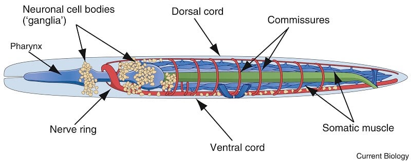
Ascaris has a well developed and complicated nervous system. It is located in the body wall like an excretory system. i.e. hypodermic. According to Goldschmidt (1908-1910) nerve cells forming the system are constant in number, position, form, and course of fibers.

Figure: Overview of nematode nervous systems. Image Source: https://doi.org/10.1016/j.cub.2016.07.044.
1. Central nervous system
- It consists of a rich ganglionated nerve ring or circumenteric ring around the pharynx.
- The ring is made of nerve fibers and some diffusely arranged nerve cells.
- The ganglia present on the nerve ring is an unpaired dorsal ganglion and close to it is a pair of sub-dorsal ganglia.
- On each side of the ring is a pair of lateral ganglia which is divided into 6 ganglia.
- On the lower side of the ring is a pair of large ventral ganglia.
- Each ganglion has an affixed number of nerve cells.
2. Peripheral nerves
- 8 nerves from the nerve ring run anteriorly to supply to the anterior parts of the body.
- Out of these, 6 supply to 6 labial papillae and each bears a papillary ganglion, near its base.
- The remaining 2 nerves are known as amphidial nerves as these supply to the amphids.
- Each amphidial nerve arises from one of the 6 lateral ganglia, called amphidial ganglion, of its side.
- Posterior parts of the body are supplied by 6 long nerves from the rings.
- These 6 nerves, one is the mid-dorsal nerve lies in the dorsal line and one is the mid-ventral nerve lies in the ventral line.
- The main nerve is the mid-ventral nerve and it is ganglionated along the anterior length, it may be called the nerve cord.
- A dorsal nerve cord runs through the dorsal epidermal chord.
- A ventral ganglionated nerve cord runs through the ventral epidermal chord. It terminates at the posterior end after forming an anal ganglion.
- The other 4 posterior nerves are thinner, they are 1 pair of dorsolateral nerves and 1apir of ventrolateral nerves, they lie on the side close to the excretory canals.
- Several transverse commissures are arranged asymmetrically along the entire length connect the ventral nerve cord with lateral and dorsal nerve cords.
3. Rectal nervous system
- In males, the posterior end has lateral nerve cords to supply to pre-anal papillae, ventral nerve cord to supply to post-anal papillae.
- Dorsal, ventral, and lateral nerve cords are interconnected. But such a complex rectal nervous system is absent in female roundworms.
Sense organs of Ascaris lumbricoides
Ascaris has well-developed sense organs which are simple elevations supplied by nerves. They are either as minute elevations or pits in the cuticle of the body. They include various papillae, amphids, and phasmids.
A. Papillae are in the form of small villi situated on different body parts.
1. Labial Papillae
- They are formed by sensory cells surrounded by many supporting cells. These are called gustatoreceptors or taste organs present on 3 lips surrounding the mouth.
- There are 4 labial papillae, 2 on the dorsal lip, and one each on the ventrolateral lips, each is a double sense organ.
2. Cervical papillae
- They are a pair of small pits situated about 2 mm behind lips dorsally.
- They are bulb-like nerve endings with supporting cells.
- These are tactile organs.
3. Anal papillae
- They are found in males.
- They consist of nearly 50 pairs of preanal and 5 pairs of postanal papillae.
- These are also formed 1-3 nerve fibers embedded in the supporting cells.
- They are present ventrally below the posterior end of males.
- They are tactile in function and help in copulation.
B. Amphids are two, situated on each, on each latero-ventral lip. These are small pits containing glandular and nerve cells supplied by amphidial nerve from the lateral or amphidial ganglia. These are gustatory sensory or chemo-receptors.
C. Phasmids are pit-like chemo-receptor. they are unicellular glands situated one on each side of the tail behind the anus.
Locomotion of Ascaris lumbricoides
- The ability to perform a change in body length is restricted due to the absence of circular muscles in the body wall.
- There is a limited change in the length of the body due to the fibers of the cuticle.
- These fibers are themselves inelastic but their arrangement in spirals and mesh allows this limited change in the body length.
- To counteract the peristaltic activity of the host’s intestine, the dorsolateral and ventrolateral muscles in the anterior end of Ascaris, contract alternately to perform undulating movements.
References and Sources
- Kotpal RL. 2017. Modern Text Book of Zoology- Invertebrates. 11th Edition. Rastogi Publications.
- Jordan EL and Verma PS. 2018. Invertebrate Zoology. 14th Edition. S Chand Publishing.
- 9% – https://www.studyandscore.com/studymaterial-detail.php?Id=ascaris-digestive-system-locomotion-respiration-excretion-nervous-system-sense-and-organs
- 7% – https://www.biologydiscussion.com/invertebrate-zoology/phylum-aschelminthe/ascaris-lumbricoides-habitat-sense-organs-and-life-history/29004
- 2% – https://www.studyandscore.com/studymaterial-detail/ascaris-digestive-system-locomotion-respiration-excretion-nervous-system-sense-and-organs
- 1% – https://www.notesonzoology.com/marine-animals/study-notes-on-ascaris-aschelminthes/1653
- 1% – https://www.differencebetween.com/difference-between-dorsal-and-vs-ventral/
- 1% – https://sites.google.com/site/suneelszoology/non-chordata/ascaris
- <1% – https://www.vocabulary.cl/Basic/Body_Parts.htm
- <1% – https://www.studyandscore.com/studymaterial-detail/ascaris-general-characters-body-wall-and-body-cavity
- <1% – https://www.healthline.com/human-body-maps/spine-nerves
- <1% – https://www.brainkart.com/article/Cockroach-(Periplaneta-americana)_33178/
- <1% – https://www.answers.com/Q/Explain_the_importance_of_considering_cuticle_scales_when_disentangling_hair
- <1% – https://www.aimisrobotics.com/condition/anus-and-rectum-diseases/
- <1% – https://quizlet.com/38010163/kin-310-strength-endurance-flash-cards/
- <1% – https://factslegend.org/how-is-food-digested/
- <1% – https://courses.lumenlearning.com/wm-biology2/chapter/parts-of-the-digestive-system/
- <1% – https://answers.yahoo.com/question/index?qid=20120628140141AAIIxRR
