Electrical potential guided by ions exists across the membrane of all body cells. Some cells, such as neuronal cells, cardiac muscle cells, etc., are excitable. The property is called excitability, guided by the stimulus that can exceed the threshold potential.
The action potential is a series of changes in the electrical potential/voltage across the membrane of these cells that causes impulses to transmit along their membrane.
Action potential in neurons is generated due to changes in cationic gradient (mainly Na+ and K+, additionally, in cardiac muscle cells, Ca2+ comes into play).
Interesting Science Videos
Action Potential Mechanism
The mechanisms that maintain the nerve impulse transmission/ action potential across neurons are as follows:
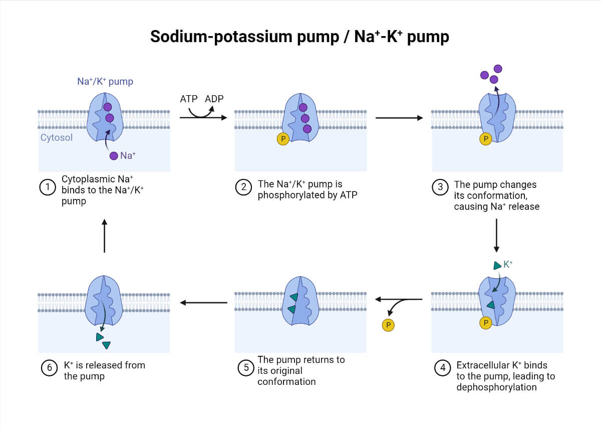
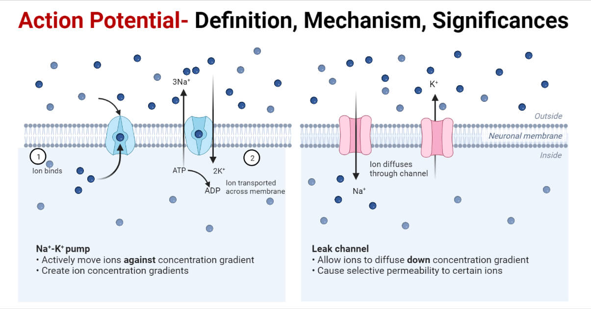
- Sodium-potassium pump/ Na+– K+ pump: This transmembrane ATPase is based on an active transport mechanism in which ions move across the membrane against their concentration gradients. These pumps maintain membrane potential with a higher concentration gradient of Na+ extracellularly and lower K+ ions intracellularly. For each ATP used, 3 Na+ are pumped out of the cell and 2 K+ into the cell.

- Leak channels: These structures are present across the membrane throughout the nerve cell. It works on the passive transport mechanism, i.e., Na+ and K+ ionic permeability depends on the concentration gradient of the ions. Ions flow freely without any impedance.

- Voltage-gated channels: These channels are transmembrane proteins that open in response to the voltage difference across the membrane. Na+ and K+ voltage-gated channels are present in the membrane of the neuronal cells.
The series of changes that occurs during the transmission of nerve impulses are as follows:
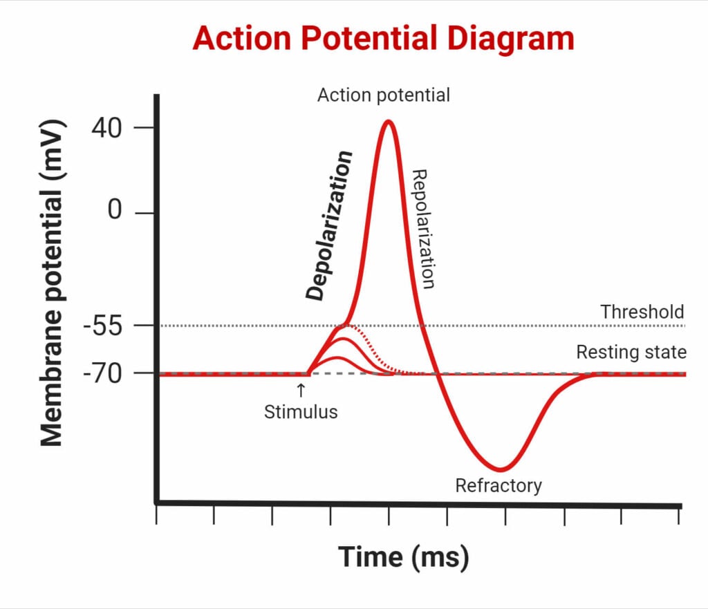
- Resting membrane potential: It comprises the membrane potential at rest when no stimulus is acting on it. For a neuronal cell, the value of the resting membrane potential lies between -50mV and -70mV.
- Depolarization: When a stimulus that can change the membrane potential beyond the threshold acts on electrically active nerve cells, depolarization occurs. The influx of Na+ ions inside the membrane occurs as voltage-gated sodium channels open. The entry of positively charged sodium ions will change the membrane potential to less negative near zero and then near +30mV. This change in membrane potential can open a voltage-gated potassium channel.
The depolarization rises to +30mV, after which the closure of voltage-gated sodium channels occurs.
- Repolarization: Voltage-gated sodium channels begin to close, and potassium-gated channels begin to open. K+ ions start to leave the cell. The membrane potential hence bounces back to negative again till it reaches the resting membrane potential.

- Hyperpolarization: A state of hyperpolarization is achieved due to the opening of voltage-gated K+ channels for a long time in which the membrane potential falls below -70mV. This prevents action potential from developing from any new stimulus for a specified time.
The period during which a new stimulus can not elicit action potential is termed a refractory period. Even a very strong stimulus is unable to generate nerve impulses, and this period lasts for about two milliseconds.
Myelin sheath
The nerve impulses originate in the neuron’s body and propagate through the axon. Most of the axons are protected by myelin sheaths formed by glial cells. The protective sheath helps in the insulation of the axonal membrane. These myelinated structures are oligodendrocytes in the central nervous system and Schwann cells in the peripheral nervous system. In between myelinated segments, there lies a gap called the nodes of Ranvier. The impulse transmission occurs between these nodes and is termed saltatory conduction.
Synapse
Synapses are the gap junction between any two neurons. The neuron sending the signal is termed a presynaptic neuron, while the one receiving it is termed a postsynaptic neuron.
Depending upon the type of signal permitted, synapses can be either chemical or electrical. Chemical synapses are mediated by chemicals called neurotransmitters in a unidirectional way. Neurotransmitters are present in synaptic vesicles released out in the synaptic cleft and bind to receptor molecules on the postsynaptic membrane.
Ions mediate electrical synapses in a bidirectional way. The voltage changes in the presynaptic membrane induce the voltage change in the postsynaptic membrane through specialized structures called gap junctions. The flow rate of impulses, in this case, is much more rapid.
Significances of action potential
- Sensory function: Propagation of information from receptors to the central nervous system.
- Motor function: Propagation of commands generated in the CNS to the periphery/ target tissue.
- Heartbeat generation due to action potential in cardiac muscle cells.
References
- https://www.sciencedirect.com/topics/neuroscience/action-potential
- https://www.kenhub.com/en/library/anatomy/action-potential
- https://open.oregonstate.education/aandp/chapter/12-5-the-action-potential
- Bezanilla F. (2005). Voltage-gated ion channels. IEEE transactions on nanobioscience, 4(1), 34–48. https://doi.org/10.1109/tnb.2004.842463
- https://sciencing.com/phases-cardiac-action-potential-6523692.html
