Plant cells are multicellular eukaryotic cells that make up a plant (a group of eukaryotes belonging to the Plantae kingdom, with the ability to synthesis their own food using water, Sunlight, and CO2). Being eukaryotic cells, they have a defined nucleus with specialized structural organelles that enable them to function in an orderly manner.
The plant cell has a well-defined cell wall made up of cellulose components, plastids that perform photosynthesis and storage of carbohydrates in form of starch, central vacuoles for regulating the cell’s turgor pressure, and a nucleus which controls the cells’ general mechanisms including reproduction of the plant cells. There are several plant cell organelles that are well defined and described in Plant cell.
Interesting Science Videos
General features of plant cell
Plants are made up of two structural systems i.e The shoot system and the root system, whereby the shoot system is made up of structures that ie above the ground including leaves, stems, fruits, flowers while the root system is made up of roots, tubers, and rhizobial structure that lie below the ground and its the origin of growth of plants.
These systems are structured differently, defined by sets of specialized mature cells that perform a wide range of functions ranging from protection, support, metabolism, reproduction enabling plant growth, and development. For example, plant cells are formed at the meristem which multiple and grows to for plant tissues. These tissues are:
- Dermal tissue – this tissue lies on the surface of plants and is made up of epidermal cells that protect the plants from losing water.
- Ground tissue – This makes up the root vascular and epidermal system majorly made up of parenchyma, collenchyma, and sclerenchyma cells responsible for plant photosynthesis, storage of water and food, and the plant support system.
- Vascular Tissue – this tissue is made up of xylem, phloem, parenchyma, and cambium cells, with its functions including transportation of water (xylem), transportation of food (phloem), minerals, hormones in the plants.s of the plant cells
Plant cells multiply by cell division, a mechanism known as Mitosis, which takes place within its nucleus. This begins at the meristem, which is found at the tip of the root and/or the shoot of vascular plants. Meristems at the tips are known as apical and lateral meristems. Apical meristems are responsible for producing the roots while the lateral meristems produce secondary growth of the stem wood and cork.
Besides cell division of the cells that leads to the formation of tissues that eventually creates a plant, there are other features of the plant cells that are of importance to plant growth and metabolisms.
- Presence of a cell wall combined with a plasma membrane. Its made up of cellulose, hemicellulose, and pectin. The cell wall gives the cell shape, cell protection, and mediation of cellular interactions.
- They have dynamic single-membranous central vacuoles that are filled with water to maintain the turgor pressure of the cell, regulate the movement of cellular molecules within the cytosol, storage of nitrogen and phosphorus, and mediate digestion of stored cellular proteins.
- They have a plasmodesmata, which is a continuous porous structure that extends from the endoplasmic reticulum, allowing cell-cell communication.
- Plant cells also have plastids. The most common plastid known as chloroplasts is made up of chlorophyll, a green pigment responsible for capturing light energy and converting it to chemical energy that is used by plants in photosynthesis. other plastids include amyloplast for storage of starch, elaioplast, for storage of fats, and chromoplasts for synthesis and storage of pigments.
- The plant cells undergo cell division by forming the phragmoplast template for building up cell plates in cytokinesis.
- Unlike animal cells, plant cells notably lack cilia, flagella, and centrioles.
List of Types of Plant Cell
- Parenchyma cells
- Collenchyma cells
- Sclerenchyma cells
- Xylem cells
- Phloem cells
- Meristematic cells
- Epidermal cells
As described above, plant cells originate from the tip of the plant roots. The development of other cells is facilitated by the initial multiplication that takes at the tip, from the undifferentiated meristematic cells to form other specialized cells and cell tissues.
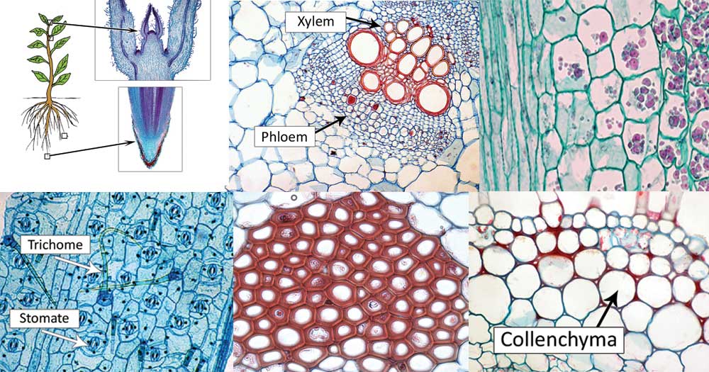
1. Parenchyma cells
Parenchyma cell definition
- These are live undifferentiated cells found in a variety of places of the plants’ bodies.
- They participate in several mechanisms of the plan including photosynthesis, food storage, secretion of waste materials.
- The experimental observation indicated that they appear green.
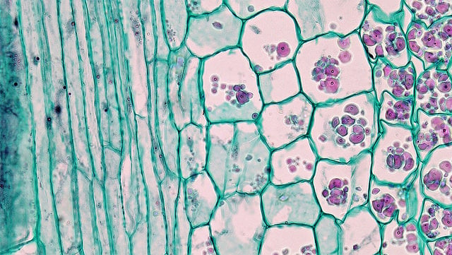
Structure of parenchyma cells
They are live thin-walled cells with permeable walls that are undifferentiated. They do not have a specialized structure hence they easily adapt and differentiate into a variety of cells performing different functions. There are two types of parenchyma cells
- Palisade parenchyma
- Ray parenchyma
Palisade parenchyma cells are columnar elongated structured cells found in a variety of leaves, lying below the epidermal tissue. Palisades are closely linked cells in layers of mesophyll cells found in leaf cells.
Ray parenchyma has both radial and horizontal arrangements majorly found within the stem wood of the plant.
Functions of the Parenchyma cells
- Parenchyma cells are closely linked to the surface epidermal cells which contribute largely to light penetration and absorption and regulate gas exchange.
- The permeable wall allows the transportation of small molecules between the cells and the cell cytoplasm.
- The palisade parenchyma combined with spongy mesophyll cells found below the layer of the epidermis tissue assists in light absorption used in photosynthesis.
- Ray parenchyma cells are found in wood rays which transport materials along the plant stem.
- The parenchyma cells are also found in good numbers within the xylem and the phloem of vascular plants, helping in the transportation of water and food materials in the plant.
- Some are also involved in the biochemical secretion of nectar and manufacturing secondary elements that act as protective materials from herbivores’ feeding.
- And those parenchyma cells found in root tubers such as potatoes, leguminous plants, help in the storage of food.
2. Collenchyma cells
Collenchyma cell definition
- They are elongated cells found below the epidermis and/or in young plants on the outer layers of their stems and leaves.
- They become alive after maturing up and are derivatives of the meristems and they are found in the vascular and/or on the plant stem corners.
- They occur in the peripheral region of the plant and are not found in the plant roots.
- In experimental observation, they appear red.
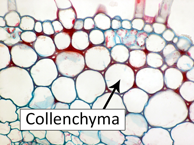
Figure: Diagram of Collenchyma cells. Source: University of Florida
Structure of collenchyma cells
- These are cells that are long with a primary thick cell wall. The cell wall is normally irregular and made up of cellulose and pectin molecules
- During maturity, at some point, they resemble the parenchyma cells which transform into the collenchyma cells. When a few cells accumulate, the Golgi bodies along with the endoplasmic reticulum come up together to form the primary cell wall. When two cells fuse, they form a thin primary wall that doesn’t differentiate to collenchyma cells.
- Therefore the more the cells accumulate and fuse, they form a strong irregular functional primary cell wall. These newly formed cells are elongated to give support for the plant to grow. However, the primary wall doesn’t have lignin, a polymeric organic complex that forms strong structural tissues of vascular plants giving it rigid support, especially in wood and bark and that it also prevents rotting.
Types of collenchyma cells
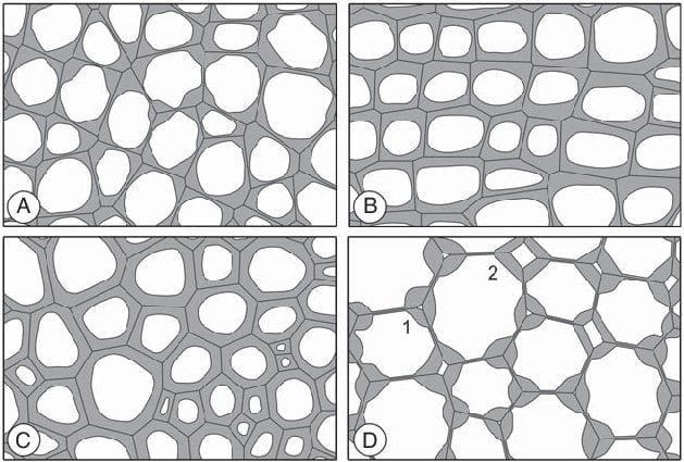
Figure: Schematic drawings of the most common types of collenchyma. (A) Angular collenchyma. (B) Tangential collenchyma. (C) Annular collenchyma. (D) Lacunar collenchyma. This type often occurs as an intermediate type with angular and lamellar collenchyma, in which the size of the intercellular spaces can vary from minute spaces (1) to large cavities surrounded by collenchymatous walls (2). Source: https://www.ncbi.nlm.nih.gov/pmc/articles/PMC3478049/
There are four types of collenchyma based on the thickness of the wall and the cell arrangement
- Angular collenchyma
- Annular collenchyma
- Lamellar collenchyma
- Lacunar collenchyma
Angular collenchyma
- The cells appear to have an angle and a polygonal shape.
- The cells are thickened at the corners of the cell
- The cells do not have intracellular spaces since they are closely packed together
- They are found below the epidermis as hypodermis
- They are the most common type of collenchyma
Annular collenchyma
- The walls are uniformly thickened.
- The cells appear to be circular in shape
Lamellar collenchyma
- The cells are thickened on the periphery making them appear tangentially arranged in rows
- They are closely packed together and therefore they don’t have intracellular spaces.
- They are commonly formed and found in the leaves petioles.
Lacunar Collenchyma
- These are cells are formed spaciously leaving intracellular spaces between each other.
- The cell wall thickens around the intracellular spaces
- They appear spherically shaped
- They are formed and found in the walls of fruits
Functions of the Collenchyma cells
- Being the living cells in plant tissues, they give support to the plant areas that are growing and maturing in length. Since the cell wall lacks lignin, it remains supple giving the plant parts like young stems, young roots, and young leaves plastic (stretchable) support.
- They offer flexibility and tensile strength to plant tissues, allowing the plants to bend.
- They also allow the plant parts to grow and elongate.
- Collenchyma can combine with the chloroplast and perform the process of photosynthesis.
3. Sclerenchyma cells
Sclerenchyma cell definition
- These are collenchyma cells that have an agent of the cell wall that plays a major role in hardening its cell wall.
- Therefore, these are mature Collenchyma cells with a secondary cell wall, over the primary cell wall.
- They are found in all plant roots and they are important in anchoring and giving support to the plants.
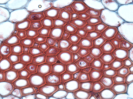
Figure: Cross-section of sclerenchyma fibers. Source: Wikiwand.
Structure of sclerenchyma cells
- They have a lignified cell wall, making them extremely hard.
- These make them more rigid in comparison to the parenchyma and the collenchyma cells.
- They also have suberin and cutin, which makes them waterproofed.
- Because of their rigidity and waterproof effect, they do not live for long since there can not exchange materials for cellular metabolisms to sustain their longevity.
- Therefore in the event of fully developing their functional maturity (a phase for cytoplasm formation), they are dead.
Types of sclerenchyma cells
There are two types of sclerenchyma cells
- Fiber sclerenchyma cells
- Sclereid sclerenchyma cells
Functions of the sclerenchyma cells
- Due to their thickened cell wall, they offer protection and support to other plants’ tissues especially the tree trunks and fibers of large herbal trees.
- The hardened cell wall discourages herbivory. Ingestion of the hard cell wall causes damage to the digestive tract of larval stage insects, especially in peach fruits.
- Sclerenchyma found fibers are used in making fabric, thread, and yarns.
4. Xylem Cells
Xylem cell definition
Xylem cells are complex cells found in the vascular tissues of plants, mostly in woody plants.
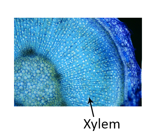
Figure: Diagram of Xylem Cells. Source: University of Florida
Structure of Xylem cells
- They have two elements for conduction: Tracheids and vessel elements
- They have tracheids which are vessels that conduct water and minerals from, the roots to the plant leaves.
- Tracheids are elongated slender vessels that are lignified, hence they have a hardened secondary cell wall, specialized to conduct water from the roots.
- The tracheid also has overlapping tap ends placed in an angel to allow a connection and communication from cell to cell.
- The vessel elements allow the transport of water. They are hollow, shorter, wider than the tracheids but lack the angeled endplates, therefore they are aligned with each other forming a continuous hollow tube, 3 meters long
- The xylem cells are also combined with fibers and parenchyma cells hence they have a primary cell wall combined with a lignified cell wall, forming rings and looped networks with pits known as bordered pits for conduction.
- The bordered pits are areas in the cell wall where primary cell wall materials are deposit, and they allow water to move between the xylem cells.
- Gymnosperms, ferns, and pteridophytes have tracheids while flowering plants have vessel elements.
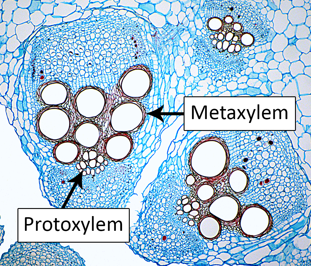
Figure: Protoxylem and Metaxylem diagram. Source: University of Florida
Functions of the xylem cells
The primary function of the xylem cells is to transport water and soluble nutrients, minerals and inorganic ions upwardly from the roots of the plants and its parts. These elements flow freely through the xylem tracheids and vessel elements with the aid of the xylem sap.
5. Phloem Cells
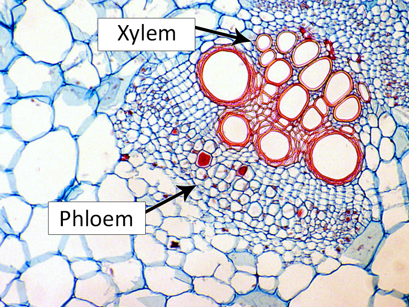
Figure: Diagram of Phloem Cells. Source: University of Florida
Phloem cell definition
- These cells are located outside the xylem layer of cells. They become alive at maturity because they need the energy to move materials.
- They function to transport food from the plant leaves to other parts of the plant.
- They also have a flaccid cell wall hence they lack tensile strength that allows them to move materials at high pressure.
Types of phloem cells
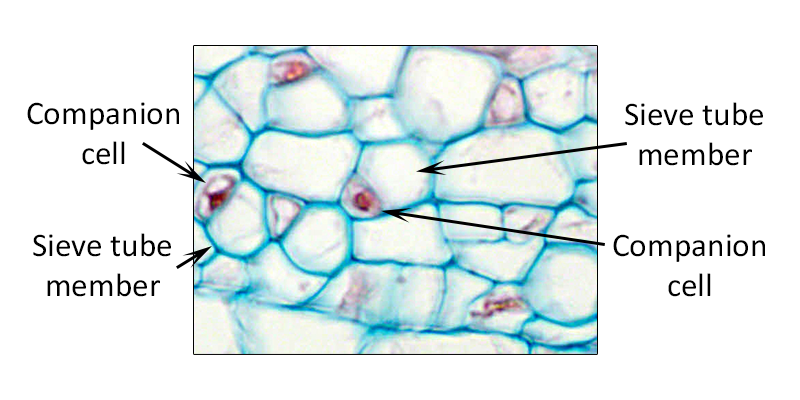
Figure: Types of phloem cells. Source: University of Florida
There are two types of phloem cells:
- Sieve tube members and Companion cells
- Sieve cells
Sieve tubes and Companion cells
- These are the cells that control the cells’ metabolism, and they are linked together with large numbers of plasmodesmata.
- Sieve tube members are shorter and wider and they are continuously arranged from one end to another into the sieve cells, where they are highly packed together.
- This concentration allows the solute materials to move faster within the sieve tubes and the sieve cells. the sieve tube members’ nucleus disintegrates, ribosomes disappear and the vacuole membrane breaks down at maturity.
- The companion cells assist in moving materials into and out of the sieve tube members. Characteristically, the sieve tubes have Phloem (P)-proteins at the cell wall and callose and together they heal injuries caused on the sieve tubes.
Sieve cells
- They are the primitive part of the phloem found in ferns and conifers.
- They are structurally long with tapered overlapping ends. They have pores all over their cell wall that is surrounded by callose (a carbohydrate that repairs the pores after an injury).
- They associate with albuminous cells to help in moving materials into the phloem.
- This is the site where dissolved food flows eg sucrose
Functions of the phloem cells
It transports dissolved foods and organic materials throughout the plants since it has the ability to move the materials in all directions of the plant, depending on the age of the plant.
6. Meristematic cells
Meristematic cell definition
- They are also known as the meristems.
- These are the cells in a plant that divide continuously throughout the life of a plant.
- They have a self-renewal ability and high metabolisms to control the cell.
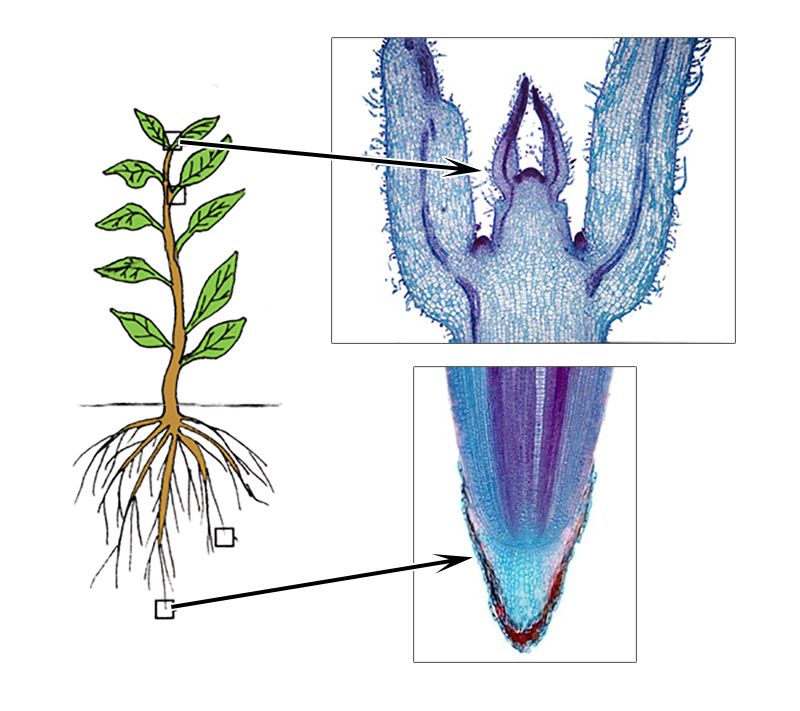
Figure: Diagram of Meristematic cells. Source: University of Florida
Structure of the meristematic cells
- These are cells that undergo cell division giving rise to the Parenchyma, Collenchyma, and Sclerenchyma cells.
- They have a thin wall and lack the central vacuole and are composed of immature plastids.
- Their protoplast is densely filled.
- They have a cubic shape with a large nucleus.
- they have high metabolic activity
- They are closely clamped together, therefore, they have no intercellular space.
- They play a major role in plant growth in width and length.
Types of meristematic cells
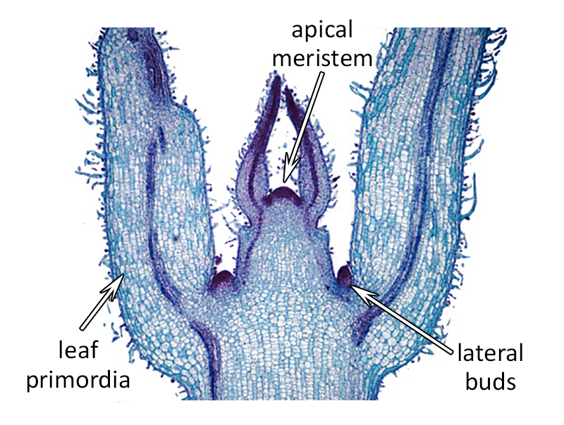
Figure: Types of meristematic cells. Source: University of Florida
There are three types of meristematic cells classified according to the tissue they exist in.
- Apical meristems – they are found at the tips of roots and stems that have started growing and they contribute to the length of the plant
- Lateral meristems – They are found in the radial part of the stem and roots and they contribute to the plant thickness
- Intercalary meristems – they are found at the base of the leaves and contribution to the size variance of the leaves.
Functions of the meristematic cells
- They play a major role in the length and width sizes of the plants
- they also give variance in the sizes of the plant leaves.
- They differentiate and mature into permanent tissues of the plants.
7. Epidermal Cells
Epidermal cell definition
- These are the external cells of the plants offering protection from water loss, pathogenic invaders such as fungi.
- They are placed closely together with no intracellular spaces.
- They are covered with a waxy cuticle layer to reduce water loss.
- These cells cover the plant stems, leaves, roots, and plant seeds.
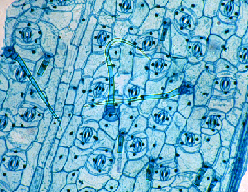
Figure: Diagram of Epidermal Cells. Source: University of Florida
Types of epidermal cells
There are three types of epidermal cells that play the primary role of protecting the plant from environmental factors such as high temperatures, pathogens, chemical exposures e.g. radiations. They include:
- Pavement cells
- Stomatal guard cells
- Trichomes
Structure and Functions of Epidermal Cells
Pavement cells
- These are the most common epidermal cells covering all plants. They are poorly specialized hence they lack a defined shape, therefore, they do not have special functions.
- The morphology of pavement cells varies from plant to plant such as the leaves of dicots appear like jigsaw pieces giving the leaves mechanical strength.
- Pavement cells found on the stem and other long plant parts appear to be rectangular with an axis running parallel to the direction of plant expansion.
- The different morphologies are associated with the functions the pavement cells perform. For example, epidermal cells are formed during the development of plant seeds by embryogenesis.
- They prevent excessive loss of water, the cells are closely packed together, making a protective lining to protect other underlying cells.
- The functions of the pavement cell include:
- maintain the plants’ internal temperature
- they act as a physical barrier from pathogens and external damages from chemicals such as radiations
- they separate the leaves’ stomata.
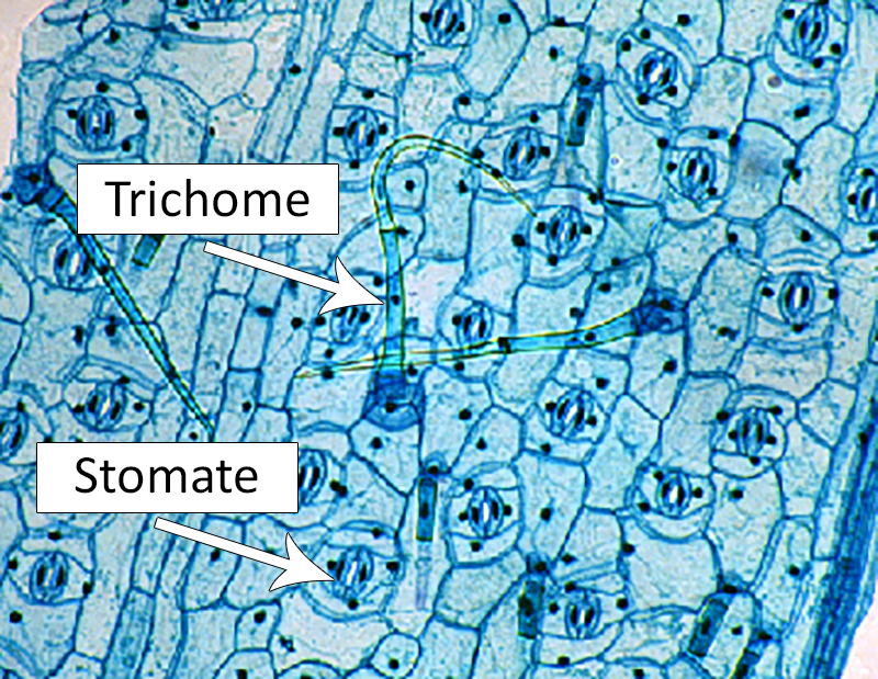
Figure: Diagram of trichomes and stomata. Source: University of Florida
Stomatal Guard cells
- Stomatal guard cells are available depending on the type of plant.
- They are highly specialized with a defined shape which allows them to perform a variety of functions.
- There are two types of guard cells defined by the structure i.e those that control water availability by opening and closing the stomata by maintaining turgor pressure and those that regulate the exchange of gases into and out of the leaves’ stomata.
- The stromal guard cells also have chloroplast. Therefore they have a functional effect on photosynthesis.
Trichomes
- These are also known as epidermal hairs found on the epidermal tissue. They are a specialized group of cells with well-defined shapes.
- They have a large size of about 300um in diameter.
- They play a major role in protecting the plants from predators and pathogens, by acting as trappers and poisoners to animal predators.
- These cells do not multiply by cell division instead they undergo endoreplication for expanding their cell population.
References and Sources
- <1% – https://www2.estrellamountain.edu/faculty/farabee/BIOBK/BioBookPLANTANAT.html
- <1% – https://www.youtube.com/watch?v=MWz4ptP_QEU
- <1% – https://www.thoughtco.com/what-is-a-plant-cell-373384
- <1% – https://www.thoughtco.com/cell-wall-373613
- <1% – https://www.studyblue.com/notes/note/n/bio-1500-study-guide-2014-15-turchyn/deck/12773783
- <1% – https://www.researchgate.net/publication/260736170_The_Plant_Vascular_System_Evolution_Development_and_FunctionsF
- <1% – https://www.oldbridgeadmin.org/cms/lib/NJ02201158/Centricity/Domain/1066/chapter-29-plant-structure-and-function.pdf
- <1% – https://www.ncbi.nlm.nih.gov/books/NBK9889/
- <1% – https://www.britannica.com/science/xylem
- <1% – https://www.britannica.com/science/parenchyma-plant-tissue
- <1% – https://www.britannica.com/science/meristem
- <1% – https://www.britannica.com/science/bacteria
- <1% – https://www.biologynotes.site/components-of-the-cell/
- <1% – https://www.bbc.co.uk/bitesize/guides/z2kmk2p/revision/3
- <1% – https://www.answers.com/Q/What_made_up_of_phloem_tissue_and_a_cork_cambium_that_protect_the_stem
- <1% – https://study.com/academy/lesson/collenchyma-cells-function-definition-examples.html
- <1% – https://sciencing.com/cells/
- <1% – https://quizlet.com/9615589/bio-exam-1-flash-cards/
- <1% – https://quizlet.com/31673504/whats-stomata-with-you-flash-cards/
- <1% – https://quizlet.com/19629366/tissues-flash-cards/
- <1% – https://quizlet.com/17179233/mastering-biology-plant-growth-flash-cards/
- <1% – https://quizlet.com/123462152/plant-kingdom-quiz-1-flash-cards/
- <1% – https://en.wikipedia.org/wiki/Tracheid
- <1% – https://en.wikipedia.org/wiki/Lignin
- <1% – https://en.wikipedia.org/wiki/Ground_tissue
- <1% – https://courses.lumenlearning.com/boundless-biology/chapter/stems/
- <1% – https://courses.lumenlearning.com/boundless-biology/chapter/leaves/
- <1% – https://bmmgizmo.wordpress.com/2013/09/21/types-of-epidermal-cells/
- <1% – https://biologydictionary.net/xylem/
- <1% – https://answers.yahoo.com/question/index?qid=20081011194035AAp3jk5
- <1% – http://www.biologyreference.com/A-Ar/Anatomy-of-Plants.html
- <1% – http://preuniversity.grkraj.org/html/3_PLANT_ANATOMY.htm
- <1% – http://facweb.furman.edu/~lthompson/bgy34/plantanatomy/plant_cells.htm
- <1% – http://brilliantpublicschool.com/files/documents/Doc-1164-XI-Biology-Support-Material-HOTS-and-OTBA-2014-15.pdf
- <1% – http://bio1520.biology.gatech.edu/growth-and-reproduction/plant-development-i-tissue-differentiation-and-function/
