Interesting Science Videos
Introduction
A well-prepared blood smear is important to produce good results on analysis after doing a Giemsa stain, in identifying blood cells or/and demonstrating the presence of parasites in a sample. Below, we discuss the procedures for preparing both thin and thick smear for Giemsa staining technique, Importance, and applications of blood smears, in detail.
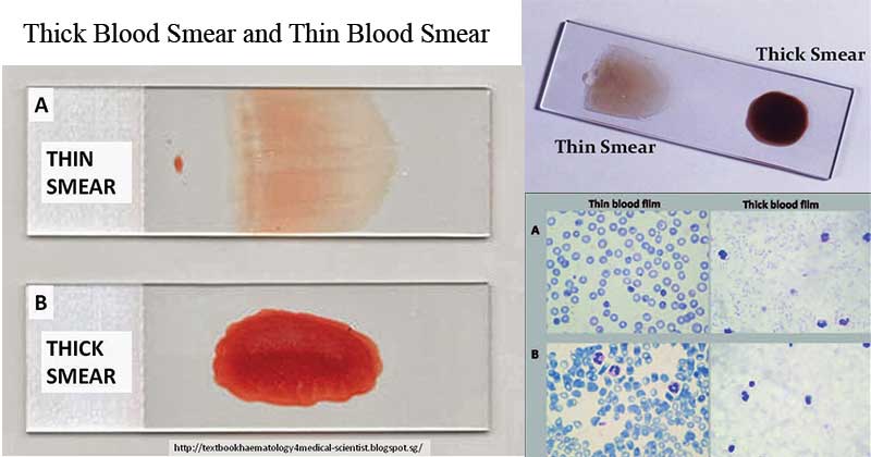
Blood smears are mostly done for Differential Leukocyte count (DLC)i.e it quantifies the white blood cells and specifies the morphologies of each leukocyte. Normally, peripheral blood is used to prepare smears and depending on the function of the smear, two types of smear can be prepared.
a. Thin blood smear – for demonstration and differentiation of leukocytes.
b. Thick blood smear – for diagnosis of blood protozoan parasites and blood abnormalities eg anemiae.

Image Source: Haematology in a NutShell, Microbiology Info, and DOI: 10.5336/caserep.2015-47850
NOTE: Dry smears are the best for staining, so ensure your smear is completely dry before applying a staining technique.
Blood smears are simple procedures to perform aiming at demonstrating and acquiring information on blood cells, qualitatively and quantitatively. Quantitative importance enables the numbering of blood cells while the qualitative function is to demonstrate and identify the cell morphologies, including types of leukocytes, erythrocytes, monocytes, and platelets.
Blood smears have also been used in detecting hematological disorders i.e by observing the morphologies and quantifying the cell numbers.
Objectives
- To prepare a thin blood smear
- To prepare a thick blood smear
Requirements
- Sterile syringe & needle
- EDTA vials
- Tourniquet
- 70% isopropyl alcohol
- Sterile Lancet
- Microscopic glass slides
Thick Blood Smear Preparation
Specimen: Venous blood sample
Principle
Thick blood smears require larger volumes of blood than the thin blood smears. this allows them to be used for the detection of blood parasites in the blood samples. A thick blood smear is made by spreading a large blood drop in a small area of about 1 cm which provides a better opportunity to detect various parasitic forms against a more transparent background.
Procedure
- Collect blood sample by venipuncture and put in a clean test tube
- Using a capillary tube collects blood from the tube and put two large drops at the center of a sterile microscopic slide.
- Holding the slide between your thumb and index finger, gently shake the slide to spread the blood about 10mm in diameter.
- Air-dry the smear for 20-30 minutes till its completely dry then apply the appropriate Romanowski stain.
Thin Blood Smear Preparation
Specimen: Peripheral Blood sample
Principle
The Thin Blood smear is prepared by making a drop of well-mixed venous blood, 2mm in diameter at the center of a sterilized microscopic glass slide. Some borders are left around the smear for easy counting and differentiating of the cells. A second glass slide is used as a spreader, streaking the blood into a thin film across the glass slide. This preparation is allowed to dry and then fixed with an appropriate Romanowski stain, depending on your objective.
Procedure
- Using a sterile pricking needle, make a prick on the index finger
- apply some pressure on the finger and put two drops of blood at the edge, leaving a margin on a sterile Microscopic slide.
- Place the edge of the sterile microscopic slide over the drops of blood, at an angle of 30-450, and make two streaks rapidly but smoothly forward from the blood sample and spread it. This will leave a thin film of blood resembling a tongue-shape.
- Allow the slide to air dry and stain with an appropriate staining technique.
Applications of blood smears
- For classification of blood disorders including types of anemia, bleeding disorders
- To characterize blood-related disorders such as leukemias
- To detect immune-mediated inflammatory disorders and infections
- To detect protozoal parasites: Plasmodium falciparum, Mycoplasma spp (Mycoplasma haemofelis and Mycoplasms haemominutus and Bacteria such as Bartonella spp.
Advantages
- It is a rapid simple technique which requires basic equipment
- It can be performed with very small volumes of blood.
Disadvantages
- Use clean slides to avoid the formation of grease spots (holes in the smear).
- Rapid air drying of smear to preserve cell morphologies
- Regular use of the technique to produce useful blood smears
References
- https://paramedicsworld.com/hematology-stainings/giemsa-staining-technique-principle-preparation-procedure-interpretation/medical-paramedical-studynotes
- https://paramedicsworld.com/hematology-practicals/preparation-peripheral-blood-smear/medical-paramedical-studynotes
- https://www.vetstream.com/treat/felis/technique/blood-smear
- https://www.cdc.gov/dpdx/diagnosticprocedures/blood/specimenproc.html

Should we not dehemoglobinize the red cells in the thick smear before applying stain?
Wow thanks alot. I’m a biomedical science student and will like to know alot from her
Please make it this notes downloa/allow me to download it
Thanks alot
got the best I needed… thanks 😊
thick smear ma de-staining nagarda pani hunchha sir ???