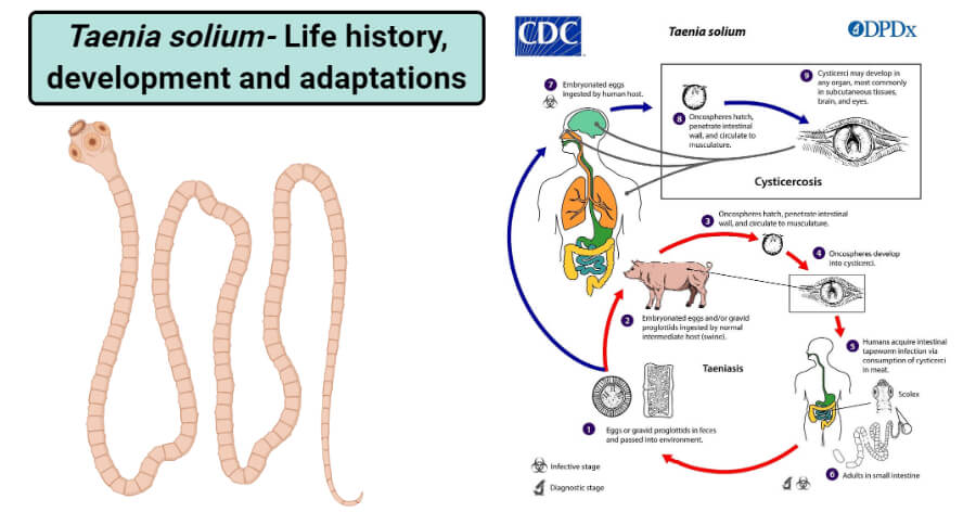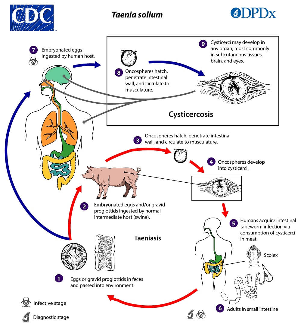
Image Source: Laboratory Diagnostic Assistance (DPDx), CDC.
Life history and Development of Taenia solium
Interesting Science Videos
1. Copulation and fertilization
- The life cycle of Taenia solium is digenetic, involving two hosts.
- But the life cycle of tapeworm is simple and without a free larval stage.
- Fertilization is preceded by copulation, inserting cirrus into the vagina of the same or another proglottid to release spermatozoa.
- Both self-fertilization and cross-fertilization occur in them.
- The cross-fertilization between different proglottids of the same tapeworm is very common.
- T. solium is protandrous, i.e., the testes mature first. Hence spermatozoa injected in the vagina are stored in the seminal receptacle till the ovary releases ova.
- When ova are released, then fertilization takes place, and zygotes are formed.
2. Capsule formation
- The zygotes or egg cells connect with yolk cells or vitelline cells in the ootype received from the vitelline glands.
- The zygote and the yolk then become enclosed in a thin shell or chorionic membrane, formed by material exuded by the yolk cell.
- The structures formed, capsule then passes into the uterus for further development.
- The passage of capsules into the uterus is lubricated by the secretion from the Mehlis glands.
- As more and more capsules pass into the uterus, it develops lateral branched-s to accommodate them.
3. Onchosphere formation
a. Cleavage
- When the capsule is in the uterus, the zygote undergoes cleavage.
- Cleavage is holoblastic and unequal.
- The zygote divides unequally resulting in a larger megamere and a smaller embryonic cell.
b. Morula
- The megamere divides further to form several similar megameres, while embryonic cells divide repeatedly producing 2 types of embryonic cells, larger mesomeres, and smaller micromeres.
- Thus, 3 types of cells result from the zygotes; small micromeres, medium mesomeres, and large megameres.
- The smaller micromeres form a rounded mass, the morula surrounded by an inner envelope of mesomeres and an outer envelope of megameres.
- The yolk or vitelline cell transfers its yolk to the megameres and gradually disappears.
- The large yolky megameres fuse to form the outer embryonic membrane which finally disappears.
- The medium mesomeres form the inner embryonic membrane or embryophore which is a hard, thick, cuticularized, and radially striated shell surrounding the morula.
- A thin basement membrane lies beneath the embryophore.
c. Hexacanth and Onchosphere
- The inner cell mass of the morula forms an embryo that develops 3 pairs of chitinous hooks at its posterior end.
- The hooks are secreted by differentiated cells called onchoblasts.
- This 6-hooked embryo, called hexacanth, possesses a pair of large penetration glands. It is surrounded by 2 hexacanth membranes.
- The hexacanth together with all the membranes surrounding it is called Onchosphere.
- By the time oncospheres are formed, the proglottids become gravid and increase in size. Its uterus forms 7-13 lateral branches on each side and contains 30,000 to 40,000 oncospheres.
- The gravid proglottids at the posterior end of strobila pass out from the host body contain embryos in the onchosphere stage.
- The eggs can survive from day to month in the environment.

Figure: Cysticercosis is an infection of both humans and pigs with the larval stages of the parasitic cestode, Taenia solium. This infection is caused by the ingestion of eggs shed in the feces of a human tapeworm carrier. Image Source: Laboratory Diagnostic Assistance (DPDx), CDC.
4. Infection to secondary host (pig)
- The secondary or intermediate host acquires infection by ingesting the oncosphere.
- The onchospheres are ingested by a pig with human feces due to its coprophagous habit.
- sometimes dogs, monkeys, and sheep are also known to get the infection by the onchosphere.
- Man himself may serve as the secondary host by ingesting oncospheres with inadequately cooked or raw vegetables.
- Auto-infection may take place in a person already serving as a primary host.
5. Migration within secondary Host
- The oncosphere loses its embryophore and basement membrane by the action of acidic juice (acid pepsin) in the stomach of secondary hosts (pig).
- The hexacanths are released from the onchospheres then passes into the small intestine, where the 2 persisting hexacanth membranes are also lost by the action of alkaline juices.
- The hexacanth, now activated by the presence of bile salts, bore the intestinal wall with the help of a pair of unicellular penetration glands found in it between the hook to reach a submucosal blood or lymph vessel.
- The hooks merely anchor the hexacanth to the intestine wall, while secretion of penetration glands dissolves the intestinal tissues.
- The entire process takes place about 10 minutes, after which the hooks, are of no further use, and shed off.
- The hexacanth are carried to the liver by submucosal blood vessels via the hepatic portal vein.
- From the liver, it reaches the heart via enters the arterial circulation,
- It finally comes to lie in the striated muscles usually of the tongue, shoulder, neck, thigh, heart, etc, where they mature into bladder-worm or cysticerci over 60–70 days.
- Cysticercus may also develop in other organs such as the lungs, liver, kidney, or brain.
- Eyes, brain, or liver (Non-muscular organs) may frequently become the sites of cysticercus formation.
6. Cysticerus or Bladderworm formation
- It is the larval stage of Taenia which has been formed by the transformation or modification of the hexacanth stage.
- The hexacanth after reaching to muscles lose their hooks, absorbs nourishment from the host’s tissues, and grows in size attaining a diameter of about 18mm.
- It is a sac filled with a fluid consisting mainly of the blood plasma of the host.
- The fluid-filled vesicle or bladder has a thin wall consisting of an outer layer of thick syncytial protoplasmic mass and an inner mesenchymal or germinal layer.
- The wall thickens at the anterior ends (i.e., opposite the side where hooks were present) and ivaginates.
- The ivagination looks like a hollow knob.
- The ivagination knob then differentiates into an inverted scolex possessing suckers, hooks, and rostellum. It is then called a proscolex.
- The embryo at this stage is known as bladderworm.
- The bladderworm is of cysticercus type in T. solium which is characterized by a large vesicle and one scolex.
- The cysticercus of T. solium is called cysticercus cellulosae.
- It takes about 10 weeks for the formation of cysticerci.
- Cysticercus develops in adult tapeworm only ingested by the human host.
- Pork contains viable cysticerci which appear white-spotted resembling something like measles known as measly pork. Thus, the pig becomes infected.
7. Infection to primary host (Man)
- Man acquires infection by eating undercooked pork containing cysticerci or measly pork.
- Cysticercus becomes active in reaching the small intestine.
- The bladder of the Cysticercus is digested in the host stomach. Proscolex evaginates and anchors to the intestinal wall.
- The neck begins to proliferate a series of proglottids from the strobila.
- In 10 to 12 weeks the proscolex is converted into adult Taenia and adult Taenia, thus, formed starts producing gravid proglottids with onchospheres within 8 to 10 weeks which is then ready for apolysis.
Parasitic adaptations of Taenia solium
The tapeworm shows several adaptative features to its internal parasitic life, in comparison with the free-living animals. some of these are as follows:
Morphological adaptations
- Taenia solium has flattened a leaf or a ribbon-like body so, that they can fit in the spaces where they have their habitat.
- The teguments out the covering of Taenia is freely permeable to water and nutrients, but it protects against digestion by the host’s alkaline digestive juice.
- It has well developed 4 suckers and hooks to anchor with the intestinal wall of the host, by which it is not dislodged from the host.
- There are no cilia and organs of locomotion since they are not needed, the host transporting the parasite.
- As the alimentary canal is absent, they absorb the digested food from the host through the general body surface. The surface area for absorption is increased by the presence of the microvilli on the outer surface of the tegument.
- They lack the special circulatory, respiratory, and sense organs and not the well-developed nervous system as these parasites do not need this system.
- Their reproductive system is well-developed and has the capability to produce a huge number of eggs (40,000 per gravid proglottid). Each mature proglottid has one complete set of male and female genitalia. The reason for the production of such a huge number of eggs is that these parasites face many challenges for survival.
- Hermaphroditism and proglottization ensure self-fertilization or cross-fertilization within another proglottid in the same worm within the same proglottids.
- The resistant covering, shell, or capsule around eggs and embryo protects from the unfavorable condition.
Physiological adaptations
- The internal osmotic pressure is higher than that of the surrounding host’s fluid or tissue, and PH tolerance is high, 4 to 11, that helps the parasite to reside conveniently in the host body.
- They live in an oxygen-free environment, as such they possess a very low metabolic rate which requires a very little amount of oxygen. Respiration is anaerobic as free oxygen is not available. The energy is obtained by the fermentation of glycogen in an oxygen-free environment, where carbondioxide and fatty acids are produced.
References and Sources
- https://www.researchgate.net/publication/236612241_A_Preview_of_Adaptations_of_Parasites_in_the_Host
- 3% – https://www.biologydiscussion.com/invertebrate-zoology/phylum-platyhelminthes/taenia-solium-habitat-structure-and-life-history/28918
- 2% – https://www.shareyouressays.com/knowledge/biology-question-bank-144-mcqs-on-animal-kingdom-answered/114611
- 2% – https://quizlet.com/37730009/microbiology-exam-4-cestodes-flash-cards/
- 1% – https://www.kenhub.com/en/library/anatomy/hepatic-portal-vein
- 1% – https://www.biologydiscussion.com/parasitology/parasitic-worm/taenia-solium-linnaeus-history-and-functions/62216
- 1% – https://learnzoology.wordpress.com/2014/05/14/life-cycle-of-taenia-solium/
- 1% – https://gurujistudy.com/bsc-1st-year-lower-non-chordates-distinctions-question-answers/
- <1% – https://www.studyandscore.com/studymaterial-detail.php?Id=taenia-taeniasis-cycticercosis-and-parasitic-adaptations
- <1% – https://www.sciencedirect.com/topics/neuroscience/osmotic-pressure
- <1% – https://www.sciencedirect.com/topics/immunology-and-microbiology/cestoda
- <1% – https://www.sciencedirect.com/topics/biochemistry-genetics-and-molecular-biology/taenia-pisiformis
- <1% – https://www.notesonzoology.com/term-paper/taenia-solium/term-paper-on-taenia-solium-platyhelminthes-microorganisms-zoology/9118
- <1% – https://www.encyclopedia.com/medicine/diseases-and-conditions/pathology/tapeworms
- <1% – https://www.cdc.gov/dpdx/diagnosticprocedures/stool/morphcomp.html
- <1% – https://www.britannica.com/animal/amphioxus
- <1% – https://www.answers.com/Q/An_oxygen_free_environment_is
- <1% – https://quizlet.com/39466394/bio-4471-parasitology-test-3-chapter-20-tapeworms-in-general-flash-cards/
- <1% – https://health.howstuffworks.com/wellness/natural-medicine/chinese/yin-organs.htm
- <1% – https://biologydictionary.net/alimentary-canal/

I like the notes the way they summarized in shot forms