
The sodium dodecyl sulfate (SDS)-based method of DNA extraction is one of the most fundamental and widely adopted approaches for isolating genomic DNA from a broad range of biological materials.
Since its early incorporation into molecular biology workflows, SDS has remained a cornerstone reagent for cell lysis due to its strong detergent properties and compatibility with downstream purification steps (Sambrook & Russell, 2001). The method has been extensively applied to plant tissues, microbial cells, environmental samples, and complex matrices such as sediments and seeds, often with protocol-specific modifications to address sample-associated inhibitors (Cota-Robles, 2020).
Historically, SDS-based lysis gained prominence alongside early phenol–chloroform extraction protocols and later evolved into hybrid systems incorporating CTAB for plant tissues rich in polysaccharides and secondary metabolites (Doyle & Doyle, 1987). Despite the emergence of commercial spin-column and magnetic bead kits, SDS-based extraction remains highly relevant due to its cost-effectiveness, flexibility, and ability to yield high-molecular-weight DNA when optimized properly (RWDST Co., 2023).
Recent studies have demonstrated the adaptability of SDS-based methods through targeted modifications. For instance, Natarajan et al. (2016) developed a modified SDS protocol incorporating bead-beating and enzymatic digestion to efficiently recover environmental DNA from seafloor sediments. Similarly, Xia et al. (2019) optimized an SDS-based method for raw soybean seeds, achieving superior DNA quality compared to CTAB-based and kit-based approaches. These advancements highlight the continued relevance of SDS-based extraction in both classical and modern molecular biology.
Key Reagents of SDS-based DNA Extraction
The effectiveness of the SDS-based method relies on a combination of detergents, buffering agents, enzymes, and purification reagents that work synergistically to lyse cells, protect DNA, and remove contaminants.
| Reagent | Typical Concentration / Amount | Purpose |
| Sodium dodecyl sulfate (SDS) | 0.5-2% (w/v) | Disrupts lipid membranes and denatures proteins |
| Tris-HCl | 10-100 mM (pH 7.5–8.0) | Maintains stable pH for DNA integrity |
| EDTA | 10-50 mM | Chelates divalent cations, inhibiting DNases |
| NaCl | 100-500 mM | Stabilizes DNA and aids protein precipitation |
| Proteinase K | 100-200 µg/mL | Digests proteins and nucleases |
| RNase A | 10-50 µg/mL | Removes RNA contamination |
| Phenol:Chloroform:Isoamyl alcohol | 25:24:1 | Removes proteins and lipids |
| Isopropanol / Ethanol | 0.6-2 volumes | Precipitates DNA |
| TE buffer | As required | DNA resuspension and storage |
Principle of SDS-based DNA Extraction
The SDS-based DNA extraction method operates on the principle of chemical lysis and protein denaturation, followed by selective DNA purification. SDS is an anionic detergent that disrupts cell membranes by solubilizing phospholipids and membrane proteins. Simultaneously, SDS denatures intracellular proteins by breaking non-covalent bonds, effectively releasing nucleic acids into solution (Sambrook & Russell, 2001).
EDTA plays a critical protective role by chelating divalent metal ions such as Mg²⁺ and Ca²⁺, which are essential cofactors for DNases. The inclusion of proteinase K further enhances protein degradation, ensuring efficient removal of histones and other DNA-binding proteins (Xia et al., 2019). Following lysis, organic extraction using phenol–chloroform separates denatured proteins and lipids from nucleic acids based on phase partitioning.
Finally, DNA is selectively precipitated using alcohol in the presence of salts, allowing recovery of high-molecular-weight DNA suitable for downstream applications. The robustness of this principle has enabled the SDS-based method to be adapted across diverse sample types over several decades (Cota-Robles, 2020).
Protocol of SDS-based DNA Extraction
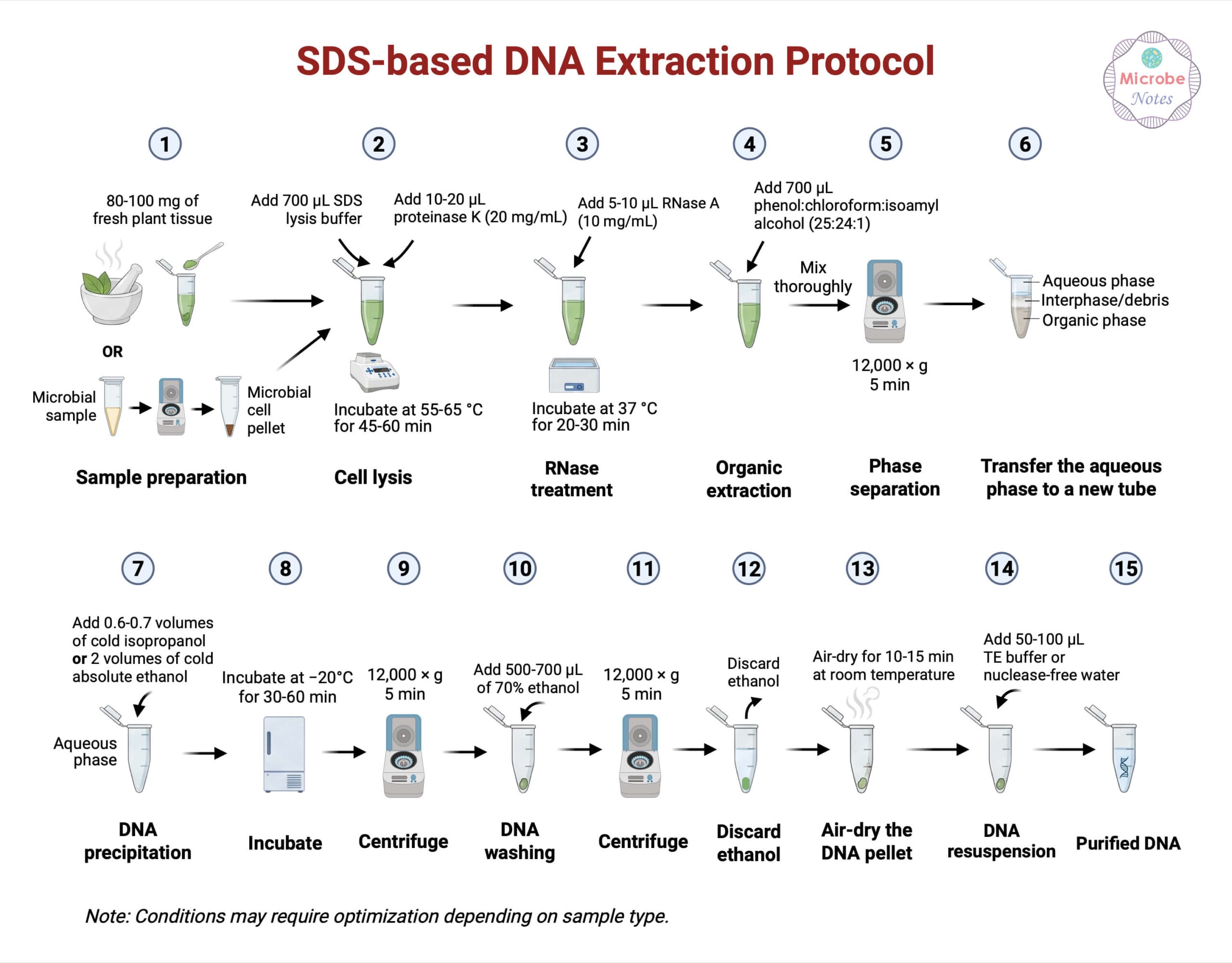
Sample Preparation
- Weigh 80-100 mg of fresh plant tissue or raw seed material.
- Transfer the sample to a pre-chilled mortar.
- Add liquid nitrogen and grind thoroughly using a pestle until a fine powder is obtained.
- Transfer the powdered sample into a 1.5 mL sterile microcentrifuge tube.
- For microbial samples, pellet 1-5 × 10⁷ cells by centrifugation (8,000-10,000 x g for 5 mins) and proceed directly to lysis without grinding (Xia et al., 2019).
Cell Lysis
- Add 700 µL of SDS lysis buffer to the tube. Composition:
- 100 mM Tris-HCl (pH 8.0)
- 50 mM EDTA
- 150 mM NaCl
- 1-2% (w/v) SDS
- Add 10-20 µL of proteinase K (20 mg/mL) to achieve a final concentration of approximately 200-400 µg/mL.
- Mix gently by inversion to avoid DNA shearing.
- Incubate the mixture at 55-65 °C for 45-60 minutes.
- Invert the tubes gently every 10-15 minutes during incubation to improve lysis efficiency.
This step ensures disruption of cell membranes and digestion of DNA-binding proteins (Sambrook & Russell, 2001).
RNA Removal
- Allow the lysate to cool to room temperature.
- Add 5-10 µL of RNase A (10 mg/mL) to the lysate (final concentration: 50-100 µg/mL).
- Mix gently by inversion.
- Incubate at 37 °C for 20-30 minutes.
Organic Extraction
- Add an equal volume (700 µL) of phenol:chloroform:isoamyl alcohol (25:24:1) to the lysate.
- Mix gently by inversion for 5-10 minutes; avoid vigorous vortexing.
- Centrifuge at 12,000 x g for 10 minutes at room temperature.
- Bottom (organic): phenol/chloroform with denatured proteins.
- Middle (interphase): white protein debris.
- Top (aqueous): DNA-containing layer.
- Carefully transfer the upper aqueous phase (~600 µL) into a new sterile microcentrifuge tube.
- Avoid disturbing the interphase containing denatured proteins.
Repeat the extraction once if protein contamination is suspected (Sambrook & Russell, 2001; RWDST Co., 2023).
DNA Precipitation
- Add 0.6-0.7 volumes of cold isopropanol (~360-420 µL) to the recovered aqueous phase. Alternatively, add 2 volumes of cold absolute ethanol.
- Gently invert the tube until the solution becomes cloudy or DNA strands are visible.
- Incubate at -20 °C for 30-60 minutes to enhance DNA precipitation.
- Centrifuge at 12,000 x g for 10 minutes.
- Discard the supernatant without disturbing the DNA pellet.
DNA Washing and Drying
- Wash the DNA pellet with 500-700 µL of 70% ethanol.
- Centrifuge at 10,000 x g for 5 minutes.
- Discard the ethanol wash carefully.
- Air-dry the pellet at room temperature for 10-15 minutes, avoiding complete overdrying.
DNA Resuspension
- Resuspend the DNA pellet in 50-100 µL of TE buffer (10 mM Tris-HCl, 1 mM EDTA, pH 8.0) or nuclease-free water.
Incubate at 4 °C for several hours or overnight to ensure complete DNA dissolution (RWDST Co., 2023).
Modifications of SDS-based DNA Extraction
- Bead-beating+SDS lysis: Mechanical disruption using beads improves cell breakage in sediments and microbial biofilms (Natarajan et al., 2016).
- Enzymatic pre-treatment (lysozyme, cellulase): Enhances lysis of rigid cell walls, particularly in bacteria and plant tissues.
- Increased salt concentration: Improves removal of polysaccharides and secondary metabolites in plant samples (Xia et al., 2019).
- SDS–CTAB hybrid systems: Combines SDS lysis with CTAB precipitation to remove polyphenols (Doyle & Doyle, 1987).
- Silica-based cleanup after SDS lysis: Improves DNA purity for sequencing applications (CD Genomics, 2025).
Troubleshooting of SDS-based DNA Extraction
| Problem | Likely Cause | Solution |
| Low DNA yield | Incomplete lysis | Increase SDS concentration or incubation time |
| DNA degradation | DNase activity | Ensure sufficient EDTA and cold handling |
| Protein contamination | Insufficient protein removal | Increase proteinase K or repeat organic extraction |
| RNA contamination | Inadequate RNase treatment | Increase RNase concentration |
| Brown or viscous DNA | Polyphenols/polysaccharides | Add a higher salt or CTAB step |
Quality Assessment of the SDS-based Isolated DNA
- Purity Evaluation: DNA purity is commonly assessed using absorbance ratios measured by spectrophotometry. An A260/A280 ratio of approximately 1.8 indicates minimal protein contamination, consistent with classical DNA purity standards.
- DNA Concentration Measurement: Fluorometric quantification methods, such as Qubit assays, are widely used to accurately measure double-stranded DNA concentration.
- DNA Integrity Assessment: DNA integrity refers to the size and fragmentation state of DNA molecules. Automated electrophoretic analysis using TapeStation systems is employed.
Safety Tips and Precautions of SDS-based DNA Extraction
- Handle SDS with care: SDS is an irritant and should be handled with gloves and eye protection.
- Use fume hoods for organic solvents: Phenol and chloroform are toxic and volatile (RWDST Co., 2023).
- Avoid vigorous pipetting: Prevents shearing of high-molecular-weight DNA.
- Proper waste disposal: Organic waste must be disposed of according to laboratory safety guidelines.
Storage and Long‑Term Stability of SDS-based Isolated DNA
Proper storage conditions are critical for maintaining DNA integrity after extraction.
- Short-term storage at −20 °C is sufficient to preserve DNA stability for routine laboratory use.
- Long-term storage is recommended at −80 °C, as it significantly reduces enzymatic degradation and chemical hydrolysis.
- Repeated freeze–thaw cycles should be avoided, as they can cause gradual DNA fragmentation and reduce sample quality.
Applications of SDS-based DNA Extraction
Plant genomics and breeding studies
SDS-based DNA extraction is widely used in plant genomics and breeding due to its effectiveness in isolating high-molecular-weight DNA from leaves and seeds. SDS and proteinase K efficiently disrupt plant cell structures and remove protein contaminants, overcoming interference from polysaccharides and secondary metabolites. Xia et al. (2019) reported higher DNA yield and purity from soybean seeds compared to CTAB-based and kit methods, supporting its use in PCR, restriction digestion, and genotyping applications.
Environmental DNA analysis
The SDS-based method is commonly applied in environmental DNA studies involving soil and sediment samples containing inhibitory compounds. When combined with mechanical disruption, SDS enhances lysis of diverse microbial cells and improves DNA recovery. Natarajan et al. (2016) demonstrated improved DNA yield and purity from seafloor sediments using an SDS-based protocol with bead-beating, enabling reliable biodiversity and metagenomic analyses.
Microbial ecology studies
In microbial ecology, SDS-based extraction provides effective lysis of mixed microbial populations, including Gram-positive and Gram-negative bacteria. Proteinase K reduces protein contamination, yielding community-representative DNA suitable for metagenomic sequencing and taxonomic analysis, with protocol flexibility allowing optimization for resistant cells (Natarajan et al., 2016; Cota-Robles, 2020).
PCR and sequencing workflows
DNA extracted using SDS-based methods is compatible with PCR, qPCR, and sequencing applications. Proper protein removal and washing minimize inhibitory residues, supporting consistent amplification and reliable sequencing results for routine and advanced molecular assays (Sambrook & Russell, 2001; RWDST Co., 2023).
Advantages of SDS-based DNA Extraction
- Cost-effective and accessible: Requires commonly available laboratory reagents.
- High DNA yield: Efficient lysis produces abundant DNA.
- Flexible and adaptable: Easily modified for diverse sample types.
- High-molecular-weight DNA recovery: Suitable for long-read sequencing (Cota-Robles, 2020).
Limitations of SDS-based DNA Extraction
- Use of hazardous chemicals: Phenol and chloroform pose safety risks.
- Time-consuming compared to kits: Multiple manual steps are required.
- Sensitivity to inhibitors: Plant metabolites and sediments may interfere with DNA purity (Green Sky Bio Works, 2024).
- Operator-dependent variability: Reproducibility may vary without strict protocol adherence.
Conclusion
The SDS-based method of DNA extraction remains a foundational and highly versatile technique in molecular biology. Despite the availability of modern commercial kits, its adaptability, affordability, and proven effectiveness ensure its continued relevance in research and diagnostic laboratories. Through strategic modifications, such as enzymatic digestion, bead-beating, and hybrid approaches, the method has been successfully applied to challenging samples ranging from plant seeds to environmental sediments.
When properly optimized and handled with appropriate safety measures, SDS-based DNA extraction provides high-quality DNA suitable for a wide spectrum of molecular applications, reinforcing its value in both classical and contemporary biological research.
References
- CD Genomics. (2025). Advances in DNA extraction: Methods, improvement and troubleshooting. https://www.cd-genomics.com/blog/dna-extraction-methods-optimization-troubleshooting/
- Cota-Robles, E. (2020). The evolution of DNA extraction methods. Biomedical Journal of Scientific & Technical Research, 27(1), 20921–20928. https://doi.org/10.26717/BJSTR.2020.27.004534
- Green Sky Bio Works. (2024). Overcoming obstacles: Troubleshooting tips for plant DNA extraction challenges. https://www.greenskybio.com/plant_extract/overcoming-obstacles-troubleshooting-tips-for-plant-dna-extraction-challenges.html
- Natarajan, V. P., Paria, A., & Anantharaman, N. (2016). A modified SDS-based DNA extraction method for high quality environmental DNA from seafloor sediments. Frontiers in Microbiology, 7, Article 986. https://doi.org/10.3389/fmicb.2016.00986
- RWDST Co. (2023). Quick guide: 8 commonly used methods for DNA extraction. https://www.rwdstco.com/quick-guide-8-commonly-used-methods-for-dna-extraction/
- Xia, Y., Chen, K., & Zhang, Y. (2019). A modified SDS-based DNA extraction method from raw soybean seeds. 3 Biotech, 9(2), Article 75. https://doi.org/10.1007/s13205-019-1593-4
- Brown, T. A. (2020). Gene cloning and DNA analysis: An introduction (8th ed.). Wiley-Blackwell.
- Kathmandu University. (2025). Phenol–chloroform extraction manual for human urine samples. Unpublished laboratory protocol.
