Interesting Science Videos
What are Gram-positive bacteria?
- These are bacteria whose classification has been based on their ability to retain the crystal violet dye after a brief wash with alcohol in the differential gram staining. Gram-positive bacteria stain purple while the Gram-negative bacteria stain Pink, after losing the purple color during the alcohol was thus taking up the safranin.
- Gram staining has been especially used because of its ability to differentiate bacteria base on their cell wall content, a major characteristic that classifies bacteria into two types namely, Gram-positive and Gram-negative bacteria.
- This classification was based majorly on the components they possess on their cell wall which enables them to stain differently. The bacterial cell was is made up of Mucin, Peptioglycan, and mucopeptides, which majorly function to give the cell wall its rigid strength.
- For the gram-positive cell wall, it has a thickness of about 20-80nm thickness made up of a thick peptidoglycan layer outside its cell membrane, unlike the thin layer of gram-negative bacteria (10-15nm) which has a very thin layer of the peptidoglycan of 2-7nm but has a thicker lipid layer making it quite complex than the Gram-positive cell wall.

Gram-positive bacteria characteristics
These bacteria have very distinct features that characterize it and differentiates it from other types of bacteria. These include:
- They lack an outer membrane
- They have a thin layer of the cytoplasmic lipid layer.
- They have a thick peptidoglycan layer
- The peptidoglycan layer has a large quantity of teichoic acid and a thin lipid layer, made up of lipoteichoic acid which plays a major role in bacterial adherence.
- The peptidoglycan layer plays a key role in maintaining the rigidity of the cell wall by crosslinking by the assistance of the DD-transpeptidase.
- They also have a thin layer of periplasm, as compared to that in the Gram-negative bacteria.
- Some have a locomotive apparatus, a flagellum that has two basal bodies for support unlike gram-negative which has four basal bodies.
- Some have a strong capsule made up of polysaccharides.
Gram-positive bacteria shape
Despite most bacteria being differentiated by the Gram staining dyes, the observation under the Microscope reveals more features that can be used to define and characterize these bacteria.
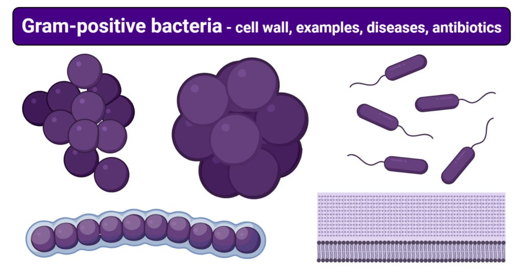
Gram-positive by definition in shape can be classified into two:
- Cocci – Singular form is known as coccus, which is round or oval shape bacteria measuring 0.5-1.0 um in diameter. They occur in pairs or in chains or clusters or as singles. For example Staphylococcus app, Streptococcus app. Special spaces of cocci include tetrads, Sarcines
- Bacilli – also rod-shaped; the singular form is known as Bacillus. They are stick-like bacteria with a round tapered, square or swollen ends. they have a size of 1-10um in length and 0.3-1.0um in width. For example the Bacillus spp
Other special shapes formed by Gram-positive bacteria include:
Tetrad- a type of cocci shape occurring in square-clusters of fours, for example, Micrococcus spp
Sarcina (Octae) – thick-walled cocci shapes, occurring in clusters of four, or cubes of eight, for example, Sarcina app
Gram-positive bacteria cell wall
The thick Gram-positive bacterial cell is made up of a large quantity of peptidoglycan, teichoic acid, a thin lipid layer below the peptidoglycan layer and glycerol polymers.
Peptidoglycan
- It is also known as murein, making up 90% of the bacterial cell wall content.
- Its major role is to provide shape and maintain cell wall strength and rigidity.
- It is a high-quality polymer made up of two identical sugar derivates, named N-acetylglucosamine and N-acetylmuramic acid and a chain of L- amino acids and three distinct D- amino acids that are rarely found in proteins i.e D-glutamic acid, D-alanine, and meso-diaminopimelic, which protect the cell wall from attack by peptidase enzymes.
- The D-amino acids and the L-amino acids connect to the N-acetylmuramic acid, L-amino acid specifically the L-lysine can replace the meso-diaminopimelic acid.
- This interconnection od peptidoglycan subunit makes the peptidoglycan Strong to maintain the bacterial shape and integrity, with the ability to be elastic and stretch.
- The peptidoglycan is also permeable allowing molecules to move in and out of the bacterial cell.
Teichoic Acid
- This is a fortified wall made up copolymers of glycerol.
- It is water-soluble making up to 50% of the total dry weight of the bacterial cell wall.
- It is either directly connected to the peptidoglycan, covalently or to the cell membrane (lipoteichoic acid). The direct link to the peptidoglycan in by the 6-hydroxyl N-acetylmuramic acid.
- It is negatively charged and they extend to the peptidoglycan surface, giving the bacterial cell wall a negative charge.
- It also contributes to maintaining the structure of the cell wall.
- It is completely absent in gram-negative bacteria.
Lipid
- They have a thin layer of lipids below the peptidoglycan, of about 2-5%, which functions to anchor the bacterial cell wall
Gram-positive bacteria examples and diseases
The table below describes various Gram-positive bacteria, their basic morphological features the diseases they cause in Humans.
Gram-Positive Bacteria |
Bacterial infection: Clinical Features |
| Staphylococcus aureus |
|
| Streptococcus pyogenes |
|
| Streptococcus pneumoniae |
|
| Bacillus anthracis |
|
| Corneybacterium diphtheriae |
|
| Clostridium botulinum |
|
| Clostridium tetani |
|
| Clostridium difficile |
|
| Enterococcus faecium and Enterococcus faecalis |
|
| Listeria monocytogenes |
|
Gram-positive bacteria antibiotics
As noted from the above table, Gram-positive bacteria are known to cause several infections which may be disastrous to humans if not treated and managed on time and properly. For this reason, scientists manufactured chemotherapeutic agents, known as antibiotics which act against the bacterial agent causing the disease, eliminating it from the system by killing it. The mechanisms of elimination of the antibiotics vary.
Gram-Positive antimicrobial agents include:
| Antimicrobial agent | Mechanism of action | Examples of Bacteria |
| ß-lactamases: Amoxicillin, methicillin, Oxacillin, Ampicillin, Penicillin G | Disruption of bacterial cell wall | Staphylococcus aureus
Streptococcus pneumonie Streptococcus pyogenes Corynebacteriun diphtheria Bcillus anthracis Clostridium botulinum |
| Vancomycin, Erythromycin, Azithromycin | Inhibits cell wall synthesis by preventing the crosslinking of the peptidoglycan peptidases. | Staphylococcus spp
Streptococcus spp Bacillus spp Clostridium difficile |
| Bacitracin | Inhibits cell wall synthesis by preventing movement of the cytoplasmic membrane and peptidoglycan precursors. | Corneybacterium spp
Bacillus anthracis |
| Macrolides: Azithromycin, Clarithromycin | Inhibit bacterial protein synthesis by preventing polypeptide elongation of 50s ribosomes | Streptococcus pyogenes |
| Cephalosporin | Inhibition of cell wall synthesis (Disruption of peptidoglycan synthesis) | Streptococcus pneumoniae
Bacillus anthracis |
| Aminoglycosides: Gentamicin | Inhibition of bacterial protein synthesis by the production of aberrant peptide chains at the 30s ribosomes | Staphylococcus aureus
Streptococcus pneumoniae Streptococcus pyogenes Enterococcus spp |
| Oxazolidinone | Inhibition of protein synthesis at the 50s ribosomes | Enterococcus spp |
| Rifampicin | It inhibits the synthesis of nucleic acids by preventing the transcription of binding DNA-dependent RNA polymerase. | Bacillus anthracis |
| Sulfonamides: sulfamethoxazole | An antimetabolite that inhibits dihydropteroate synthase and disrupts the folic acid synthesis | Listeria monocytogenes |
| Trimethoprim | An antimetabolite that inhibits dihydrofolate reductase and disrupts the folic acid synthesis | Listeria monocytogenes |
References and Sources
- Jawertz M., Alderbergs., Medical Microbiology 28th Edition.
- Prescott M. L., Microbiology. 5th Edition
- Lippincott Microbiology in review: 3rd edition
- https://en.wikipedia.org/wiki/Teichoic_acid
- https://en.wikipedia.org/wiki/Gram-positive_bacteria
- https://en.wikipedia.org/wiki/Lipoteichoic_acid
- https://en.wikipedia.org/wiki/DD-transpeptidase
- https://www.britannica.com/science/flagellum
- https://byjus.com/biology/gram-positive-bacteria/
- https://www.medicinenet.com/gentamicin-injection/article.htm
- https://www.drugs.com/drug-class/cephalosporins.html

Thanks for the help. May almighty Allah grate you knowledge