Interesting Science Videos
What are the Romanowsky Stains?
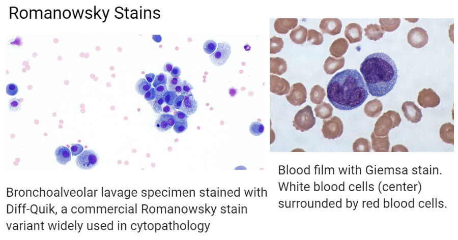
Romanowsky Stains are the stains that are used in hematology and cytological studies, to differentiate cells in microscopic examinations of blood and bone marrow samples. These stains are also applied to detect the presence of parasites in the blood such as malaria parasites. There are various Romanowsky staining types that apply the same principle. They include:
- Giemsa stain
- Wright and Wright-Giemsa stain
- May-Grunwald stain
- Leishman stain
The complex of these stains was coined after Dmitri Leonidovich Romanowsky, a Russian Physician who first identified the importance of using blood samples to make and identify effects using blood staining methodologies.

Image Source: Wikipedia.
Principle of Romanowsky Stains
- The stains are neutral, made up of oxidized methylene blue (azure) dyes and Eosin Y.
- The azures are basic dyes that bind to the acid nuclei forming a blue-purple color.
- The acid dye, Eosin binds to the alkaline cytoplasm forming red coloration.
- Romanowsky staining works principally in its ability to produce a variety of hues which makes it possible to differentiate various cellular components.
- This ability is known as the Romanowsky effect also known as metachromasia.
- The mixture of active eosin Y and active methylene blue was attributed to the formation of hues that distinguished the cell components, in that shades of purple, are formed in the cell chromatins of the nucleus and the granules in the cytoplasm of some white blood cells, to which Romanowsky defined as the Romanowsky effect or Romanowky-Giemsa effect.
- Additionally, the Romanowsky type stains can be made by oxidized unmethylated methylene blue, by the effect of oxidative demethylation. This leads to the breakdown of methylene blue into multichromatic stains some of which cause the Romanowsky effect.
- Methylene blue that has undergone oxidative demethylation is known as polychrome methylene blue, which has about 11 dyes including azure A, azure B, azure C, methylene blue, methylene violet Bernthesen, methyl thionoline, and thionoline.
Types of Romanowsky Stains
May-Grünwald-Giemsa stain
- It is a two-step staining procedure whereby the first staining is done with May-Grünwald stain and a second stain of Giemsa stain which produces the Romanowsky effect (wide range of hue/color)
Giemsa stain
- This is a special stain used for examination of blood films for parasitic infections and majorly for the diagnosis of malaria.
- It is also used as a differential stain for various blood cells (erythrocytes, platelets, leucocytes) and cellular components such as the nuclear and the cytoplasm.
- It is made up of acidic and basic dyes of eosin Y and methylene blue hence it is an azure stain.
- They produce blue-purple colored staining of the nuclear and red-pink coloration of the cytoplasm and cytoplasmic granules.
- It is used in cytogenetics and histopathology for diagnosis of:
-
- Malaria, spirochetes, and other blood parasites
- Chlamydia trachomatis inclusion bodies
- Borrelia spp
- Yersinia pestis
- Histoplasma spp
- Pneumocystis jiroveci cysts
Wright and Wright-Giemsa Stain
- The Wright stain was devised by James Homer Wright by modifying the Romanowsky stain.
- Wright stain used heated methylene blue which produces a polychrome methylene blue combined with Eosin Y.
- The heated methylene blue with Eosin Y is allowed to precipitate to form an eosinate, which is then dissolved with methanol.
- This defines the procedure for the Wright stain.
- When Giemsa stain is added to the Wright’s stain, the color brightens to a reddish-purple in the cytoplasmic granules.
- These are hematological stains that are used to differentiate blood cells from peripheral blood smears, bone marrow aspirates, and urine samples.
- Majorly they are used as differential stains for the various types of white blood cells and also they can be used quantitatively for white blood cell count in persons with parasitic-blood infections such as malaria, and disorders like leukemia.
- Use of urine samples help to diagnose for urinary tract infection and interstitial nephritis by detecting the presence of eosinophils
- In cytology and cytogenetics, it is used to detect chromosomal defects and diseases by staining the cell chromosomes.
- The stain is made up of a mixture of eosin (red) and methylene blue.
- A combination of Wright stain and Giemsa stain is known as the Wright-Giemsa stain.
- Stains that are related to Wright and Wright-Giemsa stain are buffered Wright Stain, buffered Wright-Giemsa stain. They are distinguished by the solution used for staining.
- A major difference between the Wright-Giemsa stain and May-Grünwald-Giemsa stain is the color intensity and the duration of test performance.
- May-Grünwald-Giemsa stain takes longer to perform and it produces intense color after staining.
Leishman stain
- This stain was developed by William Leishman using polychrome methylene blue and eosin Y and methanol solvent.
- The stain is used to differentiate and identify white blood cells, malaria parasites, and trypanosomes.
- The use of methanol acts as a fixative preventing perforation.
- The procedure of Leishman stain:
- Freshly prepare and rapidly air dry blood film.
- Cover the film with Leishman’s Stain (S018S) and allow it to act for 1 minute. Methanol in the stain fixes the preparation.
- Add double the volume of distilled water to the slide and mix.
- Allow the diluted stain to act for 10-12 minutes.
- Wash the film with distilled water or phosphate buffer of pH 7.0, drain and dry in air and examine
Applications of Romanowsky Stains
- Romanowsky stains are applied in several studies and diagnostics majorly in hematology and cytological diagnosis.
- Each staining technique under Romanowsky has its own applications, however, general applications include:
-
- The stains are used in hematological and cytological studies to detect for hematological disorders and chromosomal defects respectively
- Examination of blood films for blood-borne infections such as Rickettsia and rickettsia-related infections
- To detect bone marrow defects
Advantages
- The stains are readily available.
- they are simple to prepare, maintains, and use.
Disadvantages
- The morphological features of the cell may be distorted.
- The use of the required pH buffer is important to ensure maintaining the dye color.
References and Sources
- 3% – https://en.wikipedia.org/wiki/Romanowsky_stain
- 2% – https://wikimili.com/en/Romanowsky_stain
- 1% – https://www.vetstream.com/treat/felis/technique/staining-techniques-romanowsky-type-stains
- 1% – https://www.newhealthadvisor.org/Types-of-White-Blood-Cells.html
- 1% – https://wikimili.com/en/Wright’s_stain
- 1% – https://en.m.wikipedia.org/wiki/Leishman_stain
- <1% – https://www.diseasefix.com/page/diagnosis-and-tests-for-urinary-tract-infection/3479/
- <1% – https://www.answers.com/Q/What_is_the_purpose_of_Eosin_and_Methylene_blue_dyes_in_the_eosin-methylene_blue_agar_medium
- <1% – http://histology-world.com/stains/stains.htm

Thankyou so much sir you information is so important for me
Keep it up sir
Very ggod
Thank you sir to explain me the Romanowsky stain god bless you
God bless you sir
Is Romanowsky stain the only stain technique in haemoglobin stains
Wish you much success sir
Good job i like this page
Very interested
Thank you for explaining in easy way.
Hi Sonal,
Happy to hear that it was useful to you.
i think you should elaborate more on how to prepare the stain if it’s not readily available