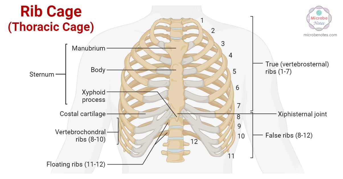
The rib cage or thoracic cage (called thorax) is located in the most medial portion of the body and protects vital organs like the heart and lungs. The rib cage is an integral part of the axial skeleton in vertebrates and it lies in the thoracic cavity.
Some organs of the respiratory, cardiovascular, nervous, immune, and digestive systems are enclosed by the rib cage as they are vital to human survival and therefore need extra protection.
Three different types of bones articulate together to form the rib cage. They are; The sternum, thoracic vertebrae, and ribs. The sternum is present anteriorly, twelve pairs of ribs form the lateral bony cage, and twelve thoracic vertebrae in the vertebral column are present posteriorly at the back.
Interesting Science Videos
Bony components of the rib cage
a. Sternum (Breast bone)
It is a flat bone that forms the middle portion of the rib cage and can be felt just under the skin in the middle of the front of the chest. It is further composed of 3 parts; manubrium, body, and xiphoid process. The cartilage joins or connects these parts in babies and children which then gradually ossifies to bone during adulthood. The ossification begins at 6 to 10 ossification centers, and fusion gets complete at about the age of 25. Before 25, the sternal body still has four separate bones whose boundaries appear as a series of transverse lines crossing the ossified sternum.
- Manubrium: The manubrium is the widest and uppermost part of the sternum. It articulates with the clavicles and the first two pairs of ribs forming the sternoclavicular joints. There is a shallow indentation on the upper surface of the manubrium which is located between the clavicular articulations and is known as jugular notch.
- Body: It is the middle tongue-shaped portion that attaches to the inferior surface of the manubrium and extends inferiorly along the midline. Individual costal cartilages from true ribs (2nd to 7th pairs) are attached to the body of the sternum.
- Xiphoid process: It is the lowermost and the smallest part and forms the pointed tip of the sternum. It gives attachment to the diaphragm, the muscles of the anterior abdominal wall (rectus abdominis), and linea alba (white line).
* linea alba is a white band of connective tissue that runs from a person’s sternum to their pubic bone. This band helps in the stabilization of the core muscles which can easily get damaged and weak from overstretching.

b. Thoracic vertebrae
12 vertebrae in the thoracic region of the vertebral column form the strong posterior portion of the rib cage. These vertebrae give rise to their corresponding ribs in the thorax. 4 peculiar features of thoracic vertebrae that distinguish them from other vertebrae are:
- The vertebral body is heart-shaped.
- Each vertebral body on its sides has demi-facets that help it articulate with the heads of the ribs.
- Transverse processes on the thoracic vertebrae (from T1- T10 only) are provided with costal facets that articulate with the tubercles of the ribs.
- Thoracic vertebrae have longer spinous processes that slant inferiorly. This provides more protection to the spinal cord and prevents any sharp and pointed object like a knife from entering the spinal canal and damaging the spinal cord.
c. Ribs
The ribs are curved, narrow, and flat bones found in all vertebrates. They are extremely light but highly resilient. The ribs are numbered accordingly from 1 to 12 based on their attachment to the corresponding thoracic vertebrae posteriorly (on the back). For example, the first pair of ribs are attached to the first thoracic vertebrae (T1).
The 12 pairs of ribs form the lateral walls of the rib cage. On the front (anterior) side, the ribs are attached to the sternum by costal cartilage forming a sternocostal joint whereas each rib articulates posteriorly with two thoracic vertebrae; by the costovertebral joint. The first rib is an exception as it articulates with the first thoracic vertebra only since there is no thoracic vertebra above it.
There are two types of ribs based on their structure; typical and atypical.
Typical ribs have a generalized structure, while atypical ribs have variations on this structure.
What are typical ribs?
A typical rib consists of a head, neck, and body:
- Head: The vertebral end of the rib articulates with the vertebral column at the head or capitulum. The articular surface of the head is divided into superior and inferior articular facets by a ridge. One of these facets articulates with the numerically corresponding vertebra whereas the other one articulates with the vertebra located just above.
- Neck: From the head, a short neck leads to the tubercle which is a small elevation that projects dorsally. The inferior portion of the tubercle contains an articular facet that contacts the transverse process of the corresponding thoracic vertebra. The neck doesn’t contain any prominent bony structure but simply connects the head and the body of the rib.
- Body: It is a flat and curved part of the rib which is also called a shaft of the rib. The costal groove present on the internal concave surface of this shaft is for the neurovascular supply of the thorax. This groove protects the vessels and nerves from damage. The outer superficial surface of the shaft is convex and provides an attachment site for the muscles of the pectoral girdle and trunk. The intercostal muscles, which move the ribs, are attached to the superior and inferior surfaces of the ribs.
What are atypical ribs?
Atypical ribs have features that are not common to all the ribs. 1st, 2nd, 10th, 11th, and 12th ribs can be described as ‘atypical’.
- The 1st rib (Rib 1) is comparatively shorter and wider than the other ribs. It articulates with its corresponding vertebra with a single facet that it has on its head. There isn’t a thoracic vertebra above it. Two grooves present on its upper surface, make way for the subclavian vessels.
- The 2nd rib (Rib 2) is thinner and longer than the 1st rib. It articulates with its corresponding vertebra by two articular facets present on its head. The Serratus anterior muscle originates from a roughened area on the upper surface of this rib.
- The 10th rib (Rib 10) has only one facet that articulates with its numerically corresponding vertebra.
- The 11th and 12th ribs (Ribs 11 and 12) each lack a neck and have only one facet, which helps them articulate with their corresponding vertebra.
Articulations of ribs
Posterior articulations
Each rib with the help of two joints is connected posteriorly with the corresponding vertebra of the spine. This way, all 12 pairs of ribs have articulations posteriorly with their corresponding thoracic vertebrae.
- Costotransverse joint: The tubercle of the rib articulates with the transverse costal facet of the corresponding vertebra to form this joint.
- Costovertebral joint: It is the joint between three structures; the head of the rib, the superior costal facet of the corresponding vertebra, and the inferior costal facet of the vertebra located just above.
Anterior articulations
The anterior attachment of the ribs varies. Based on their anterior articulations in the rib cage, ribs are of three types:
- True ribs: They are the first seven pairs (1-7) of ribs which are independently attached to the sternum anteriorly with the help of the costal cartilage and to the thoracic vertebrae posteriorly.
- False ribs: The next three pairs (8, 9, and 10) are called false ribs. They arise from the thoracic vertebrae at the back but don’t attach to the sternum directly, instead attach to the cartilage of the 7th true rib. They are also called vertebra-chondral ribs.
- Floating ribs: The lowest two pairs (11 and 12) are called floating ribs as they are attached only to the vertebral column at the back. They don’t articulate with the sternum, their anterior tips being free. The free anterior ends of such ribs are connected to the muscles in the abdominal wall. They are also called vertebral ribs.
Functions of rib cage
Protection of internal thoracic organs
The rib cage protects the internal delicate thoracic organs like the heart, lungs, thymus, aorta, etc. from damage and injuries during severe accidents.
Breathing
The ribs are quite mobile, due to their complex musculature, dual articulations at the vertebrae, and flexible connection to the sternum. The ribs curve away from the vertebral column since they are angled inferiorly and because of their curvature, the movements of the ribs change the position of the sternum and thus help in breathing.
The muscles between our ribs (external and internal intercostal muscles) help us breathe along with the help of the diaphragm. The configuration of the lower five ribs in the rib cage makes sure that the lower part of the rib cage is free to expand coordinating with the movements of the diaphragm.
When we inhale, the intercostal muscles contract to pull our rib cage both upward and outward thus expanding the chest cavity, and; when we exhale, the muscles relax to pull our rib cage both downward and inward compressing the chest cavity.
When we breathe in, our lungs expand, and the air rushes inside through our nose or mouth. This air travels down our trachea or windpipe and finally into our lungs. When we breathe out or exhale, our diaphragm and rib muscles relax, reducing the space in the chest cavity. As the chest cavity gets compressed, our lungs deflate, the same way the balloon deflates releasing the air present inside.
Providing support and stability for the vertebral column and the upper extremities
The sternoclavicular joint present in the front part of the chest helps support and move our pectoral girdle (shoulder joint: both clavicles and scapulae) and our upper extremities (upper limbs). At the same time, the articulations of ribs with the corresponding thoracic vertebrae give a supportive framework to the vertebral column and help in maintaining its position and body posture.
The bent structure of ribs and their movements can cushion shocks and absorb hits, but severe or sudden impacts can cause painful rib fractures.
Ribs serve as attachment points for muscles of the upper body part thus helping in supporting the body.
References
- Waugh A. and Grant A., Ross and Wilson Anatomy and Physiology in Health and Illness, 12th edition, 2014
- Elaine N.M, and Katja H., Human Anatomy and Physiology, 12th global edition, 2022
- Martini, Nath, and Bartholomew., Fundamentals of Anatomy and Physiology, 11th edition, 2018
- https://teachmeanatomy.info/thorax/bones/ribcage/
- https://www.kenhub.com/en/library/anatomy/thoracic-cage
- https://byjus.com/neet/ribs/
- https://www.theskeletalsystem.net/rib-cage
- https://www.nhlbi.nih.gov/health/lungs/breathing-benefits
