
The pharynx is a hollow, muscular, tubular cavity that serves as a shared passageway for the respiratory and digestive systems. It is the 12-14 cm vertical segment of the throat that extends from the oral cavity and nasal cavity to the esophagus and larynx.
It helps air pass to the larynx and food and liquid to the esophagus inside the body. Despite structural differences, it is present in both vertebrates and invertebrates.
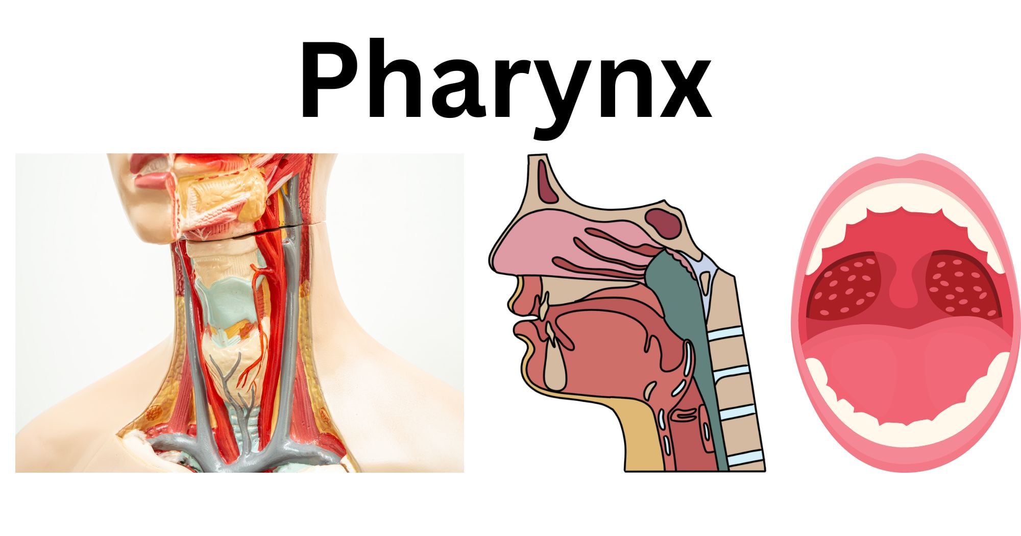
Anatomy and Structure of the Pharynx
- The pharynx begins at the posterior region of the nasal cavity, extends towards the mouth, and ends before the upper part of the esophagus. It is divided into three anatomical sections: Nasopharynx, oropharynx, and laryngopharynx (hypopharynx).
- The pharyngeal walls are made up of i) circular muscles (that propel the food down the digestive organs) and ii)longitudinal muscles (that are essential during the deglutition mechanism).
- The nasopharynx is lined with a layer of pseudostratified ciliated columnar epithelium, while the oropharynx and hypopharynx are lined with a non-keratinized stratified squamous epithelium layer.
- The pharynx is also richly supplied by arteries and innervated by nerves.

Source: https://www.cancer.gov/publications/dictionaries/cancer-terms/def/pharynx
Parts of the Pharynx
The pharynx can be divided into 3 parts based on its functional region:
Nasopharynx
- This part of the pharynx is found posterior to the nasal cavity in the respiratory system.
- It is bounded superiorly by the skull base, anteriorly by the nasal cavity, and posteriorly by the pharyngeal wall.
- It is surrounded inferiorly by the soft palate. On the lateral side, the pterygoid palates and pharyngeal constrictor muscle form the boundary.
- The roof of the nasopharynx is called the vault or the fornix. It also contains adenoid lymphoid tissue (the pharyngeal tonsil).
- Posterior choanae connect the nasopharynx with the nasal cavity.
- The nasopharynx is also connected with the middle ear cavity via the Eustachian tube. The posterior part of the eustachian tube creates a protrusion known as torus tobarius. Behind the eustachian tube and torus taborius, there is a pharyngeal cavity called the Fossa of Rosenmuller on the lateral wall. It is a common site for nasopharyngeal tumors.
- The space between the nasopharyngeal wall and the vertebral column is called the retropharyngeal space.

Source: https://standardofcare.com/nasopharynx/
Oropharynx
- It is the oral section of the pharynx, situated right behind the oral cavity.
- It is lined by the squamous stratified non-keratinized epithelium tissue.
- It consists of 4 walls: Pharyngeal part (base) of the tongue and oropharyngeal isthmus form the anterior wall, posterior wall is present opposite to the cervical vertebrae (C2-C3), while the two lateral walls on each side consist of palatine tonsils and palatopharyngeal arches.
- It also includes the soft palate on the roof, while the posterior third part of the tongue, the lingual tonsil, and the side and back wall of the throat form the floor of the oropharynx.
- Within the oropharynx, the glossoepiglottic folds act as a bridge between the posterior third segment of the tongue and the epiglottis.
- The oropharynx also contains valleculae (the space between the epiglottis and the base of the tongue).
- It functions to transport air, food, and fluids from the oral cavity.

Source: https://www.cancer.gov/publications/dictionaries/cancer-terms/def/oropharynx
Laryngopharynx
- It is the lowest pharyngeal part that lies behind the larynx. It is also called the hypopharynx.
- It elongates from the epiglottis to the lower part of the cricoid cartilage.
- It connects the oropharynx with the larynx and esophagus.
- It helps with respiration and digestion by directing air to the larynx and food/fluids to the esophagus, respectively.
- The anterior laryngopharyngeal wall consists of the mucous membrane, which covers the posterior side of the cricoid and arytenoid cartilages.
- The posterior wall is supported by cervical vertebrae (C4-C6), while the lateral wall is covered by the thyroid cartilage and membrane.
- In the anterior part of the laryngopharynx, there is a vertical opening called the laryngeal inlet. It opens to the larynx.

Functions of the Pharynx
Functions of the Pharynx in Respiration
The pharynx is a significant organ of the respiratory system. It demonstrates the following functions:
- It helps direct inspired air from the nasal cavity to the larynx and trachea via the nasopharynx, oropharynx, and laryngopharynx.
- The pharynx is lined by the mucosal membrane. The internal lining (mucosa) warms and moistens (humidifies) the inhaled air in the pharynx.
- Pharyngeal tonsils play an essential role in defending the body against inhaled bacteria, viruses, and dust.
- While swallowing food, the pharynx, along with the epiglottis, helps prevent food from entering the respiratory tract and directs it into the digestive system.
- It also allows the regulation of air pressure because it is connected to the eustachian tube via the nasopharynx.
Functions of the Pharynx in Digestion
The pharynx plays the following significant roles in digestion.
- The pharynx helps in deglutition. The pharyngeal muscles contract to propel food into the inner digestive tract.
- During swallowing, the epiglottis covers the opening of the airway and prevents food from entering the respiratory tract.
- It also closes the nasal cavity and prevents food from refluxing into the nasal passages.
Muscles and Mechanism of Swallowing (Deglutition)
Deglutition is the complex biological process of moving food from the oral cavity to the esophagus, pharynx, and finally the stomach. It is a significant mechanism of the digestive system, also known as swallowing.
In this process, the food bolus is propelled into the stomach by the combined action of more than 22 internal muscles and 30 neurons.
Muscles involved in deglutition
| S.N | Deglutition region | Muscles involved |
| Oral Cavity | Tongue: Hyoglossus, Genioglossus, Styloglossus, Palatoglossus, Mylohyoid | |
| Mastication Muscles: Masseter, Temporalis, Pterygoid (Lateral and medial) | ||
| 2. | Pharynx | Tensor and Levator palitini, |
| Suprahyoid muscles: Digastric, Stylohyoid, Geniohyoid | ||
| Infrahyoid muscles: Sternohyoid, Sternothyroid, Thyrohyoid, Omohyoid | ||
| Longitudinal pharyngeal muscles: Stylopharyngeus, Salpingopharyngeus, Palatopharyngeus | ||
| Pharyngeal constrictor muscles: Cricopharyngeus muscles | ||
| 3. | Larynx | Posterior and lateral cricoarytenoid, oblique, and transverse arytenoids |
| Aryapiglotticus |
Mechanism of Deglutition
Once food is ingested in the mouth, it is broken down into smaller pieces by chewing (mastication) and grinding. It is then converted into a soft mass known as a bolus.
The food bolus is swallowed from the oral cavity, and thereafter, it is pushed down the alimentary canal through the oropharynx, laryngopharynx, esophagus, and stomach.
There are 3 phases of deglutition:
- Oral phase
- Pharyngeal phase
- Oesophageal phase

Source: https://storymd.com/journal/mqb9raa1em-deglutition/page/58gvyvt96naa-deglutition
Oral phase:
- It is the first stage of swallowing, a complete voluntary process.
- The tongue elevates and contracts to move the bolus posteriorly towards the oropharynx.
- In the oropharynx, the presence of bolus food activates the sensory receptors of the glossopharyngeal nerve. It transmits signals to the swallowing center of the brain (pons and medulla), which then sends signals to the efferent nerves to initiate the reflex action during the pharyngeal phase.
- The oesophageal sphincter is closed in this phase.
Pharyngeal phase:
- It is the involuntary, irreversible phase of deglutition.
- After receiving the efferent signal, the pharyngeal phase begins.
- The soft palate and uvula lift to seal the nasopharynx. The tensor and levator palatini muscles are also involved to prevent the escape of food from the nasal cavity.
- The pharynx shortens and widens simultaneously to receive the bolus.
- The suprahyoid and pharyngeal muscles contract, elevating the larynx. The posterior and lateral cricoarytenoid, oblique and transverse arytenoid muscles, and arytenoid cartilage are involved in the process of glottic closure. Consequently, the epiglottis blocks the trachea to prevent the food from entering the respiratory tract (airway) and opens the passage towards the esophagus.
- During this process, the respiratory process is inhibited temporarily.
- The contraction of thyrohyoid muscles opens the upper oesophageal sphincter, as the larynx moves superiorly and anteriorly.
- When the pharyngeal muscles contract, the food bolus is moved down the esophagus by peristaltic waves.
Oesophageal phase:
- It is the final involuntary phase of deglutition.
- The pharyngeal muscle contracts and propels the bolus into the esophagus by the peristaltic movement.
- Once the bolus reaches the esophagus, the upper oesophageal sphincter contracts and closes.
- The food is further propelled into the stomach by the lower oesophageal sphincter. Afterward, this muscle contracts to prevent stomach acid from refluxing.
Blood Supply and Innervation of the Pharynx
The pharynx is one of the highly vascularized structures that receives the arterial blood supply primarily via the external carotid artery. The 4 major carotid arteries that are responsible for the blood supply to the pharynx are:
- Ascending pharyngeal artery(pharyngeal branch)
- Facial artery (palatine and tonsillar branch)
- Lingual artery
- Maxillary artery
They are responsible for supplying blood to the soft palate, pharyngeal constrictor muscles, palatine tonsils, and the eustachian tube.
In addition, the pharynx also consists of the pharyngeal nervous plexus originating from the cranial nerves. They provide the nerve supply to the pharynx. It involves:
- Vagus nerve: The pharyngeal branches provide motor innervation to the pharynx (except to the stylopharyngeus). The internal branch of the vagus nerve provides the nerve supply to the laryngopharynx.
- Glossopharyngeal nerve: It provides the sensory innervation to the majority of the pharynx (except nasopharynx) and motor innervation to the stylopharyngeus
- Maxillary nerve: It supplies the sensory innervation to the superior and anterior part of the nasopharynx.
Waldeyer’s Ring: The role of Tonsils in Immune Defense
Waldeyer’s ring is the anatomical structure composed of 4 specialized tonsillar lymphoid tissues:
- Pharyngeal tonsil
- Tubal tonsil
- Palatine tonsil
- Lingual tonsil
- It serves as the first line of the immune defense mechanism. It screens for the microorganisms entering the nasal and oral pathways and activates the immune system.
- They contain specialized WBCs (lymphocytic cells and macrophagocytic cells), which detect and trap the pathogens.
- They also function in producing antibodies (IgA) that protect the mucous membranes.
Source: https://teachmeanatomy.info/neck/vessels/tonsils-and-adenoids/
Common Diseases and Disorders of the Pharynx
Tonsilitis
It refers to the infection of the lymphoid tonsils. It is more common among children. The symptoms include: fever, headache, difficulty in swallowing, pus-filled white patches in the tonsils, swollen glands, and bad breath.
Pharyngitis
It refers to the bacterial or viral infection of the pharynx. It causes the acute inflammation of the pharyngeal mucus membrane. It is also commonly referred to as a sore throat. The symptoms include: dry, painful throat, mild cough, bad breath, and redness in the posterior part of the mouth.
Dysphagia
It is the clinical term for difficulty swallowing food, even saliva. The common causes of dysphagia are neurological disorders such as Parkinson’s disease, stroke, muscle diseases, and throat cancer. The symptoms often include choking, coughing, acid reflux, voice hoarseness, heartburn, etc.
Strep throat
It refers to the bacterial infection of the throat caused by Group A streptococcus (S. pyogenes). It is highly contagious and can be transmitted via respiratory droplets (coughs or sneezes). The symptoms include severe sore throat, dysphagia, fever, swollen tonsils, headache, nausea, vomiting, etc.
Gastroesophageal Reflux Disease(GERD)
It refers to the condition in which stomach acid flows back into the esophagus. It is caused by weak or relaxed lower oesophageal sphincter muscles. The common symptoms of GERD include: chest pain, heartburn, regurgitation of food with a sour taste, chronic cough, and difficulty swallowing.
Diagnostic Procedures: Throat Swabs, Endoscopy, and Imaging
It is important to diagnose infections or inflammation in the pharynx, identify abnormal behavior in the pharyngeal tissues, and detect tumors. There are various diagnostic procedures for detecting abnormalities in the throat. They are:
Laboratory analysis by collecting the throat swabs
When the clinician suspects a sore throat (strep throat), a throat swab is collected for laboratory culture and sensitivity testing to detect infectious germs or bacteria.
To collect the throat swab, a tongue depressor is applied to lower the tongue. Afterward, the patient is asked to roll their tongue upward, and a sterile swab is rubbed against the affected area in the throat.
Endoscopy
It is a non-surgical process in which an endoscope (a thin, flexible tube featuring a camera) is inserted into the oral cavity and gradually advanced to the throat, stomach, and the anterior segment of the small intestine to examine the pharyngeal region and make an accurate diagnosis.
The throat endoscopy (laryngoscopy) is performed to visualize and examine the larynx (voice box) and the surrounding structures, such as swelling, redness, or irritation, and to assess the severity of lesions in the throat.

Source: https://www.cancer.gov/publications/dictionaries/cancer-terms/def/upper-endoscopy
Imaging
If the endoscopy results are not sufficient to detect the medical condition or disease, medical imaging is required. X-rays, CT scans, MRI, and videofluoroscopy are essential for diagnosing pharyngeal or laryngeal cancer and evaluating mucosal lesions.
How to keep the Pharynx Healthy
It is essential to keep the pharyngeal regions healthy. Some of the ways to take care of the pharynx (throat) are:
- Avoid smoking
- Maintain oral hygiene, such as regular brushing.
- Stay hydrated to keep the mucosal lining moist and lubricate the throat.
- Protect the throat by avoiding loud shouting.
- Avoid spicy foods if you have a sensitive throat.
- Gargle with saline water if you feel slight irritation.
- Keep the surroundings clean and use a humidifier to prevent dry air in the rooms.
Conclusion
The pharynx is the hollow tubular structure in the throat, which is made up of muscles. It is divided into 3 parts: Nasopharynx, Oropharynx, and Hypopharynx. They play a significant role in the respiratory and digestive processes. Additionally, the pharynx is also involved in the food-swallowing mechanism.
References
- Dr. Ghassan. (n.d.). Anatomy of the pharynx. Retrieved from https://uomustansiriyah.edu.iq/media/lectures/2/2_2018_12_11!02_27_40_PM.pdf
- Laryngeal endoscopy – Newcastle Hospitals NHS Foundation Trust. (2023, July 10). Retrieved from https://www.newcastle-hospitals.nhs.uk/services/ear-nose-throat-service-ent/ent-speech-and-language-therapy-slt/laryngeal-endoscopy/
- Mittal, R. K. (2011). Pharynx—Anatomy, neural innervation, and motor pattern. Retrieved from https://www.ncbi.nlm.nih.gov/books/NBK54279/
- Oral cavity, oropharynx, hypopharynx, & larynx cancer prevention. (2024, November 8). Retrieved from https://www.cancer.gov/types/head-and-neck/patient/oral-prevention-pdq
- Oropharynx. (2023, October 30). Retrieved from https://www.kenhub.com/en/library/anatomy/oropharynx
- Panara, K., Ahangar, E. R., & Padalia, D. (2023, July 24). Physiology, swallowing. Retrieved from https://www.ncbi.nlm.nih.gov/books/NBK541071/#:~:text=The%20processes%20mentioned%20above%20are,protects%20the%20airway%20from%20aspiration.
- Pharynx | Anatomy and Physiology | Research Starters | EBSCO Research. (n.d.). Retrieved from https://www.ebsco.com/research-starters/anatomy-and-physiology/pharynx
- Pharynx – Knowledge @ AMBOSS. (n.d.). Retrieved from https://www.amboss.com/us/knowledge/pharynx
- Radiopaedia.org. (n.d.). Retrieved from https://radiopaedia.org/articles/nasopharynx
- Rahim, I., Napolitano, A., Burd, C., & Lingam, R. K. (2023). Imaging of pharyngeal pathology. British Journal of Radiology, 96(1149), 20230046. https://doi.org/10.1259/bjr.20230046
- Standardwp. (2021, November 24). Oropharyngeal Anatomy(Oropharynx). Retrieved from https://standardofcare.com/oropharyngeal-anatomy/
- TeachMeAnatomy. (2025, November 6). The Pharynx – Subdivisions – Blood supply – TeachMeAnatomy. Retrieved from https://teachmeanatomy.info/neck/viscera/pharynx/
- Throat swab Culture | Quirónsalud. (n.d.). Retrieved from https://www.quironsalud.com/en/diagnostic-tests/throat-swab-culture#:~:text=A%20throat%20swab%20culture%20is,caused%20by%20viruses%20and%20bacteria.
- Vinmec International Hospital. (2026, February 27). How are the throat and larynx examined? Vinmec International Hospital. Retrieved from https://www.vinmec.com
- Wikipedia contributors. (2026, February 11). Pharynx. Retrieved from https://en.wikipedia.org/wiki/Pharynx