Karyotyping is a diagnostic tool used in medical genetics to examine the chromosomes of an individual to detect any abnormalities.
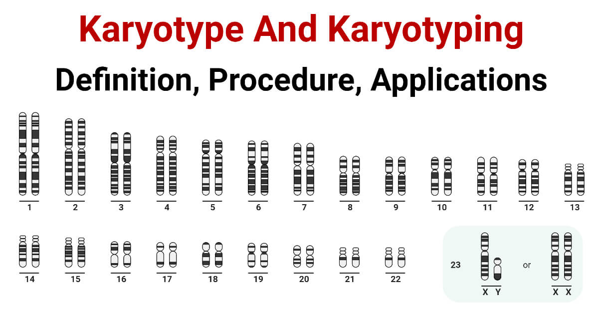
It involves arranging and analyzing the chromosomes from a cell sample to create a visual representation of the chromosome complement, known as a karyogram.

Karyotyping has been used in clinical practice for over half a century and remains an important tool in identifying genetic disorders, determining the sex of an individual, and studying evolutionary relationships between species.
This note provides an overview of karyotyping, including its history, methods, applications, and limitations.
Interesting Science Videos
History of Karyotyping
Cytogenetics, the study of chromosomes and their relationship to inherited characteristics, was developed as a result of the discovery of chromosomes in the late 19th century and its link with heredity. Giemsa staining was used by Tjio and Levan in 1956 to see the chromosomes in a human cell and produce the first karyotype. Giemsa banding, often known as the standard method for karyotyping in clinical use, was developed.

Karyotyping Procedural Steps
- A cell sample is needed for karyotyping and can be taken from a variety of places, including tissue, amniotic fluid, and blood.
- The chromosomes may be seen during metaphase, the stage of cell division when chromosomes are most condensed and visible, thanks to the laboratory-induced cell division that the cells undergo.
- Giemsa or other dyes are then used to stain the chromosomes, which results in a distinctive pattern of dark and light bands that makes it possible to identify and organise the chromosomes according to size and form.
- The resultant karyogram is a graphic depiction of the chromosomal complement, with each chromosome’s quantity and appearance representing the person’s genetic makeup.
Applications of Karyotyping
Chromosomal Abnormalities
- For identifying chromosomal anomalies that result in genetic illnesses, karyotyping is an important method in medical genetics.
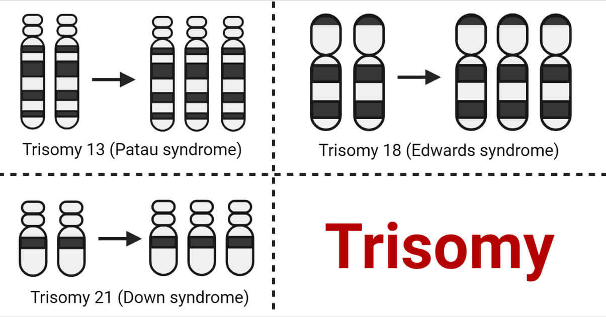
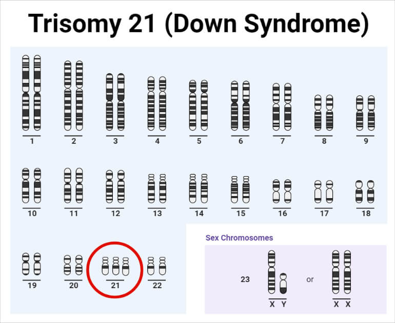
- It can pick up on numerical anomalies like trisomy 21, which causes Down syndrome, or monosomy X, which causes Turner syndrome.
- Structural abnormalities, like translocations, inversions, or deletions, which might result in diseases like leukaemia, lymphoma, or muscular dystrophy, can also be found by karyotyping.
- In order to find chromosomal abnormalities in the growing foetus, karyotyping is also employed in prenatal diagnostics, when a sample of amniotic fluid or chorionic villi is examined.

Gender Determination
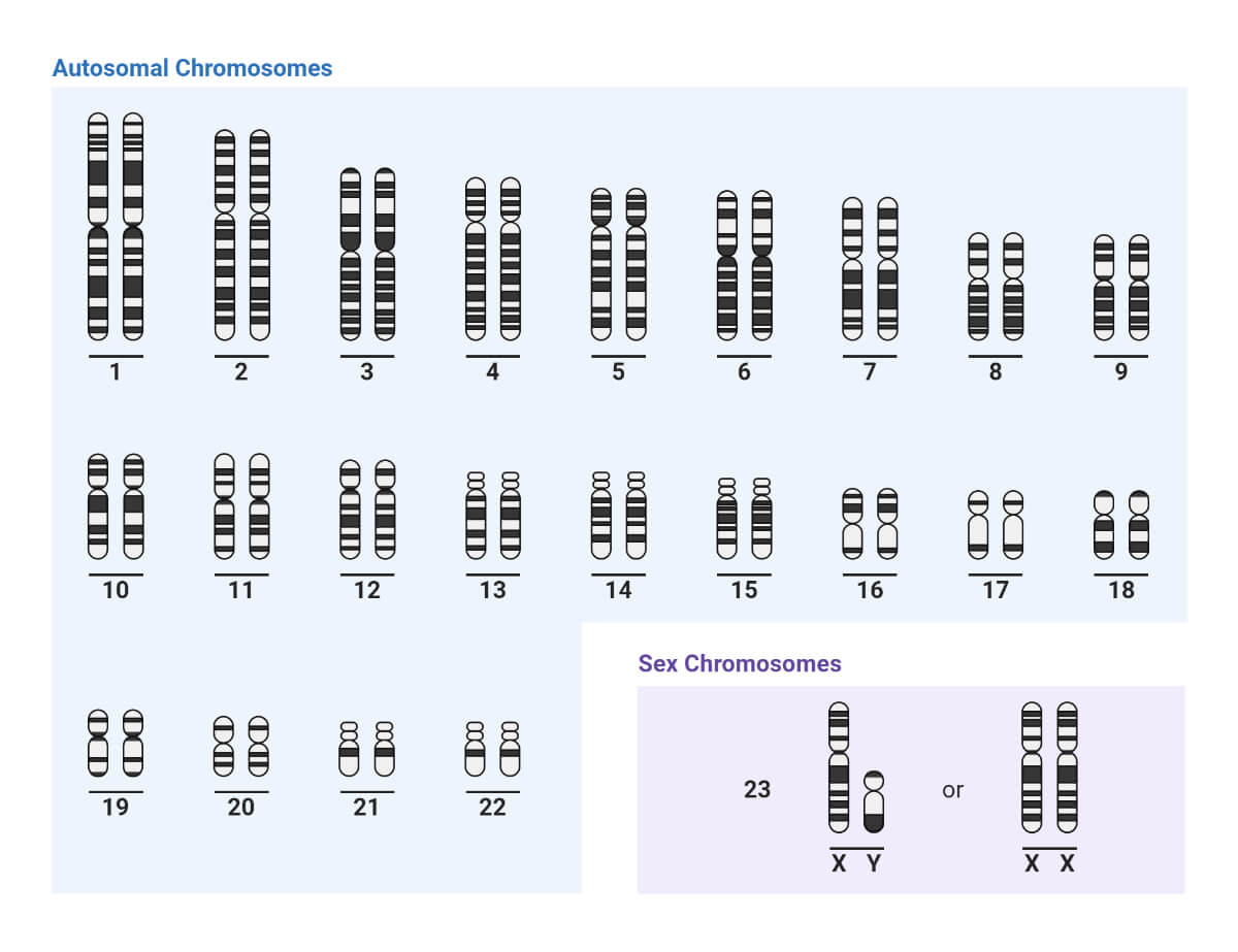
Karyotyping is also used to categorise a person’s sex. Males have one X and one Y chromosome, whereas females have two X chromosomes, which are the sex chromosomes in humans. By determining whether a Y chromosome is present or absent, karyotyping can be used to establish a person’s gender.
Evolutionary Biology
In evolutionary biology, karyotyping is also used to investigate the links between different species. Scientists can determine a species’ evolutionary history and point of divergence by comparing the karyotypes of various species. For instance, although chimps have 24 pairs of chromosomes, humans have 23 pairs. The human lineage’s fusion of two ancestral chromosomes, which took place after the species split from the chimpanzee lineage, is the cause of the discrepancy in chromosomal number.
Limitations of karyotyping
In order to find chromosomal abnormalities, karyotyping has significant limitations.
- Only a small portion of the genome, or the number or shape of chromosomes visible during metaphase, may be detected by this test.
- It is unable to find anomalies such as epigenetic changes or mutations that impact how genes are expressed.
- Karyotyping also needs enough cells to analyse, which could not be possible in some circumstances, such as when using early embryos or foetal tissues.
- Karyotyping’s limits in identifying a person’s sex are also notable. It can only tell if a Y chromosome is present or not, which is not always conclusive. Rarely, people with ambiguous genitalia or differences in the makeup of the sex chromosomes can be identified using karyotyping alone.
Additionally, there are several drawbacks to karyotyping when examining interspecies evolutionary links. It is constrained by the chromosomal bands’ resolution, which might not be able to distinguish more minute variations in chromosome structure or gene content. Additionally, it is unable to explain convergent or parallel evolution, in which related features develop independently in several lineages.
Recent advances in karyotyping
The range and resolution of karyotyping have been increased as a result of recent developments in molecular genetics and genomics.
- Using fluorescent probes, the method known as fluorescence in situ hybridization (FISH) targets certain DNA sequences and makes them visible on the chromosomes.
- FISH has a better sensitivity for abnormalities than traditional karyotyping, and it can pick up on minor deletions or duplications that bands would miss.
- Another method that employs microarrays to compare the DNA content of two samples, such as healthy and cancerous cells, is comparative genomic hybridization (CGH).
- By detecting copy number variations (CNVs) that karyotyping could miss, CGH can offer a genome-wide perspective of genetic alterations.
Whole Genome Sequencing
One’s genome, comprising all of their chromosomes and genes, may be fully and precisely seen using whole-genome sequencing (WGS), a potent technology. WGS may identify point mutations, structural alterations, and other kinds of genetic changes in addition to chromosomal abnormalities with a better level of resolution than karyotyping or FISH. WGS is currently more expensive and time-consuming than karyotyping, and it might be difficult to understand the results.
Karyotypes from Mitotic cells
- Karyotyping entails examining the quantity, make-up, and arrangement of chromosomes within a cell.
- Mitotic cells, or cells that are actively dividing, are used most frequently to prepare karyotypes.
- Cells are first administered a chemical to stop them in metaphase, a stage of cell division where the chromosomes are most densely packed and visible, in order to create a karyotype.
- After that, the cells are stained with a dye like Giemsa that preferentially binds to the AT-rich regions of the chromosomes to produce a distinctive banding pattern.
Banding Patterns and Chromosome Structure
The placement of centromeres, telomeres, and other structural elements is reflected in the banding pattern on the chromosomes. Each chromosome also has a distinct banding pattern that makes it possible to recognize and separate them from one another. The banding pattern is made up of bands, sub-bands, and interband that are formed by the differential staining of several chromosomal regions. G-banding, R-banding, C-banding, and Q-banding are a few examples of the various banding patterns that may be produced by staining methods.
Chromosome Organization in Karyograms
- After being stained and photographed, the chromosomes are cut out and paired together based on their size, shape, and banding pattern.
- The final configuration is known as a karyogram or karyotype.
- The visual examination of the chromosomes made possible by the karyogram might identify anomalies or changes in chromosomal number or shape.
- With the sex chromosomes (XX or XY) at the end, the human karyogram normally consists of 23 pairs of chromosomes, organized in order of size from biggest to smallest.
Detection of Abnormalities Using Karyogram
Karyotyping may identify chromosomal abnormalities linked to a number of genetic illnesses and birth malformations, making it a crucial diagnostic technique in medical genetics. Aneuploidy, or an abnormally high number of chromosomes, such as in Down syndrome (trisomy 21) or Turner syndrome (monosomy X), structural abnormalities, such as deletions, duplications, inversions, or translocations, and mosaicism, or the coexistence of normal and abnormal cells, are a few examples of chromosomal abnormalities that can be identified by karyotyping.

Conclusion
Karyotyping is a crucial tool in evolutionary biology and medical genetics that enables the visualization and study of a person’s or a species’ chromosomes. For over 50 years, it has been used to identify chromosomal anomalies, ascertain a person’s gender, and investigate evolutionary links. Karyotyping has certain limits when it comes to identifying the sex of people who have ambiguous genitalia or differences in the sex chromosome makeup. It also has limitations when it comes to detecting genetic alterations that do not influence the chromosomes visible during metaphase. The precision and range of karyotyping have increased as a result of recent developments in molecular genetics and genomics, but they also present new obstacles to interpretation and application.
Overall, karyotyping continues to be a useful and adaptable method for figuring out the genetic causes of both health and disease. Chromosomal visualization and analysis are made possible by the method known as karyotyping. A karyogram, which groups chromosomes into pairs, is constructed by treating and staining mitotic cells to produce a banding pattern that represents the structural specifics of the chromosomes. Karyotyping can identify chromosomal anomalies linked to hereditary diseases and birth problems, making it a useful technique in medical genetics. Early detection of these problems allows medical practitioners to better care for and treat afflicted patients.
References
- Karyotyping | Learn Science at Scitable – http://www.nature.com/scitable/topicpage/karyotyping-for-chromosomal-abnormalities-298
- KARYOTYPE – https://www.genome.gov/genetics-glossary/Karyotype
- KARYOTYPES, CHROMOSOMES, AND TRANSLOCATIONS – http://www.informatics.jax.org/silver/frames/frame5-2.shtml
- Gardner, R. J. M., and G. R. Sutherland. “Down syndrome, other full aneuploidies, and polyploidy.” Chromosome abnormalities and genetic counseling. Oxford University Press, New York, 2004. 249-256.
- Liehr, T. (Ed.). (2014). Fluorescence in situ hybridization (FISH) – Application guide. Springer.
- Gu, Wenli, Feng Zhang, and James R. Lupski. “Mechanisms for human genomic rearrangements.” Pathogenetics 1 (2008): 1-17.
- Ford, C. E., and J. L. Hamerton. “A colchicine, hypotonic citrate, squash sequence for mammalian chromosomes.” Stain technology 31.6 (1956): 247-251.
- Speicher, Michael R., Stephen Gwyn Ballard, and David C. Ward. “Karyotyping human chromosomes by combinatorial multi-fluor FISH.” Nature genetics 12.4 (1996): 368-375.

Such amaizing interesting educative info ..