Interesting Science Videos
What is an Inverted Microscope?
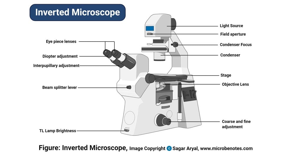
Invented in 1850 by a faculty member of Medical College of Louisiana, named J. Lawrence Smith, this microscope just like it sounds is a light microscope that has its components placed in an inverted order, this means, light source and condenser lens are placed above the specimen stage, pointing down, while the objectives and the turret are found below the stage pointing upwards. This is a reverse of the normal construction of a microscope, where the objective lenses are found above the stage while the condenser and the light source are below the stage. Hence the word, ‘inverted’. And therefore, instead of viewing the image from up, downwards, with the inverted microscope you view the image from down, upwards.
Principle of the Inverted Microscope
The working principle of the inverted microscope is basically the same as that of an upright light microscope. They use light rays to focus on a specimen, to form an image that can be viewed by the objective lenses. However, in the inverted microscope, the light source and the condenser are found on top of the stage pointing down to the stage. The condenser lens above the specimen stage functions primarily to concentrate the light on the specimen. The specimen is placed on a large stage that can be able to hold. With the objectives located below the stage and pointing upwards, it collects light from the condenser magnifying the image, which is then sent to the ocular lens. Light is reflected by the ocular lens through a mirror. The cells can be viewed and observed through the bottom part of the cell culture apparatus, where total optical points are reached, with the assistance of an ibidi Polymer Coverslip and ibidi Glass coverslip.
Parts of the Inverted Microscope
However inverted the microscope is, it still has similar components to those of other microscopes, the difference is the arrangement of these components, which are placed in inverted positions.
It has:
- Stage – they have a large fixed stage able to hold large vessels like Petri plates.
- Objective lens – movable lenses that are 4- 6 in the number of different magnification powers, they move on a vertical axis for viewing of the specimen.
- Dual concentric knobs – fine and coarse adjustment knobs for fine-tuning and focusing the objectives to the specimen.
- Nosepiece – this is a rotating turret that holds the objectives.
- The condenser lens is used to concentrate light on the specimen.
- Removable cameras, fluorescent illuminators, scanners can also be temporally attached.

Uses of the Inverted Microscope
- It is used in diagnostics fungal cultures, for example, detection of Phytophthora spp in cultures.
- Used for diagnosis of nematology extraction specimens to observe nematodes such as Vermiform nematodes.
- Used to observe living microbial cells found at the bottom of lab vessels such as tissue culture flasks and Petri plates.
Advantages of the Inverted Microscope
- The inverted Microscope has a wide stage that favors it to view specimens in glass tubes and Petri plates and therefore, it is commonly used to study live cells, by viewing the cells from the bottom of the cell culture apparatus.
- It can also be used to view and study cells in large amounts of the medium.
- It can be used to view the cell tissues in their original vessel, which are larger than microscopic slides, which makes it better than the upright microscope which only views specimens in small microscopic slides.
- It can be used to view cells in large quantities of the medium than in small specimen quantities on a glass slide under a coverslip.
- Viewing of cells can also be accessed from both the top and bottom sections of the holding apparatus because most cells will sink to the bottom of the apparatus such as the Petri plate
- The specimen remains uncontaminated by touch with the objective lens, hence maintaining the sterility of the sample.
Limitations
- Generally, these microscopes are very expensive to acquire.
- They are manufactured by very few companies because they are expensive to manufacture.
- They are rarely found in the market for purchase and usage.
- It is difficult viewing the specimens through thick glass vessels such as a Petri plate hence they require very high optical quality.
Because of the difficulty and the cost of manufacturing the Inverted microscope, very few companies are known to manufacture the microscope. Some of the companies include:
- Zeiss Axio Vet Al
- Olympus
- Nikon
- Kruss Germany
- echoLAB
- Metkon Technology
- Horiba Scientific
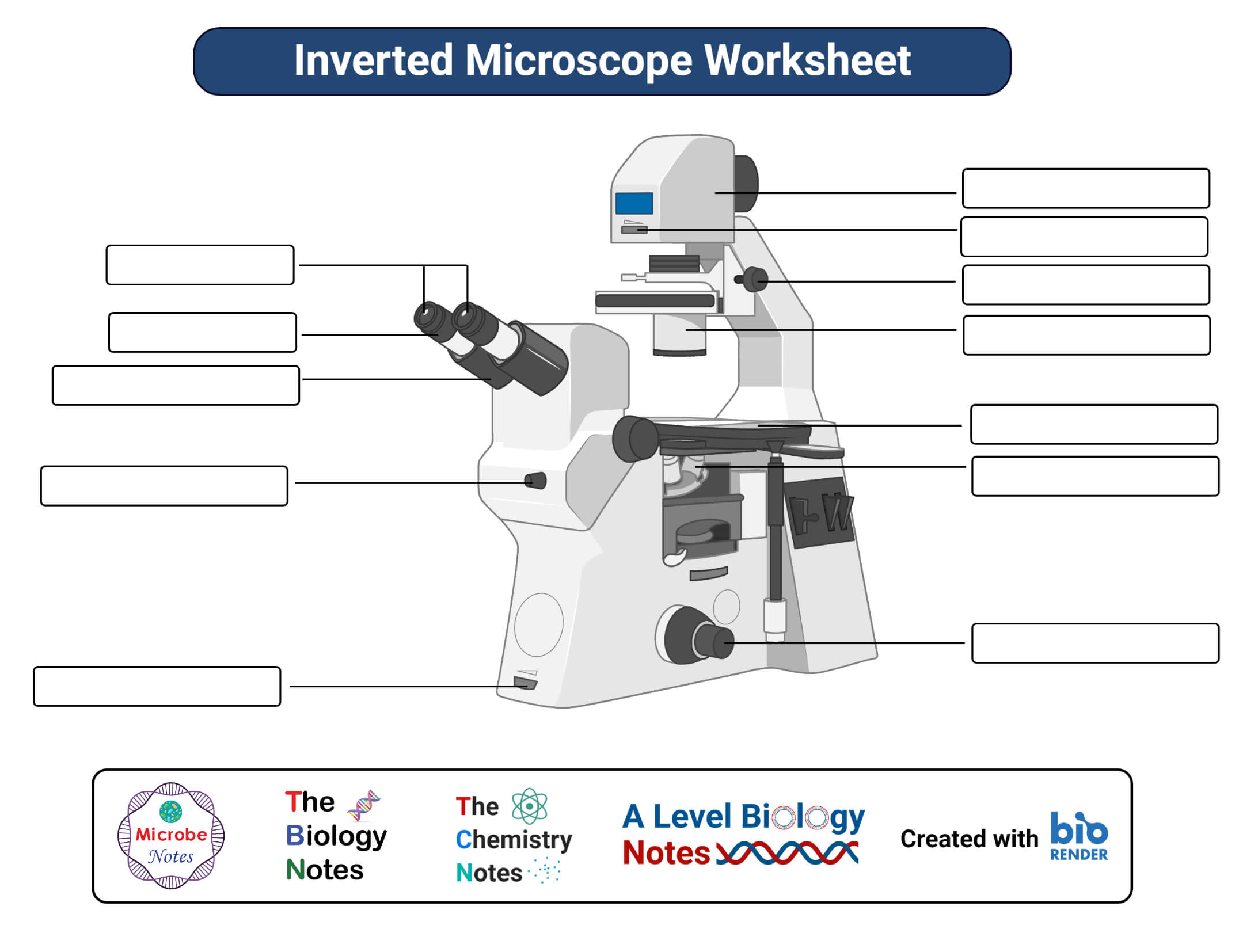
Inverted Microscope Free Worksheet

References and Sources
- https://ibidi.com/content/212-inverted-and-upright-microscopy
- https://www.quora.com/How-does-an-inverted-microscope-work
- https://en.wikipedia.org/wiki/Inverted_microscope
- https://www.microscopemaster.com/inverted-microscope.html
- https://wiki.bugwood.org/Diagnostic_Uses_for_the_Inverted_Microscope

Thanks
Very useful informations