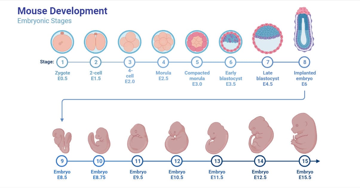
The embryo is an early stage in the development of a multicellular being.
This stage occurs in those multicellular organisms which involve in sexual reproduction.
Embryo formation happens after the fertilization process (where the male and female gametes fuse together) is completed in an organism.
The stage that is formed after the fusion of male and female gametes is called the zygote. The zygote divides multiple times to form different stages of development, such as morula, blastula, and gastrula, followed by neurulation and organ formation, eventually leading to the development of an embryo. The process of development of an embryo is called embryogenesis.

The embryo formation and structure are different in different organisms. Plants and animals that reproduce sexually possess embryos at their developmental stage, which varies in them.
Interesting Science Videos
Plant Embryo
The embryogenesis in the dicots, monocots, and gymnosperms is different. The angiosperms, which are flowering plants, produce embryos after fertilizing a haploid ovum with pollen. DNA from the egg and pollen combine to form a single-celled diploid zygote that finally develops into an embryo. The general summarized process of development of the embryo in plants is as follows:
In Dicot Plants
- At first, the zygote divides transversely, and a basal layer along with terminal cell formation occurs.
- Further division of the basal cell occurs transversely to form a six to the ten-celled structure called a suspensor.
- The terminal cell at the start divides longitudinally and, after that, transversally to finally form a globular structure of embryo which later on changes to a heart-shaped structure. This development is simple and primitive, shown by the Capsella bursa and hence called the crucifer type.
- The main axis in the embryo of plants is called the tigellum.
- Dicot embryo consists of two cotyledons.
In Monocot Plants
- The development in the monocots is almost similar to the embryogenesis in dicots.
- The basal cell, in this case, does not go into division and results in the formation of a single-celled vesicular suspensor.
- Monocot embryo consists of only one cotyledon.
The embryo derives the nutrition from the tissues of the endosperm, perisperm, and chalazosperm.
Plants that do not produce seeds but spores which include bryophytes and ferns, are involved in the formation of the embryo differently than monocots and dicots.
Polyembryony in Plants
It is the condition in which the number of embryos formed in one seed is multiple. It is very common in gymnosperms such as Pinus and Citrus. Leeuwenhoek in citrus plants in the year 1719 first observed it.
Animal Embryo
Fertilization takes place in the fallopian tube (in the ampulla region) of the female reproductive system. Before reaching the uterus, the embryo undergoes a series of divisions that help in its growth and development. Based on the location of the development of the embryo, embryogenesis can be categorized into two types:
- Viviparity: It is the type in which the fetus grows in the mother’s uterus. The animals that show viviparity are called viviparous. It results in the birth of young ones directly from the mother. For eg. In the case of humans, dogs, etc.
- Oviparity: It is the type in which the fetus grows outside the mother’s uterus into the eggs. The organisms that show oviparity are called oviparous. It results in the birth of young ones from the eggs expelled out from the mother. For eg. in the case of the chicken, ducks, etc.
The general embryogenesis process in mostly viviparous animals occurs in the following way:
- The fertilized cell or zygote in the fallopian tube further divides into two cells, followed by four, and eventually to 16-celled stages. This results in the formation of a ball-like or mulberry-shaped structure. This ball contains many cells inside it. This ball-shaped structure is called the morula.
- The morula further divides to form another structure called the blastula. In the blastula stage, the cells are arranged in such a way that they form a hollow mass. In fact, the cells align themselves to the ends and leave a cavity within, which gets filled with fluid.
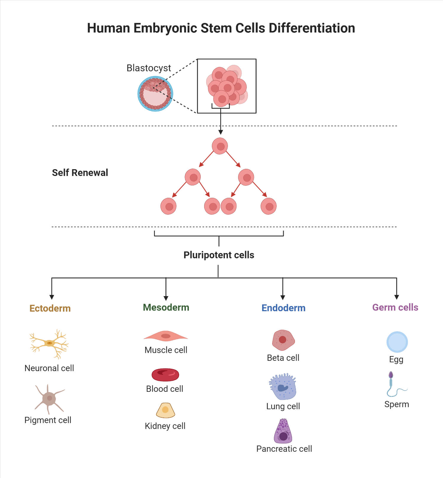
- The next stage of development after that will be the blastocyst. In this stage, the embryo will have cells that have differentiated and will even differentiate further. The differentiation process is that which makes it possible for a single cell to develop into an embryo and then into a complex organism. Differentiation is a process in which a cell gets differentiated from the other cells around it, and we get a whole new type of cell after multiple successive divisions.
- The Blastocyst stage is a mass containing differentiated cells. As it grows further, the body of the embryo begins to develop.
- The next stage is the implantation stage. As the blastocyst growing into an embryo requires an ample amount of nutrition, it needs to get attached to the walls of the uterus. This process of getting attached to the walls of the uterus is called the implantation process. This process is aimed at deriving nutrition from the mother, removing wastes, and exchanging gases. This is served through a barrier called the placenta.
- After that, the embryo starts to grow further when the organs of the baby’s body start to develop is called the fetal stage. The embryo can then be called the fetus.

References
- Moore, Keith L. (1988) Essentials of Human Embryology. Toronto: B.C. Decker Inc, p.2
- Sadler, T.W. (1995) Langman’s Medical Embryology. 7th edition. Baltimore: Williams & Wilkins.
- Larsen, William J. (1197) Human Embryology. 2nd edition. New York: Churchill Livingstone
- Plant Development: Embryogenesis. Accessed from: https://biology.kenyon.edu/courses/biol114/Chap12/Chapter_12A.html. Accessed on: 11/29/2022
- Keshari A.K. and Ghimire K.R. 2010. Development of Embryo. Vidyarthi Pustak Bhandar. 4th ed. Pg, 723-735
