- The nematode worm Caenorhabditis elegans is a small (1 mm long), unsegmented, vermiform, free-living soil nematode.
- It is a relatively simple, and precisely structured organism, extensively used as a model organism for molecular and developmental biology.
- The body of an adult C. elegans hermaphrodite contains exactly 959 somatic cells, whose entire lineage has been traced through its transparent cuticle.
- Its genome has also been entirely sequenced, the first-ever for a multicellular organism.
Interesting Science Videos
Reasons for Selection as Model Organism
- It has a rapid period of embryogenesis (about 16 hours), which it can accomplish in a petri dish.
- It has relatively few cell types.
- The predominant adult form is hermaphroditic, with each individual producing both eggs and sperm.
- Roundworms can reproduce either by self-fertilization or by cross-fertilization with the infrequently occurring males.
- Unlike vertebrate cell lineages, the cell lineage of C. elegans is almost entirely invariant from one individual to the next.
- C. elegans also has a small number of genes for a multicellular organism about 19,000.
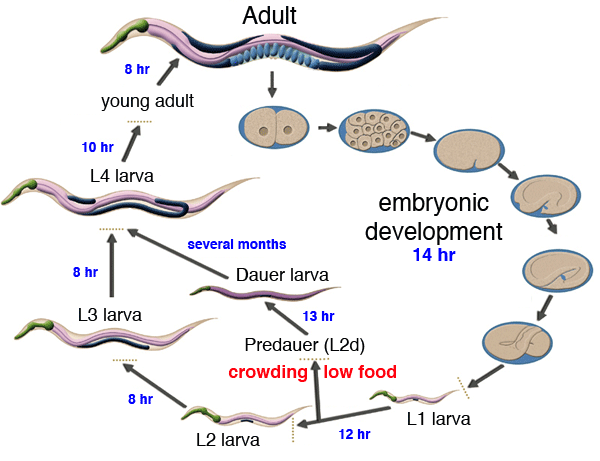
Development of Caenorhabditis elegans

Image Source: Simon Fraser University
1. Pre- Embryonic Development of Caenorhabditis elegans
The embryo of the nematode Caenorhabditis elegans progresses through several distinctive phases in developing towards the first larval stage, when the embryonic worm first emerges from the eggshell.
These phases/stages can be subdivided as follows:
Fertilization
- The moment when the haploid oocyte and haploid sperm combine to create a diploid, a single-cell embryo occurs within the spermatheca of the adult hermaphrodite.
- For a brief period, the fertilized embryo becomes covered by a membrane that may prevent polyspermy which is followed by the formation of a harder eggshell consisting of three layers secreted by the egg.
- The newly fertilized embryo exits prophase arrest and leaves the spermatheca to continue its development in the uterus.
Proliferation
- After fertilization, the single-cell embryo begins a series of highly stereotyped cell divisions.
- During this phase, all embryonic cells look similar in their cytoplasmic structures, and many cells also begin to make short-distance migrations away from their sister cells, through a process that has been termed “global cell sorting”.
- Early proliferation events span from 0 to 150 min post-fertilization (at 22°C) and take place within the uterus.
- Proliferation events continue in later stages, including gastrulation and morphogenesis and these occur after the embryo is laid.
- Coincident with proliferation, certain daughter cells always undergo immediate programmed cell death, or apoptosis, while their sister cells continue to proliferate and develop.
- Starting at the 30 cell stage, gastrulation encompasses the process by which single cells begin to become internalized and migrate into the center of the embryonic mass to eventually create separate ectodermal, endodermal and mesodermal compartments.
- Continued global cell sorting during this phase results in many functional cell groups becoming established by proximity rather than by sisterhood.
Morphogenesis
- This phase overlaps with the end of gastrulation when most cells have ended proliferation and are joining tissue subgroups.
- Cells become structurally specialized to adopt shapes and cell contents that reflect their eventual cell fates within specialized tissue compartments.
- Much directed cell migration occurs where certain cells extend processes or sheet-like arms to enfold their neighbors or penetrate through narrow passages inside the developing tissues.
- Some tissues begin to form syncytia sharing multiple nuclei, with the disappearance of intervening cell membranes.
- Terminal differentiation occurs without many additional cell divisions.
- In rare cases, specific cells undergo delayed apoptotic cell death in the embryo or in the L1 larva after fulfilling their role in tissue morphogenesis.
Elongation
- Once the embryo’s developing tissues begin to form a longer worm-like shape, they become folded within the eggshell.
- Early elongation events begin around 350 min after the first cleavage and involve both microfilaments and microtubules.
- Moving through the comma, two-fold and finally the three-fold stage, the embryo decreases in circumference and increases in length until it is ready for hatching.
- Elongation occurs coincidentally with later stages of morphogenesis but will be considered separately.
Quickening
- The first signs of muscle movement are seen around 430 minutes, and by the three-fold stage, the worm can move in a coordinated fashion within the egg.
- A series of squeezing events, and later, active wriggling motions, precedes hatching.
- These squeezing activities surely aid in further elongation of the animal, though they occur quite late.
- Quickening occurs coincidentally with the last stages of morphogenesis but will be considered separately.
Hatching
- When the embryo contains about 600 cells and many immature tissues, the young L1 larva breaks through the eggshell to emerge into the world.
- At this point, the L1 larva will begin postembryonic development where it will grow ten-fold in length and ten-fold in breadth before achieving adulthood.
2. Post-embryonic development of Caenorhabditis elegans
- Under environmental conditions favorable for reproduction, hatched larvae develop through four larval stages – L1, L2, L3, and L4 – in just 3 days at 20 °C.
- When conditions are stressed, as in food insufficiency, excessive population density or high temperature, C. elegans can enter an alternative third larval stage, L2d, called the dauer stage (Dauer is German for permanent).
- Dauer larvae are stress-resistant; they are thin and their mouths are sealed with a characteristic dauer cuticle and cannot take in food. They can remain in this stage for a few months.
- The stage ends when conditions improve favor further growth of the larva, now moulting into the L4 stage, even though the gonad development is arrested at the L2 stage.
- Each stage transition is punctuated by a molt of the worm’s transparent cuticle.
References
- https://www.wormatlas.org/embryo/introduction/EIntroframeset.html
- Alberts B, Johnson A, Lewis J, et al. Molecular Biology of the Cell. 4th edition. New York: Garland Science; 2002. Caenorhabditis Elegans: Development from the Perspective of the Individual Cell. Available from: https://www.ncbi.nlm.nih.gov/books/NBK26861/
- Gilbert, S. F. (2000). Developmental biology. Sunderland, Mass: Sinauer Associates.
- https://www.sciencedirect.com/topics/neuroscience/caenorhabditis-elegans
- http://www.wormbook.org/chapters/www_celegansintro/celegansintro.html
