Cartilage is a connective tissue found in the skeletal system, characterized by its avascular nature and supportive function. It contains high amounts of chondroitin, a substance that provides elasticity and flexibility to the tissue. Cartilage acts as a protective cushion, reducing friction and preventing bone-on-bone contact when we move our joints.
Cartilage cells, also known as chondrocytes, are specialized cells found in cartilage tissue that are responsible for maintaining the structure and function of cartilage.
Cartilage cells are dispersed within the cartilage matrix and are essential for tissue homeostasis, repair, and adaptation to mechanical stresses. Their activities ensure the proper growth, development, and maintenance of cartilage, contributing to overall joint health.
Interesting Science Videos
Types of Cartilage Cells
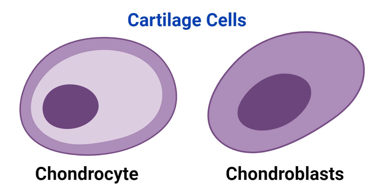
Chondrocytes are the main type of cartilage cells. However, during cartilage development and maintenance, other types of cells may also play important roles. The formation and maintenance of cartilage involve the deposition of matrix by cartilage-forming cells known as chondroblasts and chondrocytes. Cartilage can also be removed by multinucleated cells called chondroclasts.
- Chondrocytes are the main cells found within cartilage tissue, responsible for synthesizing the components essential for cartilage structure and function. These cells play a vital role in the development and maintenance of cartilage throughout the body. Chondrocyte activity is regulated by various signaling pathways and extracellular factors. Dysregulation of these pathways can lead to abnormal cartilage development and joint diseases.

- Chondroblasts are precursor cells that mature into chondrocytes. They are found in the perichondrium, a connective tissue layer surrounding cartilage. These cells are involved in the formation of cartilage. Chondroblasts produce and secrete extracellular matrix components, such as collagen and proteoglycans, essential for cartilage formation and growth.
- Chondroclasts are multinucleated cells that are involved in the remodeling and resorption of cartilage tissue. The process of resorption or degradation of the deep surface of joint cartilage is observed in various pathological conditions affecting the cartilage. Chondroclasts have been identified at these sites. However, the characterization of these cells is limited and remains poorly defined.
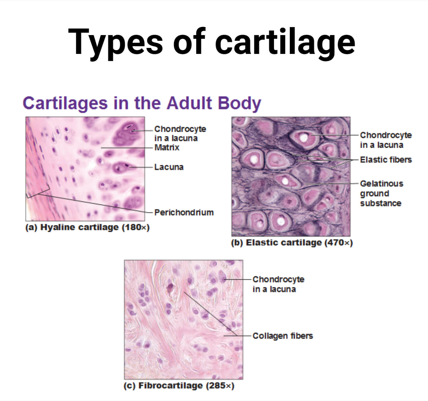
There are three types of cartilage:
- Hyaline cartilage, the most common type of cartilage, covers the ends of bones. It is mostly made up of proteoglycans and type II collagen. It has a pale blue-white appearance and smooth texture. The smooth surface allows smooth movement between bones within the joints. It is present in the ends of bones that form joints, the spaces between ribs, and the nasal passages.

- Elastic cartilage is the most flexible type of cartilage that provides support to body parts requiring bending and movement for proper function. Elastic cartilage can return to its previous shape even after enduring high pressure. Elastic cartilage is found in structures like the larynx, eustachian tubes, and external ear that require both support and flexibility.
- Fibrocartilage is composed of dense fibers and is the strongest yet least flexible among the three types of cartilage. It provides strength and support to different parts of the body. It is rich in type I collagen and has lower levels of proteoglycans compared to hyaline cartilage. It is commonly found in structures, such as tendons, ligaments, intervertebral discs, and the meniscus within the knee.
Structure of Cartilage
- The structure of cartilage includes the outermost perichondrium layer and the inner extracellular matrix.
- The perichondrium is a layer of connective tissue that contains an outer layer with fibrous connective tissue and blood vessels, as well as an inner layer containing chondroblasts that are responsible for secreting proteins that form the extracellular matrix of cartilage. Chondroblasts become trapped within the matrix and transform into chondrocytes.
- The gel-like extracellular matrix contains chondrocytes along with protein fibers like collagen and elastin, water, and proteoglycan aggregates. Chondrocytes are found within small holes called lacunae.
Structure of Chondrocytes
- Immature chondrocyte initially has an elliptical shape. As it develops and shifts inward, its shape transitions to a rounder form.
- Chondrocytes may organize into isogenous groups, comprising up to eight cells. This grouping occurs as a result of mitotic cell division, leading to cellular clustering within the tissue.
- During histological development, both chondrocytes and the surrounding matrix undergo shrinkage. This process results in the irregular shape characteristic of cartilage tissue.
- Chondrocytes are located in oblong spaces known as lacunae within the cartilage tissue.
Functions of Cartilage Cells
- Chondroblasts play a crucial role in the initial development of cartilage. They transition into chondrocytes, which form the structural framework of cartilage tissue.
- Chondroblasts synthesize key components of the extracellular matrix which provides support to the developing cartilage.
- Chondroblasts contribute to appositional growth of cartilage which involves the addition of a new matrix along existing surfaces. Chondroblasts promote lateral growth and increase the thickness of the tissue.
- Chondrocytes secrete new matrix within the cartilage contributing to the interstitial growth of cartilage.
- Chondroblasts and chondrocytes are involved in the regulation of cartilage development and growth, particularly during embryonic and adolescent stages.
- Chondrocytes play a crucial role in maintaining and repairing the extracellular matrix of cartilage. They continue to secrete extracellular matrix components to maintain the tissue.
- Chondroclasts are involved in endochondral ossification which involves the gradual replacement of cartilage with bone tissue during skeletal development.
Endochondral Ossification
- Bone ossification is the process of bone formation that begins during embryonic development, typically around the sixth to seventh week, and continues until early adulthood. This process occurs in two ways: intramembranous and endochondral ossification.
- Intramembranous ossification is the formation of bone directly from the mesenchymal cells.
- Endochondral ossification involves bone formation from cartilage. This process occurs mainly in long bones like in the arms and legs.
- Endochondral ossification starts with mesenchymal cells differentiating into chondrocytes, which make cartilage.
- Chondrocytes in the middle of the cartilage model add proteins to the matrix allowing the cartilage to calcify. This calcification cuts off nutrients to the chondrocytes, causing them to die and leaving holes in the cartilage.
- Blood vessels come in and make the voids bigger, forming a cavity. It also carries in osteoblasts which turn the membrane around the cartilage into bone creating the primary ossification center. After birth, a similar process happens at the ends of the bone, creating secondary ossification centers.

Diseases and Disorders of Cartilage Cells
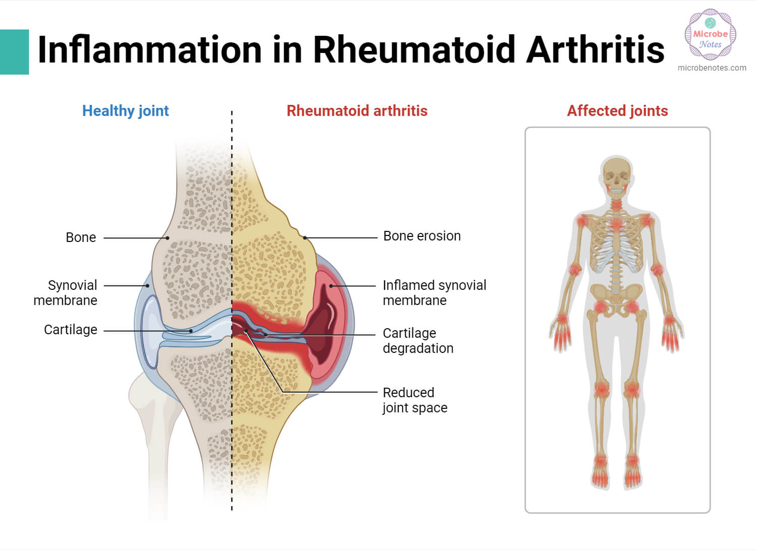
There is a wide range of clinical conditions involving cartilage that result from different degenerative, inflammatory, and congenital causes. These conditions include osteoarthritis, spinal disc herniation, traumatic rupture, achondroplasia, costochondritis, and various others.
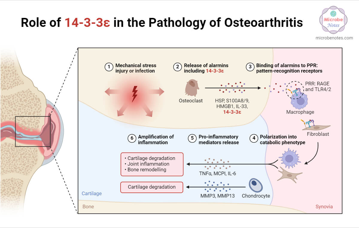
- Osteoarthritis results from the thinning of the articular cartilage, leading to decreased joint movement and pain. With aging, cartilage in the joints may degrade, causing pain and inflammation. Initial treatment typically involves anti-inflammatory medications and corticosteroid injections to relieve inflammation. Lifestyle changes like exercise, weight loss, and joint stress reduction also provide relief. In more serious cases, joint replacement surgery might be required.

- Spinal disc herniation, another common degenerative condition, arises from changes in the annulus fibrosus, a fibrocartilage surrounding intervertebral discs. Trauma and lifting injuries can weaken the annulus fibrosus, predisposing it to disc herniation. While some cases may require surgery, most can be managed with anti-inflammatory medications and lifestyle changes.
- Achondroplasia is a genetic disorder affecting cartilage formation and is the leading cause of dwarfism. This condition arises from a mutation in the fibroblast growth factor receptor 3 (FGFR3) gene, disrupting cartilage growth and development. It is typically diagnosed during prenatal ultrasound examinations. There is currently no cure for achondroplasia, and treatment options are limited.
- Costochondritis is the inflammation of the costal cartilage that connects ribs to the sternum. It causes chest pain which may worsen with movement or deep breathing. It is usually treated with rest and over-the-counter pain medications. However, severe pain may require medical treatment.
- Trauma, especially in sports, can result in cartilage damage. Sports injuries such as a torn meniscus or a separated shoulder can harm the cartilage, leading to conditions like osteochondritis dissecans.
References
- 6.2A: Structure, Type, and Location of Cartilage – Medicine LibreTexts
- Biologydictionary.net Editors. (2019, April 25). Cartilage. Retrieved from https://biologydictionary.net/cartilage/
- Breeland G, Sinkler MA, Menezes RG. Embryology, Bone Ossification. [Updated 2023 May 1]. In: StatPearls [Internet]. Treasure Island (FL): StatPearls Publishing; 2024 Jan-. Available from: https://www.ncbi.nlm.nih.gov/books/NBK539718/
- Britannica, T. Editors of Encyclopaedia (2024, February 1). cartilage. Encyclopedia Britannica. https://www.britannica.com/science/cartilage
- Cartilage: What It Is, Function & Types (clevelandclinic.org)
- Chang LR, Marston G, Martin A. Anatomy, Cartilage. [Updated 2022 Oct 17]. In: StatPearls [Internet]. Treasure Island (FL): StatPearls Publishing; 2024 Jan-. Available from: https://www.ncbi.nlm.nih.gov/books/NBK532964/
- Chondroblasts: What Are They, Function, and More | Osmosis
- Hall, B. K. (2015). Vertebrate Skeletal Tissues. Bones and Cartilage, 3–16. doi:10.1016/b978-0-12-416678-3.00001-x
- Knowles, H.J., Moskovsky, L., Thompson, M.S. et al. Chondroclasts are mature osteoclasts which are capable of cartilage matrix resorption. Virchows Arch 461, 205–210 (2012). https://doi.org/10.1007/s00428-012-1274-3
- Nahian A, Sapra A. Histology, Chondrocytes. [Updated 2023 Apr 17]. In: StatPearls [Internet]. Treasure Island (FL): StatPearls Publishing; 2024 Jan-. Available from: https://www.ncbi.nlm.nih.gov/books/NBK557576/
