
The human skeleton is categorized into 2 parts. One is the axial skeleton, and the other is the appendicular skeleton.
The axial skeleton is the bony structures that form the central core of the human body. The axial skeletal system serves as the structural framework that makes up the longitudinal (vertical) axis of the body and protects the major vital internal organs, such as the heart, lungs, and the central nervous system.

Components of Axial Skeleton (Skull, Vertebral Column, and Thoracic Cage)
The axial skeleton comprises three major anatomical components: the skull, vertebral column, and the thoracic cage. Each of them is distinct in its structural alignment and supportive functions.
- Skull bones: They form the foundational framework of the head and face. They protect the brain as well as the spinal cord. There are altogether 22 bones in the skull (8 cranial bones and 14 facial bones), excluding the bones of the inner ear and the hyoid region.
- Vertebral column: Also known as the spinal column/vertebrae, the vertebral column consists of 24 bones piled on top of each other, including the sacrum bone fused with the tailbone. They function to protect the spinal cord.
- Thoracic cage: There are 25 bones (12 pairs of ribs and 1 sternum) that align together to form the thoracic cage/ribcage. They primarily protect essential internal organs, such as the lungs and heart. In addition, they also play an essential role in the breathing mechanism.
Furthermore, 6 tiny bones of the middle ear, known as the auditory ossicles, and 1 hyoid bone of the neck region are also included in the axial skeleton system.
Therefore, the axial skeleton system is a complex organization of the following 80 bones.
| Bones | Shape of bones | Number of bones |
| Skull | ||
| Cranial | Flat | 8 |
| Facial | Irregular | 14 |
| Auditory ossicles | Short | 6 |
| Hyoid bone | Sesamoid (small and round) | 1 |
| Vertebral column | ||
| Cervical bones | Irregular | 7 |
| Thoracic bones | Irregular | 12 |
| Lumbar | Irregular | 5 |
| Sacrum | Irregular | 5 |
| Coccyx | Irregular | 4 |
| Thoracic cage | ||
| Ribs | Flat | 24 |
| Sternum | Flat and long | 1 |
Anatomy and Structure of Axial Skeleton
The axial skeleton predominantly supports the head, neck, and the trunk region of the body. It is composed of the following anatomical structures:
Skull bones
Cranial bones: They enclose and protect the brain inside the cranium cavity. The cranium consists of 8 bones.
| Left and right parietal bones (2) | They form the lateral and superior part of the cranium. |
| Left and right temporal bones(2) | They house the ear structures and form a part of the lateral wall of the cranium. |
| Frontal bone(1) | It forms the anterior portion of the skull and the superior surface of the orbits |
| Occipital bone(1) | It makes up the posterior and inferior surface of the skull. |
| Ethmoid bone(1) | It makes up the anterolateral wall of the skull and the roof of the nasal cavity. |
| Sphenoid bone(1) | It forms the floor of the skull and connects the cranial bones and the facial bones. |
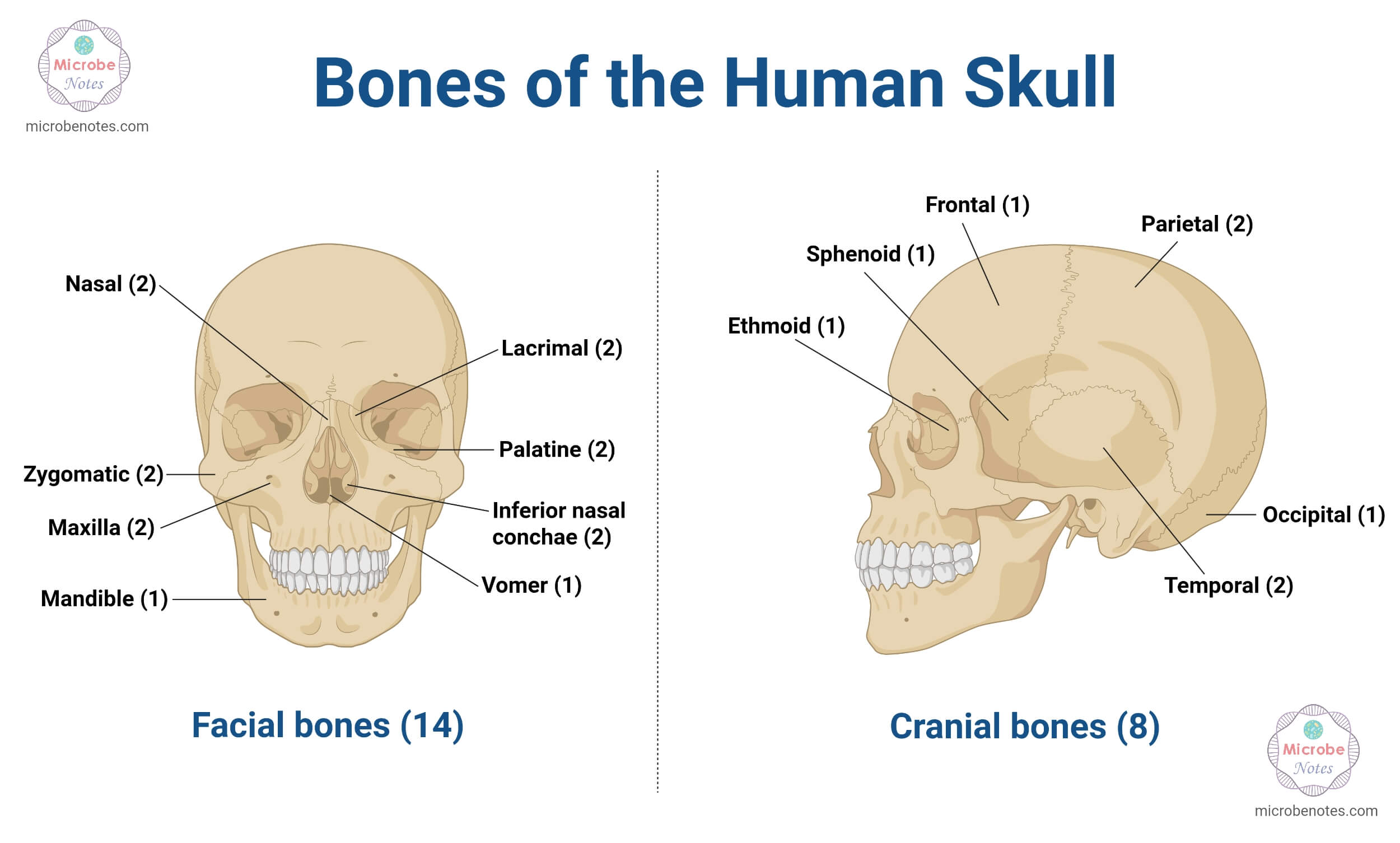
Facial bones: There are 14 facial bones that form the shape and structure of the face.
In addition, they also support the entry point of the digestive and respiratory tracts. All the facial bones are found in pairs except the vomer and the mandible.
| Maxilla bones | They form the upper jaw and hard palate. |
| Zygomatic bones | They form the cheekbones. |
| Nasal bones | They form the nasal septum. |
| Palatine bones | They form the posterior hard palate and orbital floor. |
| Nasal Concha bones | Inferior nasal concha articulates with the maxilla and palatine bones, whilst the middle and superior conchae are part of the ethmoid bones. |
| Lacrimal bones | They form the medial orbital wall. |
| Mandible bone(1) | It is the one and only movable bone in the skull, which forms the lower jaw. |
| Vomer bone (1) | It forms the inferior nasal septum. |
Vertebral column
| Cervical vertebra | There are 7 cervical vertebrae, from C1 to C7, located in the neck region. C1 (atlas) and C2 (axis) are necessary for the movement of the head. |
| Thoracic vertebra | There are 12 thoracic vertebrae (T1-T12), located in the chest region. They articulate with the ribs. |
| Lumbar | They are the 5 (L1-L5) large- kidney shaped bone, located in the lower back |
| Sacrum | It comprises 5 fused vertebrae (S1-S5), located in the pelvis region. |
| Coccyx | They are also called the tailbones. There are 4 tailbones that help to attach the muscles as well as the ligaments of the pelvic floor. |
Thoracic cage (rib cage)
| Ribs | They are curved bones, present in the thoracic region. There are 12 rib bones found on the left and right sides of the thoracic cavity. They have been categorized as True ribs, false ribs, and floating ribs. |
| Sternum | It is the single bone present in the midline of the thoracic wall. It is also known as the breast bone. |
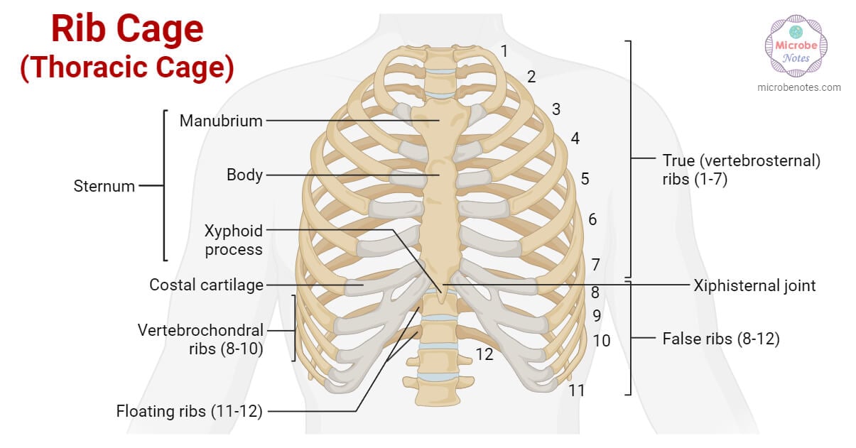
Based on their structural shape, the bones of the axial skeleton are classified as flat, irregular, sesamoid, and short bones. Cranial bones, ribs, and sternum are generally thin and flat. The spinal column and the facial structures vary as they have complex, specialized irregular shapes. On the other hand, the auditory ossicles are short and provide limited mobility, while the hyoid is a small, U-shaped, round (sesamoid) bone.
Functions of the Axial Skeleton
The axial skeleton system serves as an integrated support system of the body. The crucial roles of the axial skeleton system are as follows:
Supportive function
The axial skeleton not only creates the central core of the body, but also supports the overall body weight against gravity. It distributes the weight from the head, neck, and trunk region towards the lower limbs and thereby assists in maintaining an upright posture and gives the body its characteristic structure.
Protective function
The axial skeleton houses most of the vital internal organs. The heart and lungs are protected within the thoracic cage, while the brain is encased inside the skull. Also, the vertebral column acts as a shield for our spinal cord. Thus, the ribs and sternum of the thoracic cage, the skull bones, and the vertebrae prevent possible damage to the vital organs and provide respiratory, cardiac, as well as neurological protection.
Likewise, the facial bones also protect and define the shape of the sensory organs, such as the eyes, ears, nose, etc.
Site of Attachment
The axial skeleton acts as an attachment site for the core muscles. Skeletal muscles, as well as the respiratory muscles, attach to the broad surfaces of the axial skeleton.
Stability and Movement
The axial skeleton maintains our body’s centre of gravity and aids in stabilising our body during movements. It coordinates with the appendicular skeleton and allows the flexible movement of the body via the vertebral joints.
Breathing
Ribs and sternum expand and contract during the breathing mechanism. Consequently, they increase and decrease the volume of the thoracic and pulmonary cavities. This action facilitates the passage of air from the external environment to the lungs and vice versa.
Development and Ossification of the Axial Skeleton
The formation of the axial skeleton begins at an early embryonic stage. The mesoderm layer and neural crest cells play a primary role in dividing and forming the bones of the axial skeleton system.
The paraxial mesoderm (also known as the presomatic or somatic mesoderm) differentiates into somites, which further differentiate into sclerotome, myotome, and dermatome. Out of these, the sclerotome forms the skull bones, vertebral column, and rib bones. Additionally, the lateral plate mesoderm serves to develop the sternum, whilst the neural crest cells gradually form the facial bones and part of the cranium.
Ossification refers to the biological process of forming bones and the skeleton from the cartilaginous fibrous tissues known as osteoblasts. It is also called osteogenesis. In the human body, bones develop via 2 kinds of ossification processes: Intramembranous ossification, and Endochondral ossification.
In both processes, mesenchymal tissue (embryonic connective tissues developed from mesoderm) serves as the initial precursor. It gradually transforms into bone tissue.
Intramembranous ossification
It is the process of osteogenesis in which bones are formed from fibrous membranes directly on the mesenchyme. This process results in the formation of flat bones, the top and side parts of the cranium.
Mesenchymal cells differentiate to form osteoblasts, which produce an unmineralized collagen-proteoglycan matrix called ‘osteoid’. Osteoids surround the osteoblast cells and the blood vessels to form the spongy bone. Gradually, the continuous differentiation of the mesenchymal cells results in the development of compact bones.
As the name suggests, the development of bone tissues takes place within the periosteum membrane of the ossification centre.
The series of events that occur during the intramembranous ossification process are:
Mesenchymal cells → Osteoblasts → osteoblasts cluster and form ossification centers →Secretion of osteoid → Osteoid binds to the calcium → Osteoid encapsules and traps the osteoblast → Osteoblast transforms to osteocytes → Osteoid further surrounds the blood vessels and forms of spongy bone → mesenchymal cells forms the periosteum membrane → Continuous differentiation of mesenchymal cells to the osteoblasts within the membrane → formation of many layers → The layers are called the compact bone.
Endochondrial ossification
It is the bone formation process that initially develops the intermediate cartilaginous tissues prior to the osteoblasts. This mechanism for bone development produces the floor of the cranium and the facial bones, vertebral column, and thoracic cage.
First, the mesenchymal cells differentiate to form chondrocyte cells and form the hyaline cartilage. The chondrocyte cells near the cartilages mineralize the extracellular matrix and undergo the following transformations to form bone tissues.
Mesenchyme → Chondrocytes → Extracellular matrix → Cartilage model(Hyaline cartilages surrounded by perichondrium membrane) → Addition of collagen X and fibronectin to the matrix → Calcification of extracellular matrix →Apoptosis →Invasion of blood vessels → Formation of the medullary cavity with the osteogenic cells →Perichondrium transform into periosteum → Formation of a primary ossification center by the osteoblast cells in the diaphysis region of the periosteum → Proliferation of cartilage at the ends of bone that increases the bone length → Proliferative region transforms into epiphyseal plates → Secondary ossification process begins in epiphyseal plates after birth.
Joints and Ligaments of the Axial Skeleton
The axial skeleton requires specialised joints and ligaments to facilitate its movement and flexibility. Joints of the axial skeleton can be grouped into 4 types based on their site.:
Joints of Skull
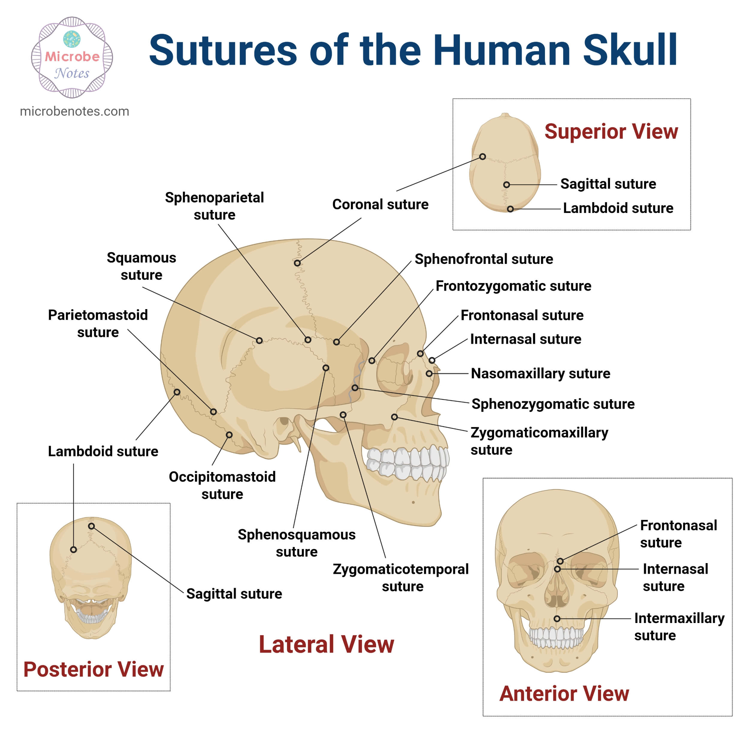
All the skull bones are interlocked by the immovable fibrous joints called the synarthroses, except the mandible. The mandible is attached to the skull by the movable temporomandibular joints (TMJ). In other terms, synarthroses are also called the sutures.
| Sutures | Location |
| Coronal suture | Present between the parietal and frontal bones |
| Sagittal suture | Occurs between the parietal bones |
| Lambdoid suture | Between the parietal and occipital bones |
| Squamous suture | Between the temporal and parietal bones |
| Metopic/Frontal suture | Connects the frontal bone at the forehead |
Joints of the vertebral column
The vertebral column consists of two types of joints: the intervertebral joints and the facet joints.
| Intervertebral joints | They are the cartilaginous joints present between the vertebral bodies, which play an essential role by absorbing external shock. They facilitate slight movement of the bones. |
| Facet joint | Also known as zygapophaseal joints, these types of connective tissues occur between the articular facets of the adjacent vertebrae. |
Additionally, there are two synovial joints that interconnect the skull with the spine. They are called the craniovertebral joints, and they include:
- Atlanto-occipital joint: It is a type of condyloid joint present between the atlas and occipital bone. It facilitates the nodding motion of the head.
- Atlanto-axial joint: It is a type of pivot joint located between the atlas and axis bones. It allows rotation of the head in the ‘no’ motion.
Joints of the thoracic cage
The joints of the thoracic cage allow the expansion and contraction of the ribcage during the breathing process.
| Sternocostal joints | They connect the ribs with the sternum. Rib I forms the cartilaginous joint, while Rib II-VIII form the synovial joint (which allows movement). |
| Costovertebral joints | They are the synovial joints that occur between the rib heads and the thoracic vertebrae. |
| Costotransverse joints | They are also the synovial joints that occur between the rib tubercle and the transverse process of the vertebrae. |
Furthermore, the axial skeleton is stabilised and supported by several ligaments.
| Skull ligaments | |
| TMJ ligament | Consists of lateral, sphenomandibular, and Stylomandibular joints |
| Stylohyoid ligament | Bridges the styloid process of the temporal bone with the hyoid bone |
| Vertebral column ligaments | |
| Longitudinal ligaments | Anterior longitudinal ligament passes along the front of the spine and prevents hyperextension.Posterior longitudinal ligament passes along the back of the spine and prevents hyperflexion. |
| Segmental ligaments | Ligamentum flavum is the yellow elastic tissue present between the laminae of the adjacent vertebrae.Interspinous ligament is the thin connective tissue present between the spinous processes of adjacent vertebrae.Supraspinous ligaments cover the tips of spinous processes.Intertransverse ligaments are present between the transverse processes. |
| Cervical Ligaments | Nuchal ligament is a thick fibroelastic ligament located at the back of the neck.Cruciform ligament is present at the craniovertebral junction, and it stabilizes the atlanto-occipital and atlanto-axial joints.Alar ligaments connect the axis bone to the occipital bone and prevent excessive head rotation |
| Thoracic cage ligaments | Costotransverse ligaments stabilize the costotransverse jointsInterchondral ligaments connect the costal cartilages of the adjacent ribs.Radiate ligaments of the rib’s head attach to the two adjacent vertebrae and the intervertebral disc |
Common Disorders and Injuries of the Axial Skeleton:
There are various kinds of disorders related to the axial skeleton system. They include:
Spinal Disorders
- Herniated Disc: When the jelly-like centre of the spinal disc bulges out / herniates and presses the neighbouring nerves, the condition is called a herniated disc. In this disorder, the pain often radiates towards the lower limbs.
- Scoliosis: The condition in which the spine abnormally curves sideways is called scoliosis.
- Spinal stenosis: It is a condition in which the spinal column gradually narrows.
- Compression fractures: These types of fractures are most common in the cervical vertebrae and lumbar zones. It is usually caused by certain forceful trauma,
Metabolic disorders
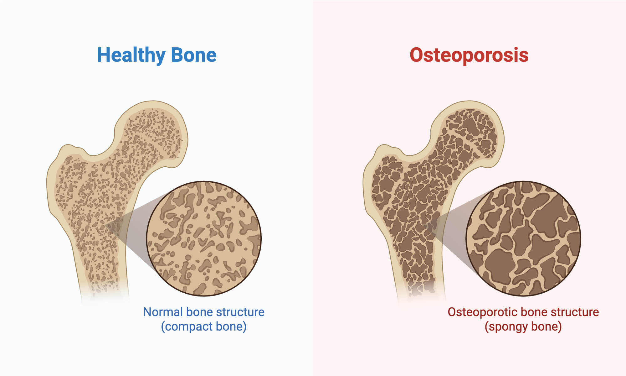
- Osteoporosis: It is caused by low bone mass/density.
- Osteopenia: It is a condition in which the bone density is lower than the normal range.
Traumatic injuries/ Fractures
Severe accidents or trauma may often cause fractures in the cranium, skull bones, bones of the thoracic cavity, and often the spinal column.
Clinical Significance of the Axial Skeleton
The axial skeleton is clinically significant because of its crucial role in housing, shielding, and protecting the vital internal organs, along with preventing disorders such as organ failure or paralysis, diseases often associated with neurons, respiratory organs, or the heart. Axial bones also contain red bone marrow, which is capable of producing red blood cells.
Conclusion
To sum up, the axial skeleton is a complex framework organised by a total of 80 bones. It comprises the skull bones, spinal bones of the vertebral column, rib bones and the sternum. The bones of the auditory ossicles and the hyoid bone are also included in the axial skeleton system. These structures provide support and maintain the posture of the body while protecting the vital organs, facilitating respiration process, and serving as the site of attachment for the muscles.
References
- Axial Skeleton – definition and list of bones with labeled diagram. (2022, November 1). TheSkeletalSystem.net. https://www.theskeletalsystem.net/axial-skeleton
- Axial skeleton – Structure, Function, Diagram, Significance. (n.d.). https://anatomy.co.uk/axial-skeleton
- Axial Skeleton (80 bones) | SEER Training. (n.d.). https://training.seer.cancer.gov/anatomy/skeletal/divisions/axial.html
- Breeland, G., Sinkler, M. A., & Menezes, R. G. (2023, May 1). Embryology, bone ossification. StatPearls – NCBI Bookshelf. https://www.ncbi.nlm.nih.gov/books/NBK539718/
- Carragee, E. J. (2012). Overview of axial skeleton injuries. Journal of the American Academy of Orthopaedic Surgeons, 20, S18–S22. https://doi.org/10.5435/jaaos-20-08-s18
- Dr, D. N. S., Dr, R. L., & Butler, J. L. (2015). Hole’s Human Anatomy & Physiology. McGraw-Hill Education.
- Embryonic development of the axial skeleton | Anatomy and Physiology I. (n.d.). https://courses.lumenlearning.com/suny-ap1/chapter/embryonic-development-of-the-axial-skeleton/
- Human Axial Skeleton | Biology for Majors II. (n.d.). https://courses.lumenlearning.com/wm-biology2/chapter/human-axial-skeleton/
- Joints and ligaments of the vertebral column. (2023, October 30). Kenhub. https://www.kenhub.com/en/library/anatomy/joints-and-ligaments-of-the-vertebral-column
- MasteringA&P. (n.d.). The axial skeleton (p. 121). https://www.pearson.com/content/dam/one-dot-com/one-dot-com/us/en/higher-ed/en/products-services/course-products/marieb-10e-info/pdf/marieb-lab-manual-exercise9.pdf
- OpenStaxCollege. (2013, March 6). Embryonic development of the axial skeleton. Anatomy & Physiology. https://pressbooks-dev.oer.hawaii.edu/anatomyandphysiology/chapter/embryonic-development-of-the-axial-skeleton/
- Professional, C. C. M. (2026a, January 14). Axial skeleton. Cleveland Clinic. https://my.clevelandclinic.org/health/body/22344-axial-skeleton
- Professional, C. C. M. (2026b, April 27). Lumbar spine. Cleveland Clinic. https://my.clevelandclinic.org/health/articles/22396-lumbar-spine
- Skull. (2023, November 21). Kenhub. https://www.kenhub.com/en/library/anatomy/the-skull
- Wikipedia contributors. (2025a, December 14). Ossification. Wikipedia. https://en.wikipedia.org/wiki/Ossification
- Wikipedia contributors. (2025b, December 14). Ossification. Wikipedia. https://en.wikipedia.org/wiki/Ossification
- Wikipedia contributors. (2026, June 5). Spinal column. Wikipedia. https://en.wikipedia.org/wiki/Spinal_column
