
The human skull is one of the body’s most essential and complex structures. It is a hard, rigid bony framework that encloses and protects the brain. It is also the uppermost part of the axial skeletal system, providing structural support to the face and protecting the delicate sensory organs in the head.
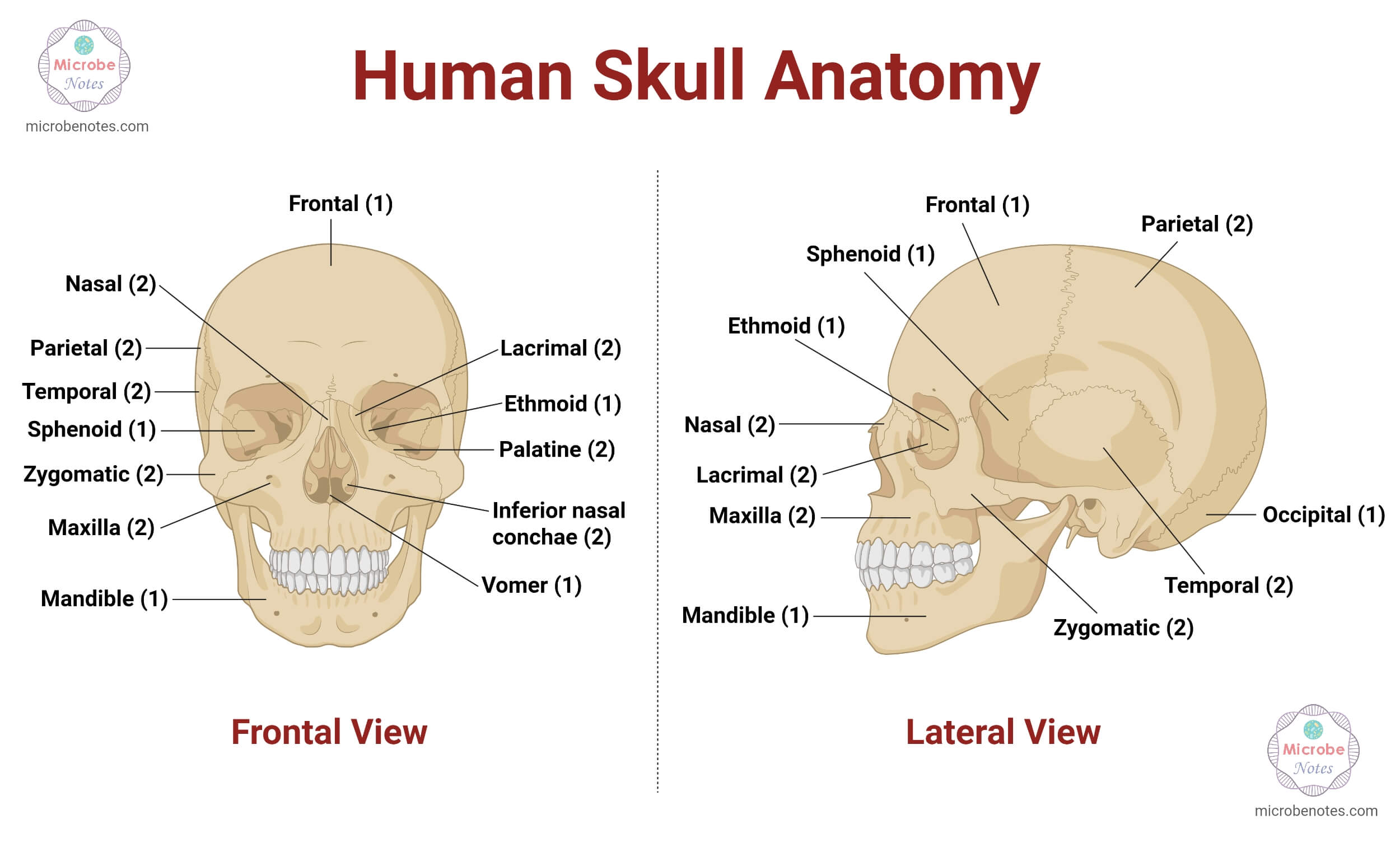
Anatomy and Structure of the Human Skull
Anatomically, the human skull consists of 2 major parts: the cranial part and the facial part. The cranial structure of the skull is called the ‘neurocranium,’ while the facial skeletal structure is termed the ‘viscerocranium.’ These parts of the skull are joined by immovable fibrous joints known as ‘sutures.’
Neurocranium/Calvarium
It is the superior-most part of the skull that encases and protects the brain, meninges, and the network of blood vessels surrounding the brain. It can be subdivided into 2 parts: a roof and a base.
| Cranial roof | Cranial base |
| It is formed by the membranous part of the skull. | It is formed by the cartilaginous part of the skull. |
| It is also known as the calvarium or the skullcap. | It is also known as the chondrocranium. |
| It mainly consists of flat parts of the bones, namely the frontal, occipital, and two parietal bones. | It consists of irregular portions of the frontal, sphenoid, ethmoid, occipital, temporal, and parietal bones. |
The cranial roof is built of 3 layers: an outer layer of dense compact bones, a middle layer of diploe (the cancellous spongy bone), and an inner layer of hard compact bone. The diploe, sandwiched between the compact bones, contains the channels for venous drainage.
Viscerocranium
It refers to the facial skeleton that provides structural support and shapes the soft tissues of the face. There are altogether 14 bones in the face. They include the nasal bones, vomer, maxilla, mandible, lacrimal bones, palatine bones, and zygomatic bones. Except for the vomer and the mandible, all other facial bones are present in pairs.
In addition to these 22 bones, the skull also consists of 6 auditory ossicles (malleus, incus, and stapes), three in each ear. It is comprised of the connective tissues called sutures and fontanelles, and there are a few cavities known as foramina through which the neurovascular tissues pass between the skull and the rest of the body. Likewise, it also consists of anatomical features such as eye sockets, nasal cavities, and paranasal sinuses.
Bones of the Human Skull (Cranial and Facial Bones)
The human skull is comprised of 8 bones in the cranial region, while the remaining 14 bones are the facial bones.
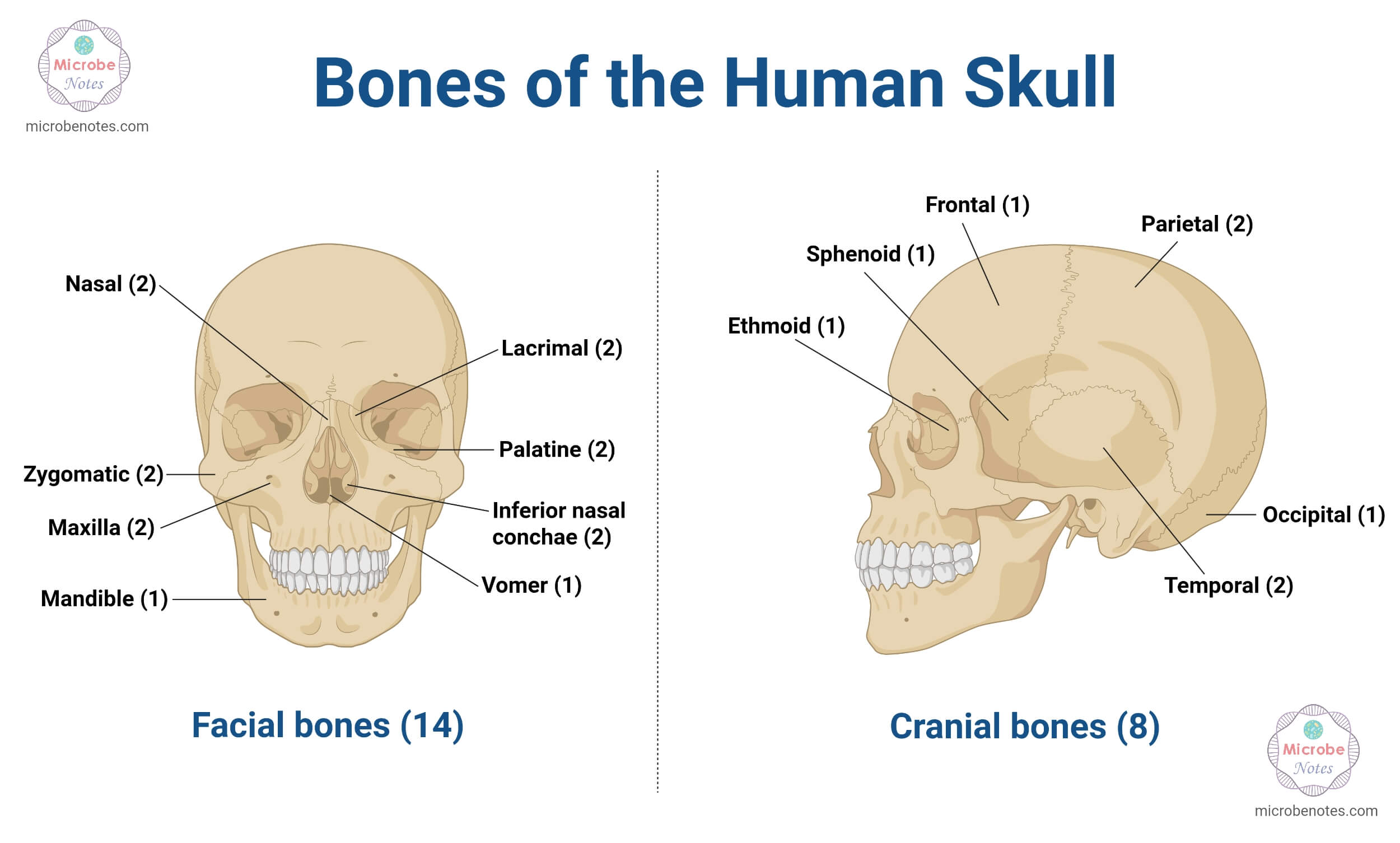
| Cranial bones (8) | Numbers | Facial bones (14) | Numbers |
| Frontal bone | 1 | Nasal bones | 2 |
| Sphenoid bone | 1 | Inferior nasal conchae | 2 |
| Ethmoid bone | 1 | Vomer | 1 |
| Temporal bones | 2 | Lacrimal bones | 2 |
| Parietal bones | 2 | Palatine bones | 2 |
| Occipital bone | 1 | Zygomatic bones | 2 |
| Maxilla | 2 | ||
| Mandible | 1 |
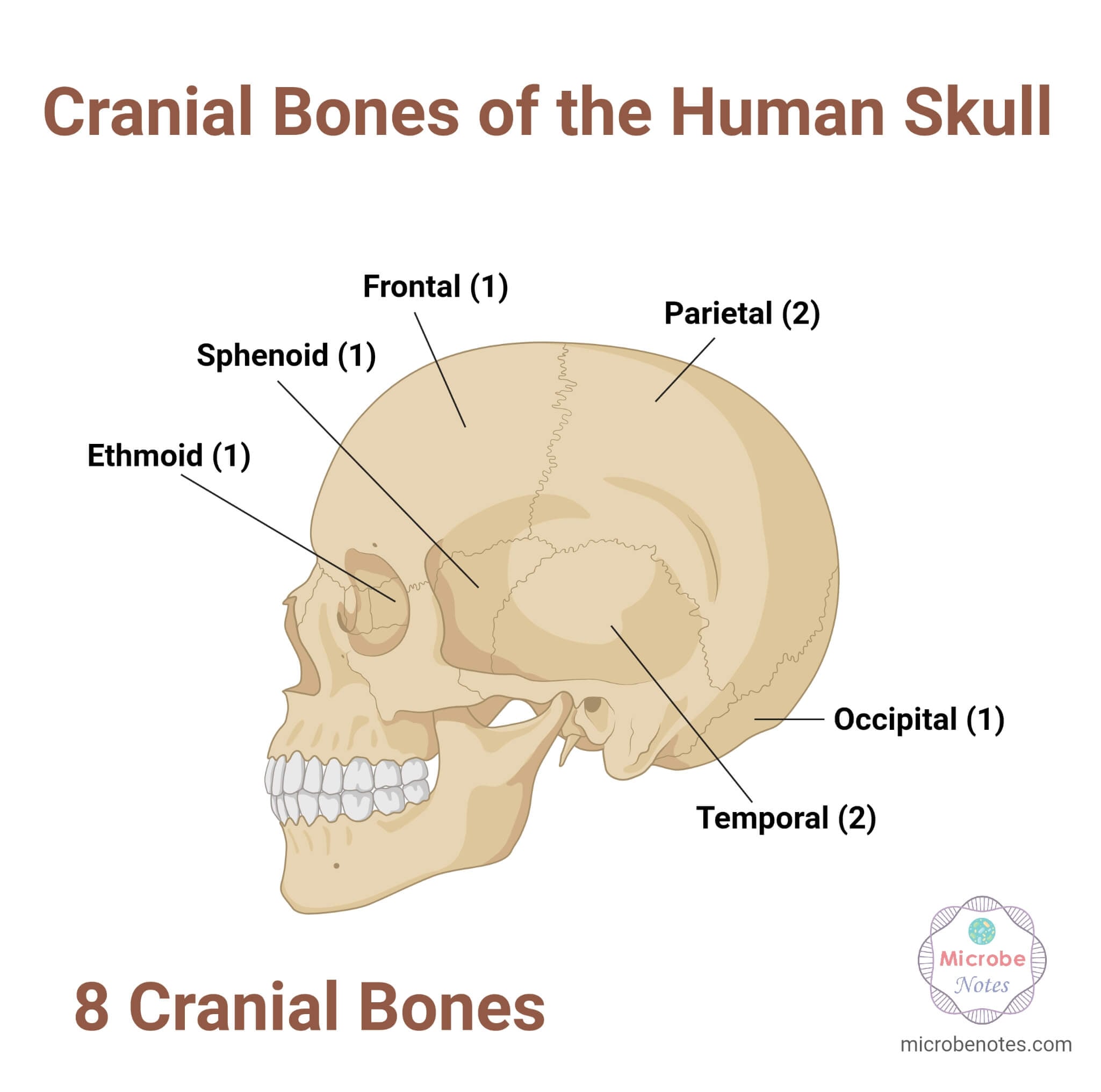
Cranial bones of the Human Skull
- Frontal bone: The flat bone that forms the forehead and upper part of eye sockets
- Sphenoid bone: It is the bone at the center of the skull.
- Ethmoid bone: An irregularly shaped, light, spongy bone below the frontal bone
- Occipital bone: A flat bone that forms the posterior side and the base of the skull
- Temporal bones: A pair of irregular bones located under each parietal bone
- Parietal bones: A pair of flat bones present behind the frontal bone on either side of the head
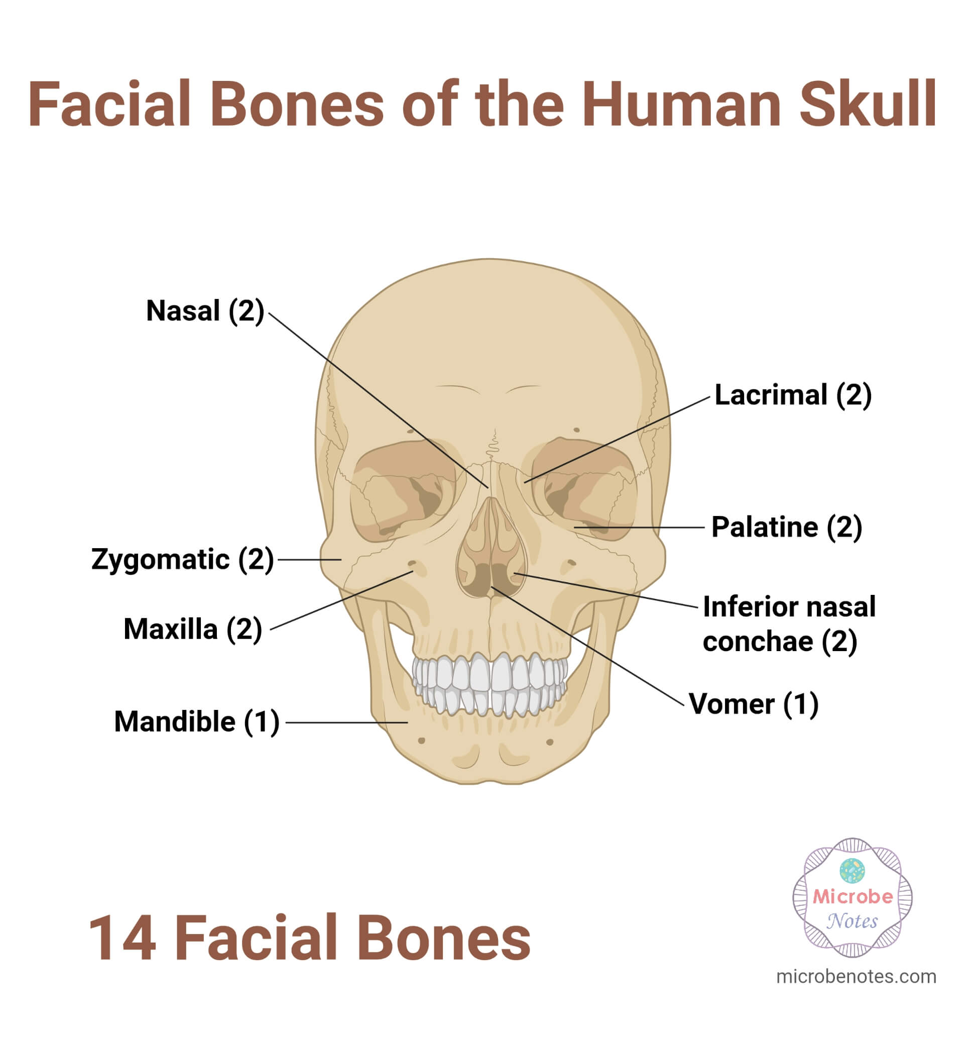
Facial bones of the Human Skull
- Nasal bones: A pair of small bones that form the nose
- Inferior nasal conchae: The thin curved bones that project into the nasal cavity
- Vomer: A single, thin, and flat-shaped bone that forms the posterior and inferior side of the nasal septum.
- Lacrimal bones: The smallest bones of the face
- Palatine bones: The L-shaped bones that form the hard palate of the oral cavity and the floor of the eye sockets.
- Zygomatic bones: They are also called the cheekbones.
- Maxilla: They are also called the upper jaw bones, which form the central face and hold the upper teeth and hard palate.
- Mandible: Also known as the lower jaw, it is the one and only movable bone in the skull that holds the lower teeth.
Functions of the Skull in the Human Body
Protection: The human skull not only houses the brain and brainstem inside the head (cerebrum, cerebellum, meninges, and orbits), but also protects the essential sensory organs in the face. It protects the central nervous system, oral cavity, nasal cavity, ears, and eyes.
Structural support: The skull supports the essential facial structures, such as the jaws, nose, teeth, eye orbits, and provides the structural shape of the head and the face frames.
Attachment site for muscles: The skull acts as an important site for muscle attachment. It supports the facial muscles and tendons, which aid in the regular daily movement of the head, chewing food, facial expressions, and even normal speech.
Openings and cavities: There are certain openings or cavities in the skull that are necessary for the passage of blood vessels and neurons, and perform regular physiological processes. For instance, the skull consists of a pair of paranasal sinuses that are crucial for warming and moistening the inhaled air.
Production of blood cells and mineral storage: The bones of the skull are also the essential sources of blood cells and minerals.
Sutures and Fontanelles of the Human Skull
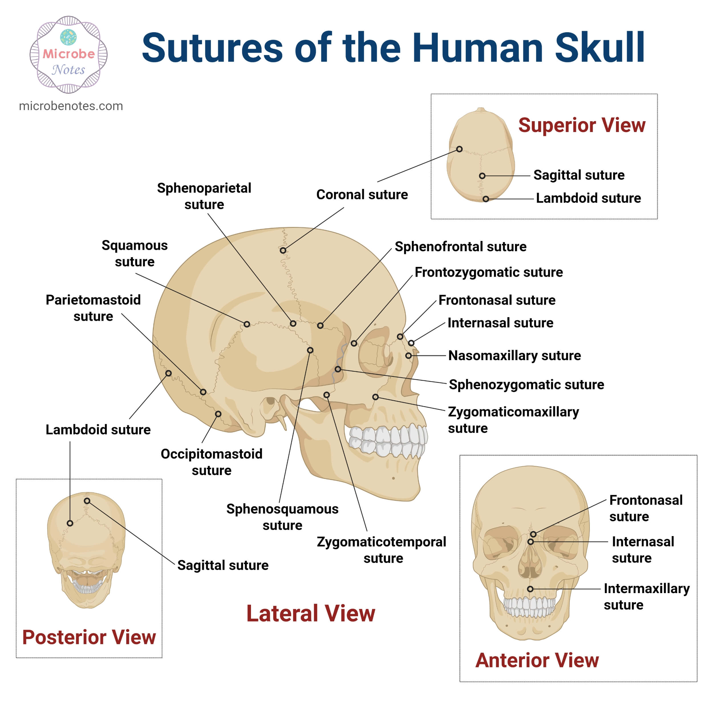
The bones of the human skull are connected by several joints called the ‘sutures.’ They are the articulation points of bones made up of fibrous connective tissues. These joints are immovable and are categorized under synarthrosis joints. However, during birth, they allow the skull bones to overlap, which aids in passing the infant’s head through the birth canal. In adults, they also act as mechanical stress absorbers.
Types of Sutures
There are 2 types of sutures present in the human skull. They are:
Common Cranial Sutures
- Coronal suture: A junction between the frontal and 2 parietal bones.
- Sagittal suture: Joins the parietal bones along the midline of the skull.
- Lambdoid suture: Connects the parietal bones with the occipital bone.
- Occipitomastoid suture: Connects the occipital bone with the mastoid process of the temporal bone.
- Squamous suture: Point of articulation between the temporal and parietal bones.
- Metopic suture (Frontal suture): A vertical suture that connects the frontal bones at the forehead.
- Sphenofrontal suture: Joins the frontal and sphenoid bones.
- Sphenozygomatic suture: Joins the greater wing of the sphenoid with the zygomatic bone.
Common Facial Sutures
- Frontozygomatic suture: Joins the frontal and zygomatic bones.
- Frontomaxillary suture: Connects the frontal process of the maxilla to the frontal bone.
- Frontonasal suture: Connects the frontal and two nasal bones.
- Frontolacrimal suture: Connects the frontal bone to the lacrimal bone inside the eye socket.
- Zygomaticotemporal suture: Connects the zygomatic process of the temporal bone to the temporal process of the zygomatic bone.
- Zygomaticomaxillary suture: Joins the maxilla to the zygomatic bone.
- Nasomaxillary suture: A small suture that articulates the nasal bone with the frontal process of the maxillary bone.
- Internasal suture: Connects the left and right nasal bones.
- Intermaxillary suture: Connects the right and left maxillary bones.
- Lacrimomaxillary suture: Connects the lacrimal bone and the maxilla.
Additionally, the human skull is also comprised of several soft spots called the fontanelles. Fontanelles are the most prominent anatomical feature of an infant skull. In newborn babies, the individual bones of the cranium region are not fully formed and are unfused at the beginning. As a result, there are certain gaps present in the skull.
Fontanelles refer to those soft membranous gaps in the baby’s skull where several bones connect. They are covered by a layer of tough membranes; therefore, even though they are considered the soft spots, they have a hard, rigid structure. They provide the required elasticity and allow deformation of the calvarium during delivery and after birth.
Types of fontanelles
There are 2 major types of fontanelles present in the human skull. They are:
Anterior fontanelle
It is the largest, diamond-shaped soft spot located right on top of the baby’s head. On average, it is approximately 2.1 cm in diameter. It usually closes between 12 and 18 months.
Posterior fontanelle
It is a triangular-shaped soft spot present at the back of the head. It is comparatively smaller than the anterior fontanelle (~0.5 cm). It closes within 2-3 months.
Furthermore, there are 4 additional 4 fontanelles (2 sphenoidal and 2 mastoid) present on the sides of the head. They are called the lateral fontanelles, and they typically close between 6-18 months after birth.
Foramina of the Human Skull
The human skull is provided with numerous openings or holes that allow the neurovascular tissues (nerves and blood vessels) and lymphatic tissues to pass through the cranium. These kinds of cavities are known as foramina (sing. foramen). They are predominantly present at the base of the skull.
Some of the major foramina in the human skull are as follows:
| Foramen | Structures that pass through them |
| Supraorbital foramen | Supraorbital artery, vein, and nerves |
| Foramen caecum | Nasal emissary veins |
| Optic canal | Optic nerve II and ophthalmic artery |
| Foramina of Cribriform plate | Olfactory nerve(I) |
| Olfactory foramina | Olfactory nerve (I) |
| Ethmoidal foramen | Ethmoidal artery, veins, and nerves |
| Foramen rotundum | Maxillary nerve (V2) |
| Foramen lacerum | Greater petrosal nerve |
| Foramen ovale | Mandibular nerve (V3) |
| Foramen spinosum | Meningeal vessels |
| Jugular foramen | Cranial nerves IX, X, XI and the jugular vein |
| Hypoglosseal canal | Cranial nerve XII |
Anatomical landmarks of the human skull
Apart from sutures and foramina, the skull also features certain essential anatomical landmarks that serve as significant reference points for nerves and blood vessels during radiology, surgery, and other clinical operations. They are further categorized into the following parts:
Cranial vault landmarks
- Bregma: Junction of coronal and sagittal sutures
- Lambda: Junction between lambdoid and sagittal structures
- Pterion: H-shaped junction where the frontal, parietal, temporal, and sphenoid bones connect.
- Asterion: The converging point for lambdoid, occipitomastoid and parietomastoid bones
Facial and Bony Landmarks
- Glabella: Midline landmark of the frontal bone, situated above the nose and right between the eyebrow ridges.
- Nasion: The point where the frontal bone meets the two nasal bones
- Inion: A bony bump located at the back of the skull
- Zygomatic arch: Formed by the joining of the temporal bone and the cheekbone.
Development and Ossification of the Human Skull
The skull develops from the lateral plate mesoderm, paraxial mesoderm, and neural crest cells.
There are two distinct mechanisms responsible for the formation of skull bones.
- Intramembranous ossification: In this process, the undifferentiated mesenchyme layer is directly converted into the bony structure. The flat bones of the top and sides of the calvaria and most of the bones of the viscerocranium ossify via this mechanism.
- Endochondral Ossification: It is the mechanism of bone formation in which a hyaline cartilage model developed from a mesenchyme layer is replaced by bone. This process ossifies the bones that form the base of the skull, ear ossicles, hyoid bone, and styloid process.
There are 6 pairs of pharyngeal arches in humans. The first two arches build and develop the skull, face, and the jaw. The maxillary bones in the skull are formed from the first pharyngeal pouch. The same pharyngeal pouch also develops the mandibular process and the second pharyngeal pouch. Different bony structures of the head and neck are derived from them.
Skull bones, such as the maxilla, temporal bones, palatine bones, lacrimal bones, zygoma, vomer, nasal bones, and inferior nasal conchae, have been derived from the maxillary arch.
On the other hand, the mandible, sphenomandibular ligament, malleus, and incus have developed from the mandibular process. Likewise, the second pharyngeal arch develops the styloid process, hyoid bones, and the stylohyoid ligament.
Blood Supply, Innervation, and Associated Structures of the Human Skull
The human skull features significant associated structures apart from the sutures and fontanelles. It includes the temporomandibular joint (TMJ), paranasal sinuses, and the foramina, along with the neurovascular system of the skull.
Vascular Supply of the Skull
- Primarily, the vascular supply of blood to the skull is carried by the common carotid arteries and vertebral arteries.
- The common carotid artery divides into the external and internal carotid arteries.
External carotid artery: It is the major source of blood supply to the superficial regions of the human skull. It further branches out to the following arteries and supplies blood to other parts of the skull and the meninges.
- The superficial temporal artery supplies blood to the scalp, lateral skull bones, frontal, temporal, and the temporo-parietal regions.
- The occipital artery supplies blood to the posterior region of the skull, the occipital bones, and the muscles.
- The facial artery contributes to the blood supply of the anterior scalp and frontal regions
- The maxillary artery is one of the prominent arterial branches. It further branches out from the middle meningeal artery that enters the skull via the foramen spinosum and runs between the dura mater, inner surface of the cranial bones, and diploic vessels.
Internal carotid artery: It provides the vascular supply to the skull base and intracranial structures. It not only supplies blood to the brain and meninges, but also branches out to supply the orbit and the cranial fossa.
Vertebral arteries: They supply blood to the cranial cavity. They enter via the foramen magnum in the skull, travel along the medulla oblongata, and unite with the basilar artery to form the vertebrobasilar system. The vertebrobasilar system supplies blood to the cerebellum, brainstem, and the posterior brain.
Nerve Innervation of the Skull
The bony structures in the human skull have a limited supply of neurons. However, the nerve innervations to the scalp, muscles of the face, and the associated skull structures are primarily derived from the trigeminal nerve (CN V) anteriorly and the cervical nerves posteriorly.
- The ophthalmic nerve (CN V1) branches into the supraorbital and supratrochlear nerves and innervates the anterior cranial fossa and the forehead.
- The zygomaticotemporal nerve (CN V2) provides sensory innervation to the temporal region.
- The auriculotemporal nerve (CN V3) supplies nerve innervation to the lateral temporal scalp.
- The posterior aspect of the skull is innervated by the nerve fibers derived from the cervical nerves (C2 and C3). The occipital nerve innervates the scalp region.
- The facial nerve (CNVII) originates from the brainstem and innervates the muscles of the face
Common Disorders, Fractures and Congenital Abnormalities of the Human Skull
The skull might get affected due to trauma, infections, or even certain genetic conditions at birth. Some of the common abnormalities or disorders associated with the skull include:
- Fractures: Skull fractures can be defined as a break in one or more bones of the skull. They may be caused by severe accidental trauma, sports injuries, or falls. There are different types of fractures of skull bones, ranging from simple breakage, called a linear fracture, to the fracture of the entire skull base, known as the basilar fracture.
- Meningiomas: A tumor that begins to grow in the meninges and slowly surrounds other structures of the skull is called a meningioma.
- Osteomyelitis: It is caused by bacterial or fungal infection of the skull bones.
Congenital Disorders:
- Craniosynostosis: It is the condition in which the cranial sutures of an infant’s skull fuse prematurely, leading to an unusual skull shape. Normally, the cranial sutures of infants are flexible, facilitating the expansion of the developing brain. However, in these cases, normal brain growth is restricted.
- Hydrocephalus: When cerebrospinal fluid accumulates within the brain ventricles, it gradually enlarges the cranial vault. This type of condition is called hydrocephalus, and usually, it affects infants.
- Encephalocele: When the neural tube fails to close entirely during early developmental stages of a fetus, it protrudes out of the brain and its coverings. This type of rare neural tube defect is known as encephalocele.
- Microcephaly: It refers to the disorder where the brain and skull of an infant are comparatively smaller than the usual dimensions. It is caused by prenatal infections.
Clinical Significance of the Human Skull in Medicine and Surgery
The human skull is essential to protect the brain and meninges while providing structural support to the facial organs. Any kind of injury to the skull and internal organs may result in serious consequences. In clinical science, the skull bones and associated structures, like sutures and foramina, holds its significance in medicine and surgery. Surgeons use the skull landmarks to plan craniotomy. Additionally, the shape of the human skull even guides clinicians to detect and diagnose congenital conditions like craniosynostosis. Therefore, the human skull is one of the clinically important anatomical organs, and knowledge of the skull and its associated structures is crucial for diagnosing, evaluating, treating diseases, and operative planning.
Conclusions
The human skull is a complex, specialized biological organ that shields the brain, its meninges, and the surrounding neurovascular system, and provides a facial framework for the face’s sensory organs. It is comprised of 8 cranial bones and 14 facial bones and a few anatomical structures that play an essential role in neurological functions and clinical diagnosis.
References
- Anderson, B. W., Kortz, M. W., Black, A. C., & Kharazi, K. a. A. (2023, November 9). Anatomy, head and neck, skull. StatPearls – NCBI Bookshelf. https://www.ncbi.nlm.nih.gov/books/NBK499834/
- Andrews, S., & Andrews, S. (2022, August 26). Studying the skull in Complete Anatomy | Complete Anatomy. Complete Anatomy | Advanced 3D Anatomy Platform. https://3d4medical.com/blog/studying-the-skull-in-complete-anatomy-2022
- Clinic, C. (2024, May 17). What To Know About Baby’s Fontanelles (aka Soft Spots). Cleveland Clinic. https://health.clevelandclinic.org/fontanelle-baby-soft-spot
- Cranial sutures: MedlinePlus Medical Encyclopedia. (n.d.). https://medlineplus.gov/ency/article/002320.htm
- Development of the skull. (2023, October 30). Kenhub. https://www.kenhub.com/en/library/anatomy/development-of-the-skull
- Foramina and fissures of the skull. (2023, August 10). Kenhub. https://www.kenhub.com/en/library/anatomy/foramina-and-fissures-of-the-skull
- Hegazy, A.A.M. (2021). Skull Sutures as Anatomical Landmarks. In: Turgut, M., Tubbs, R.S., Turgut, A.T., Dumont, A.S. (eds) The Sutures of the Skull. Springer, Cham. https://doi.org/10.1007/978-3-030-72338-5_10
- Libretexts. (2025, October 14). 7.1A: General features and functions of the skull. Medicine LibreTexts. https://med.libretexts.org/Bookshelves/Anatomy_and_Physiology/Anatomy_and_Physiology_(Boundless)/7%3A_Skeletal_System_-_Parts_of_the_Skeleton/7.1%3A_The_Skull/7.1A%3A_General_Features_and_Functions_of_the_Skull
- Lipsett, B. J., & Alsayouri, K. (2023, April 3). Anatomy, head and neck, skull foramen. StatPearls – NCBI Bookshelf. https://www.ncbi.nlm.nih.gov/books/NBK546621/
- Lipsett, B. J., Reddy, V., & Steanson, K. (2023, July 24). Anatomy, head and neck: Fontanelles. StatPearls – NCBI Bookshelf. https://www.ncbi.nlm.nih.gov/books/NBK542197/
- Professional, C. C. M. (2026, July 7). Skull anatomy. Cleveland Clinic. https://my.clevelandclinic.org/health/body/skull-anatomy
- Radiopaedia.org. (n.d.-a). Radiopaedia. https://radiopaedia.org/articles/cranial-foramina
- Radiopaedia.org. (n.d.-b). Radiopaedia. https://radiopaedia.org/articles/skull-landmarks
- Raits, J. Š. K. (n.d.). Cranial Sutures | Suturae cranii. https://www.anatomystandard.com/ossa-et-juncturae/cranium/suturae-cranii.html
- Seladi-Schulman, J., PhD. (2018, May 25). Cranial bones overview. Healthline. https://www.healthline.com/health/cranial-bones#anatomy-and-function
- TeachMeAnatomy. (2025a, November 6). The scalp – layers – innervation – blood supply – TeachMeAnatomy. https://teachmeanatomy.info/head/areas/scalp/
- TeachMeAnatomy. (2025b, December 15). Overview of the bones of the skull | Cranial and facial anatomy. https://teachmeanatomy.info/head/osteology/skull/
- Understanding skull base conditions. (n.d.). UC Health. https://www.uchealth.com/en/media-room/health-innovations/understanding-skull-base-conditions
- Wikipedia contributors. (2024, May 17). Calvaria (skull). Wikipedia. https://en.wikipedia.org/wiki/Calvaria_(skull)
- Wikipedia contributors. (2025a, June 17). External occipital protuberance. Wikipedia. https://en.wikipedia.org/wiki/External_occipital_protuberance
- Wikipedia contributors. (2025b, November 24). List of foramina of the human body. Wikipedia. https://en.wikipedia.org/wiki/List_of_foramina_of_the_human_body
- Wikipedia contributors. (2026a, January 16). Fontanelle. Wikipedia. https://en.wikipedia.org/wiki/Fontanelle
- Wikipedia contributors. (2026b, January 16). Fontanelle. Wikipedia. https://en.wikipedia.org/wiki/Fontanelle
- Wikipedia contributors. (2026c, February 7). Neurocranium. Wikipedia. https://en.wikipedia.org/wiki/Neurocranium
- Wikipedia contributors. (2026d, May 24). Vertebral artery. Wikipedia. https://en.wikipedia.org/wiki/Vertebral_artery
- Wikipedia contributors. (2026e, June 29). Skull. Wikipedia. https://en.wikipedia.org/wiki/Skull
