Various structures in the human body play a significant role in providing support and flexibility, which are essential for movement and locomotion. The human musculoskeletal system is the locomotor system, composed of two major biological systems: the skeletal system and the muscular system.
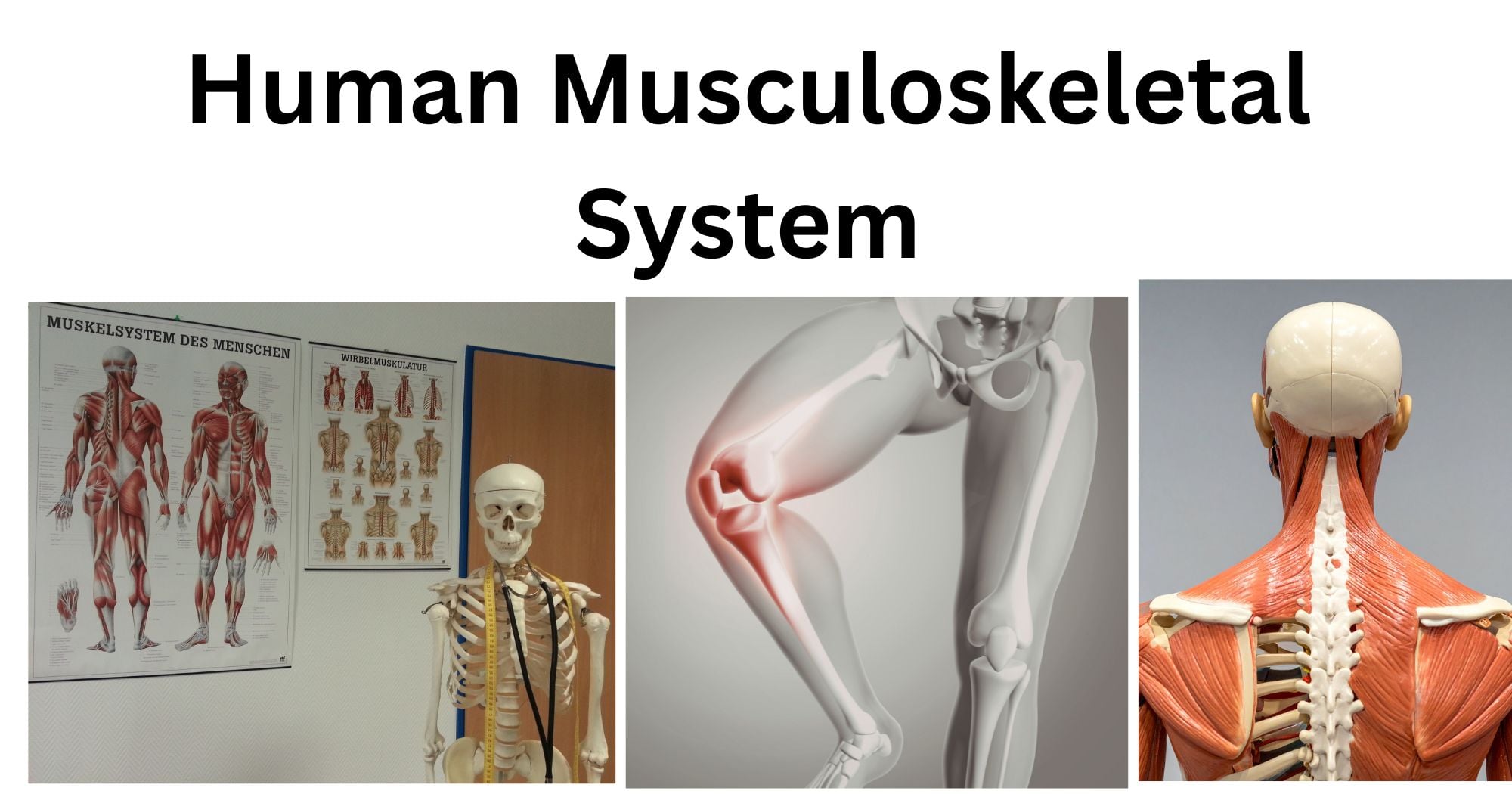
The human musculoskeletal system is a complex biological system comprising bones, the skeleton, muscles, and several types of connective tissues, and it provides the key framework for voluntary and involuntary movements. They also help protect vital internal organs, such as the lungs, heart, and brain.
Structure and Components of the Human Musculoskeletal System
The human musculoskeletal system consists of osteological structures (bones and skeleton) and muscles interconnected by joints and connective tissues.
Bones and skeleton
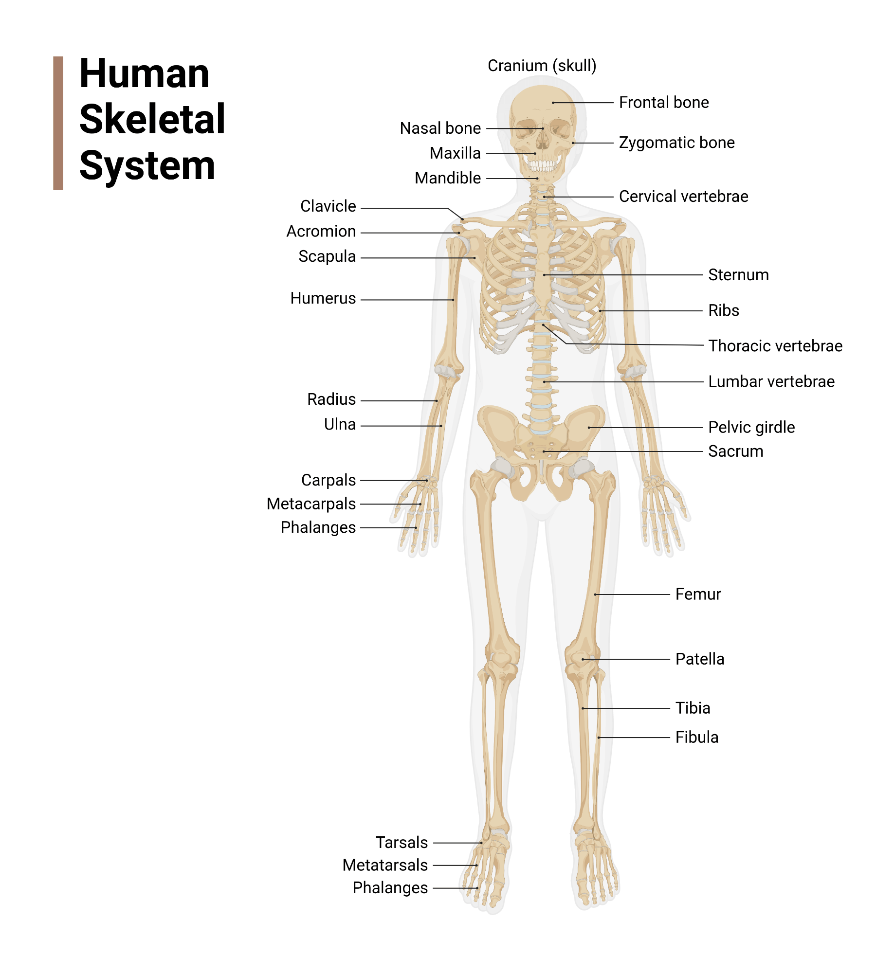
Bones are the hard, living tissues that are made up of calcium and minerals like Vitamin D. There are 206 bones in an adult human body, which have distinct shapes, sizes, and unique functions. These bones are the building blocks of the skeleton.
- Bones specifically contain a soft, fatty, spongy tissue known as ‘bone-marrow’. These bone marrows contain the stem cells that produce blood cells (RBCs and WBCs) and blood platelets.
- The term ‘skeleton’ refers to the collection of all the bones present in the body.
- The human skeleton can be divided into the axial skeleton (comprising 80 bones) and the appendicular skeleton (comprising 126 bones).
- The axial skeleton is primarily responsible for maintaining the upright posture of the body. On the other hand, the appendicular skeletal system mainly functions to facilitate mobility.
- These structures play major roles in providing structural support, homeostasis, and allowing locomotion.
Muscles
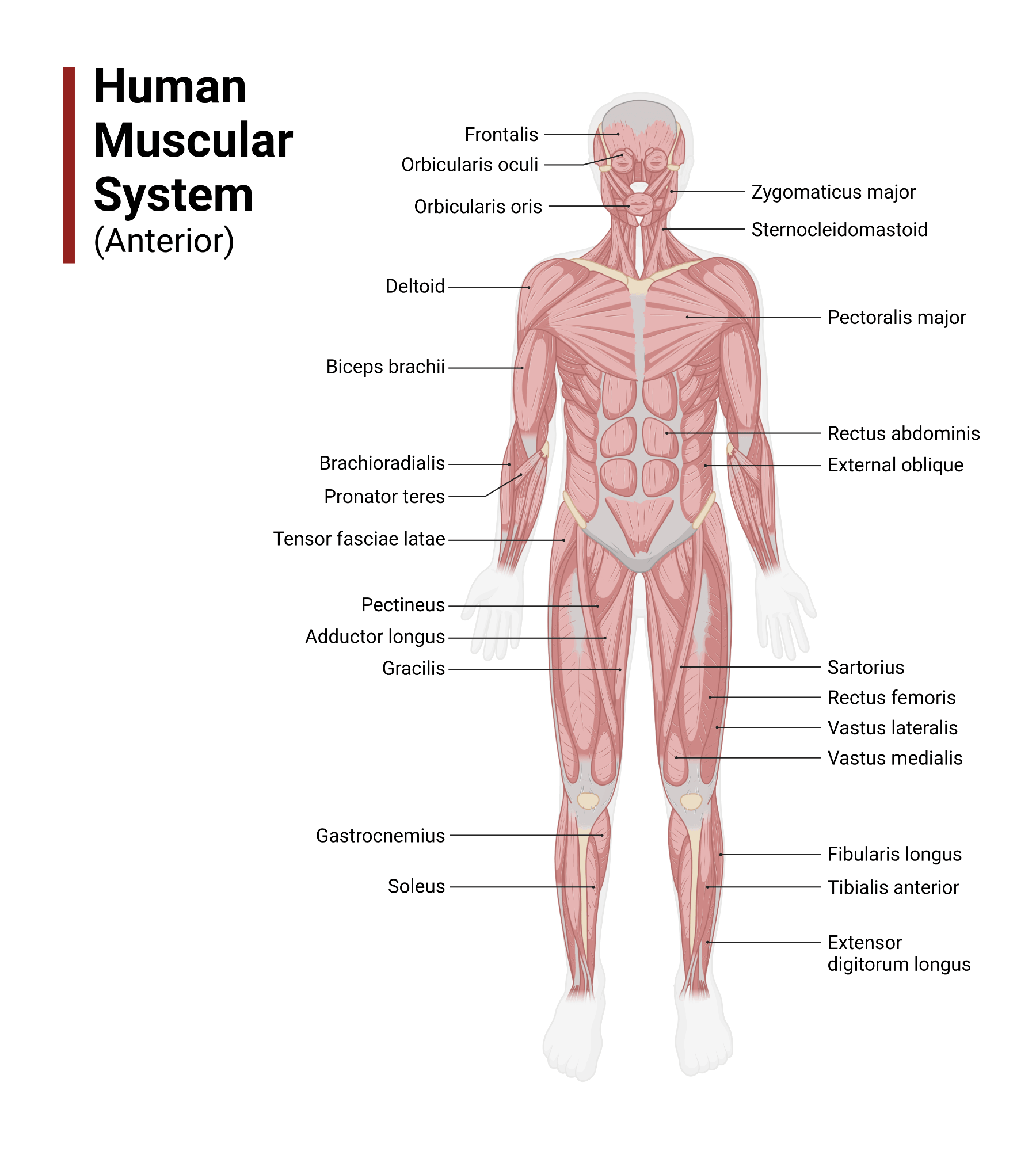
Muscles are soft, specialized tissues that are made up of protein fibers, called actin and myosin, surrounded by a group of connective tissues. The main feature of these fibers is their contractile nature: their ability to contract. The human body consists of more than 650 muscles, which together control all voluntary and involuntary movements.
- Each muscle has a distinct shape based on its primary roles. They may be thick in the middle and tapered at both ends (Spindle shaped-like biceps), long and thin (strap -like), such as sartorius, feather-shaped like deltoid, or even triangular-like pectoralis major.
- They help to maintain the body’s posture and stability and facilitate movement within and outside the body.
Joints
They are the points where two or more bones meet or connect. The key function of the joints is to provide support and facilitate flexibility and articulation during movement.
Connective tissues
The tissues that bind other tissues and organs in the body are termed connective tissues. They are primarily made up of elastin and collagen fibers. The major types of connective tissues that are significant for regulating the human musculoskeletal system are
- Ligaments: They connect the ends of bones and form joints.
- Tendons: They connect muscles to bones.
- Cartilage: They are the fibrous connective tissues, which serve as a cushion that helps to reduce the friction in joints during the movement of bones.
Functions of the Human Musculoskeletal System
The key functions of the musculoskeletal system include the following roles:
Support, balance, and posture
The musculoskeletal system provides the basic foundation for the human body. Bones and muscles coordinate to provide the required structural stability, balance, and maintain the upright posture.
Movement
This system also allows the voluntary and involuntary movement of the body, internally and externally. Controlled actions like locomotion, raising a hand, writing, etc., and involuntary actions such as breathing, heartbeat, etc., demand the movement of contractile fibres, actin, and myosin, as well as the strength of bone tissues.
Protection
The human musculoskeletal system also plays a significant role in protecting the internal organs. For example, skulls are essential for protecting the brain, and ribs and the sternum protect our heart and lungs. The cardiac muscle of the heart and the cartilaginous connective tissues between the joints also act as cushions.
Maintain body temperature
Continuous movement of muscle fibers generates heat, which significantly helps maintain body temperature.
Storage of calcium and minerals
Bones are made up of important minerals such as calcium, phosphorus, and vitamin D. Therefore, they serve as the reservoir or the storehouse of minerals.
Production of blood cells
Bones consist of an essential component at their center, called bone marrow. They play a vital role in producing red blood cells, white blood cells, and blood plasma: the components essential for regulating the overall human system.
Types of Bones
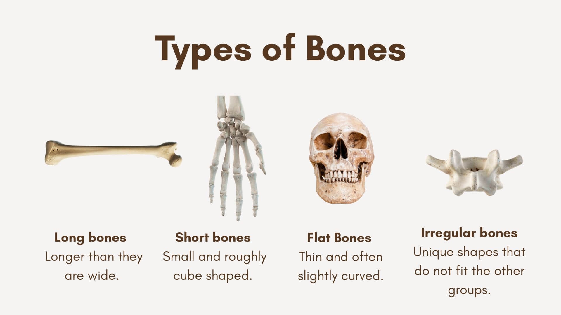
Bones are divided into 5 types based on their shape and structure.
| S.N. | Bone Type | Description | Functions | Examples |
| 1. | Long Bones | Tube-like bones, long length, and short width. Usually, the bones of the appendicular skeleton are long | To facilitate support and locomotion. | Femur (the longest bone), Humerus, Clavicle, Tibia, Ulna |
| 2. | Short Bones | Cube-shaped, thin bones are usually situated in the wrists and ankles. | To help in complex movements, provide support and stability. | Tarsals, Carpals |
| 3. | Flat Bones | Flattened and wider compared to other types | To serve as a shield for internal organs. | Skull bones (Occipital, Nasal, Frontal, Parietal, Vomer, Lacrimal) |
| 4. | Sesamoid Bones | Round-shaped small bones held by tendons | To protect the tendons from stress. | Patella |
| 5. | Irregular Bones | No specific shape or structure | To protect internal organs. | Pubis, Ilium, Ischium in the pelvis region, skull bones |
Types of Muscles
Muscles have been categorized into 3 types, each having a distinct function.
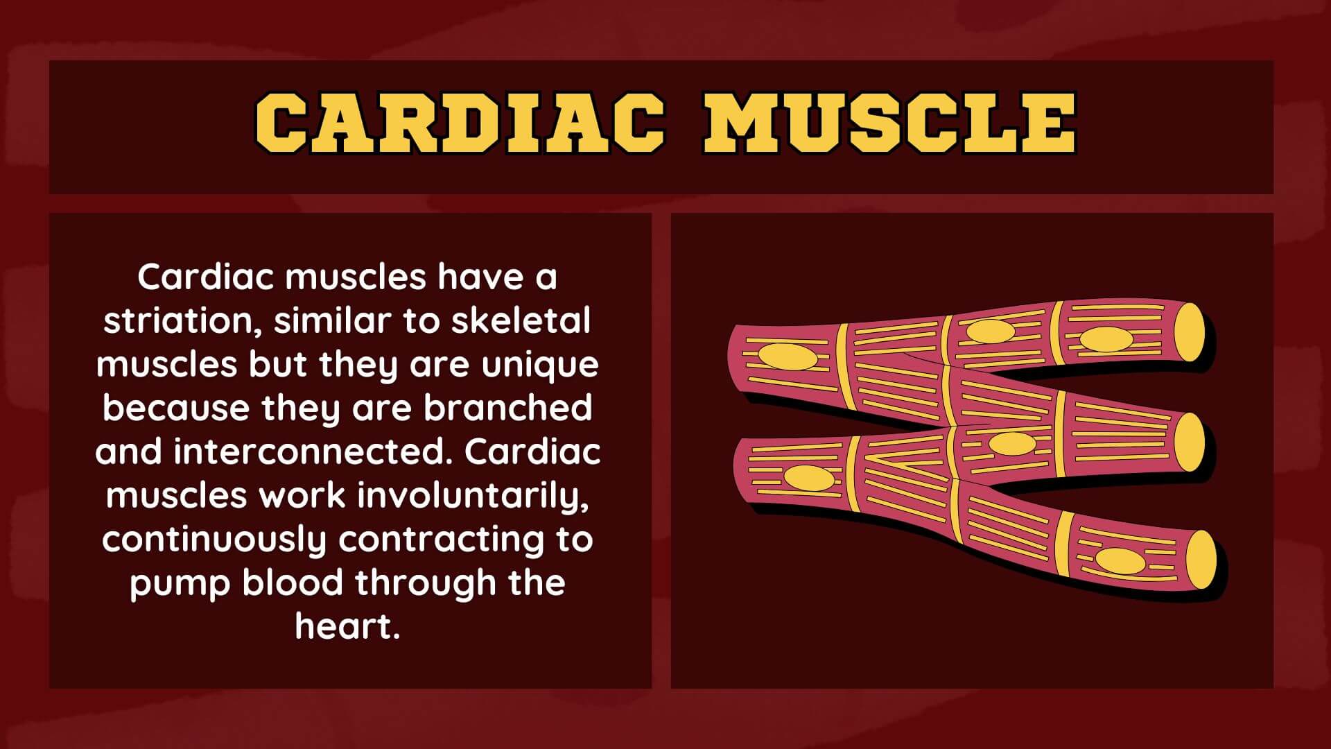
Cardiac Muscles
The muscles present in the heart are called cardiac muscles. They are also termed the myocardium. Their primary function is to protect the heart from external shocks. Contraction of these types of muscles facilitates the circulatory system by pumping the blood from the heart.
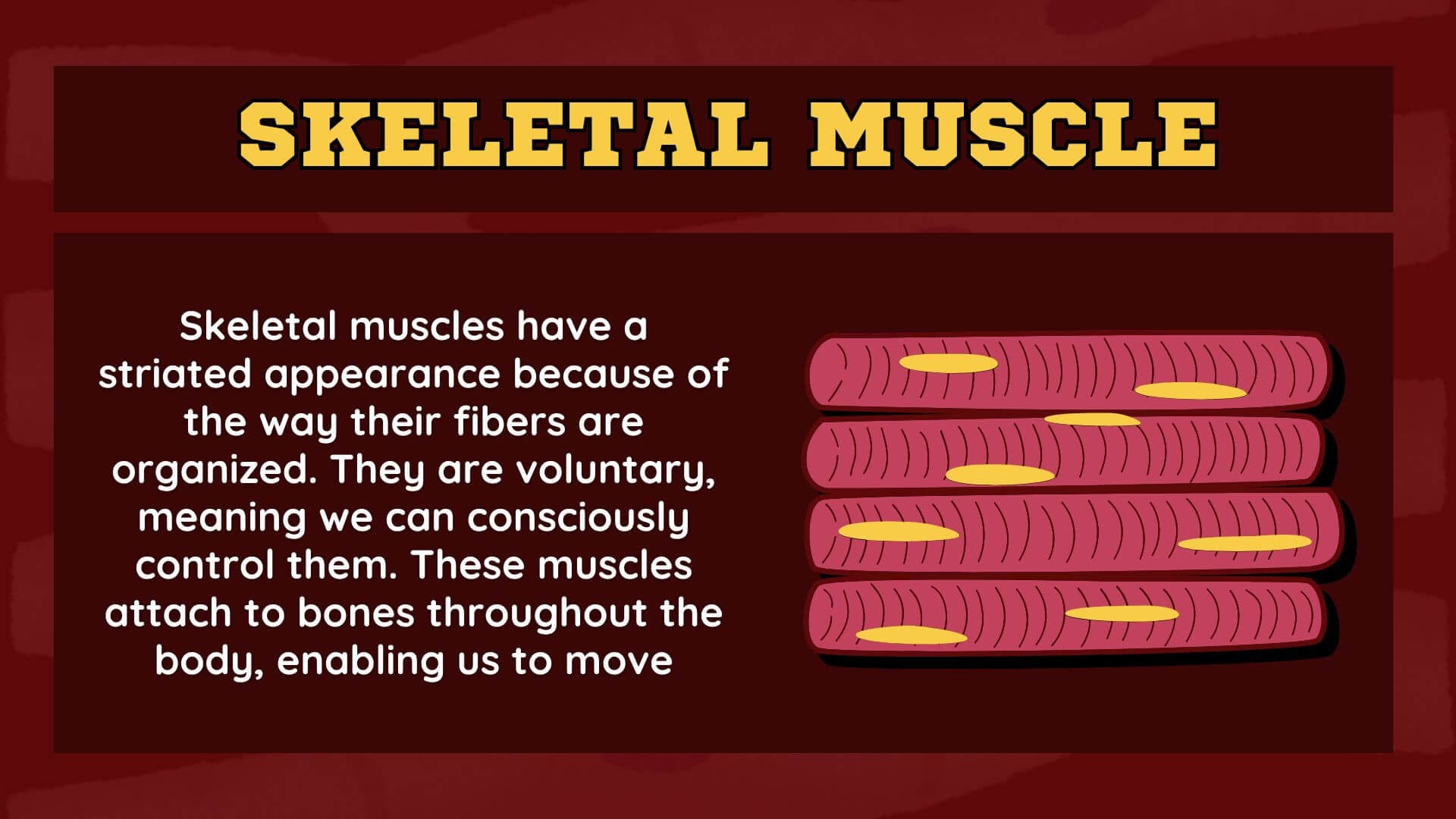
Skeletal Muscles
They are the striated muscles that provide the weight to the body and facilitate movement, such as walking, running, etc. These muscles are attached to the bones via the tendons.
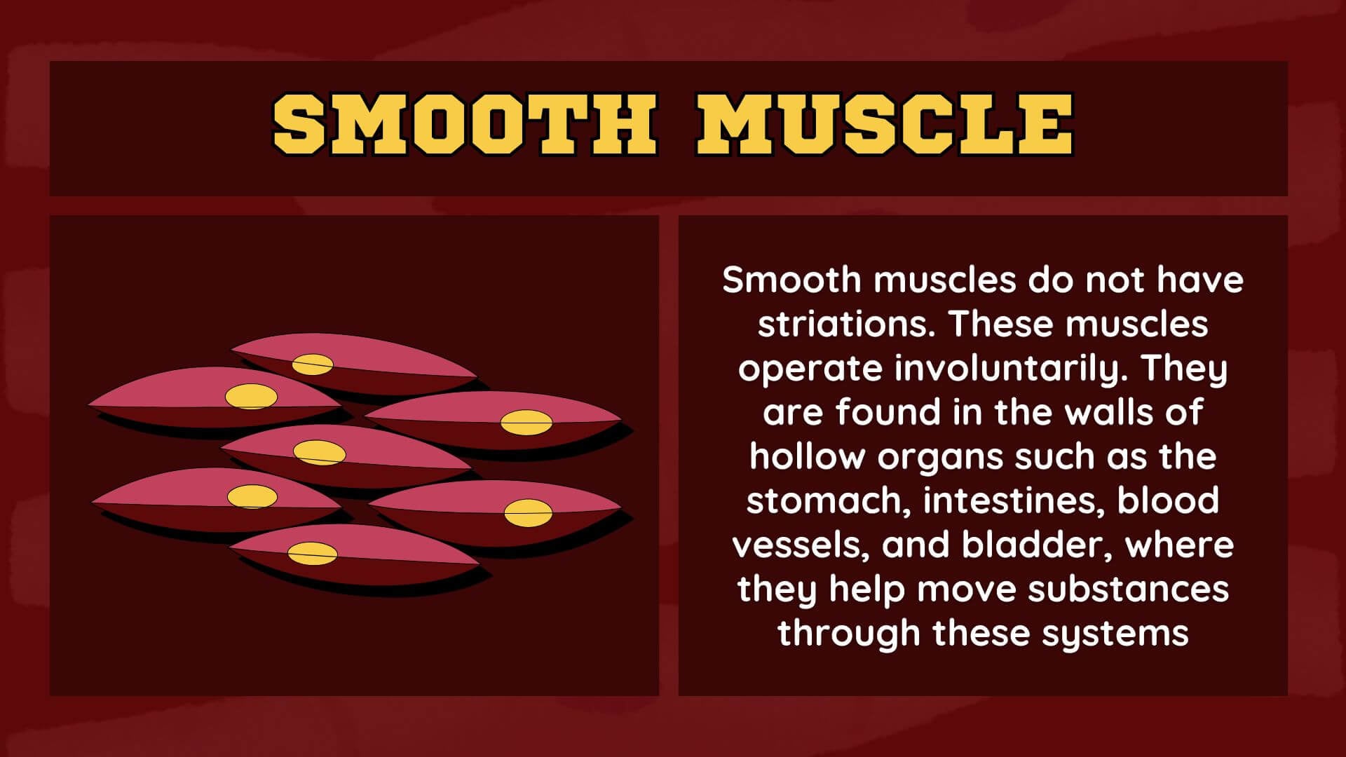
Smooth Muscles
They are the uncontrolled muscles that occur as the internal linings in the hollow organs, such as the stomach and intestines. They demonstrate strong ability to expand and contract, and are usually present in the urinary bladder and uterus.
Types of Joints
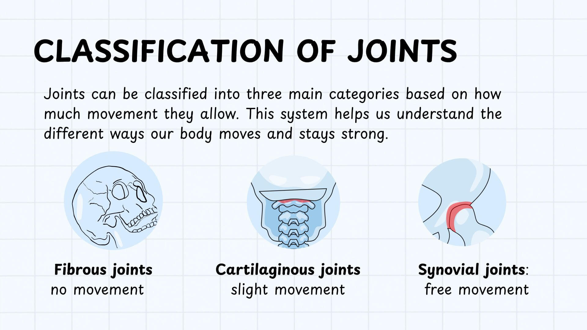
There are the following types of joints based on their structures:
Fibrous Joints
They are the thick bones that are not actively involved in generating motion. They are subdivided into 3 kinds:
- Sutures: Immovable joints that hold the skull plates together.
- Gomphoses: Immovable joints that hold the mandibular teeth together in the jaw.
- Syndesmoses: Slightly movable joints where bones are connected via ligament. For example: Joints in the tibia and fibula.
Cartilaginous Joints
They are the joints where cartilage unites the bones. They are comparatively more flexible than fibrous joints. Eg: Pubic symphysis joint is a cartilaginous joint that connects the left and right pelvic bones.
Synovial Joints
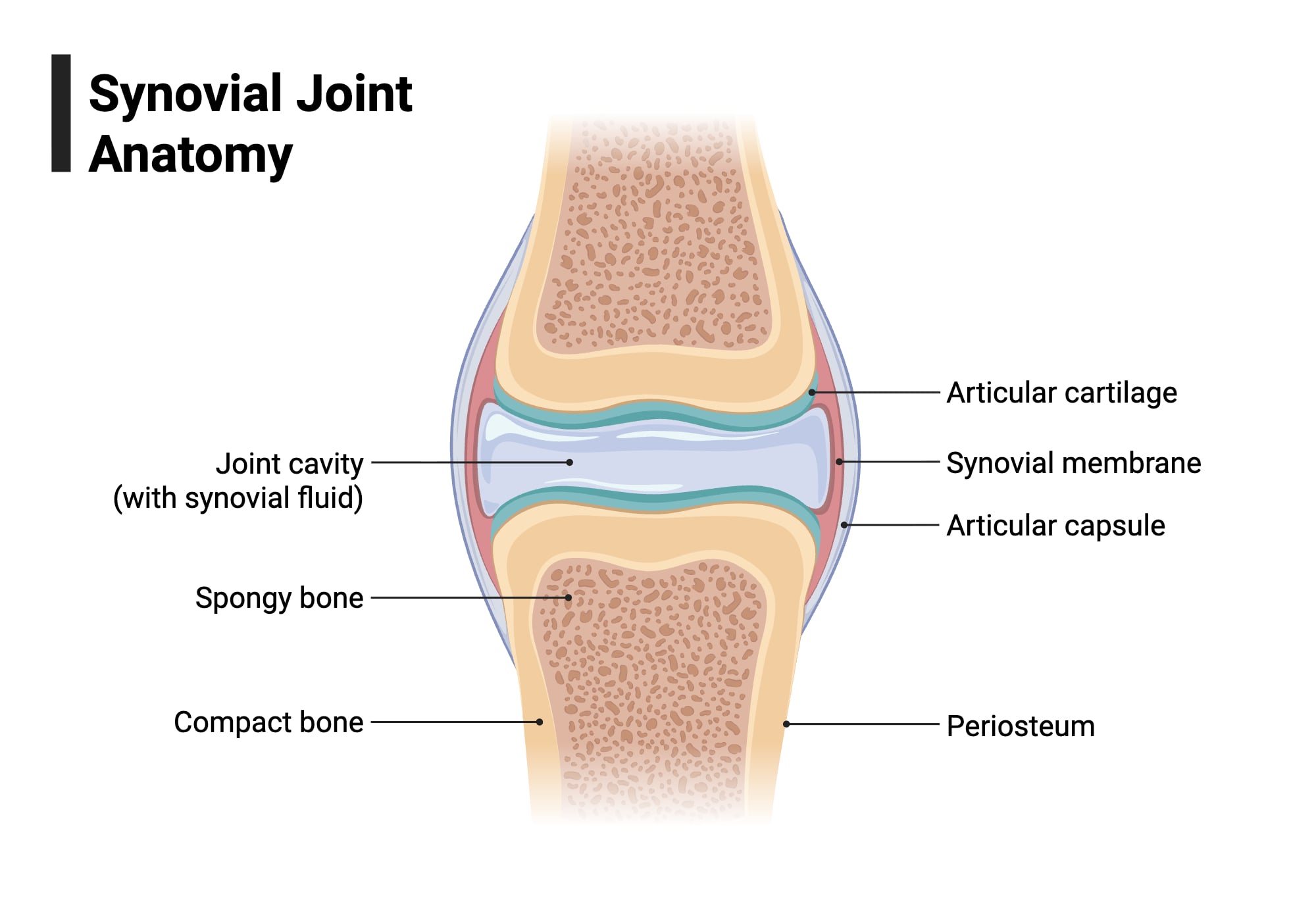
These joints are the most flexible and common joints that actively function to generate movement.
- They contain a synovial membrane, which is filled with fluid that lines the region between the bones. The joints are lubricated and protected by these membranes.
- There are 6 types of Synovial Joints: Gliding Joint, Hinge Joint, Pivot Joint, Ball and socket joint, Saddle joint, Condyloid Joint, and Planar Joints.
- These joints facilitate flexibility and articulation. Eg: Knees, elbows, shoulders.
How the Human Musculoskeletal system works (Movement and support)
The human musculoskeletal system comprises the skeletal and muscular systems, each providing structural support and facilitating specific movements. The complex structure and organization of this system act similarly to a ‘Lever’ system. Bones and the skeleton act as the lever, joints represent the fulcrum, while muscles generate the force required for movement.
- When the nervous system receives external stimuli, nerves send electrochemical signals to the muscles via motor neurons. This eventually causes the muscles to contract.
- Each skeletal muscle contains long, cylindrical fibers called myofibrils. Myofibrils further consist of thick and thin myofilaments known as Actin and Myosin. These myofilaments are composed of a structural-contractile unit termed a ‘sarcomere’.
- When the myosin head binds to the actin filaments, it changes its conformation and bends. This activity drags both filaments in opposite directions. As myosin-actin bonds are released, myosin relaxes and repeats the process.
- This combined activity of muscle contraction, actin, and myosin generates a sliding motion over the sarcomere, due to which the length of the sarcomere gets shortened while keeping the filaments’ length constant. This particular action contracts the sarcomere.
- The coordinated contractions of sarcomeres within a myofibril lead to the contraction of the muscle as a whole.
- As the muscle contracts, it pulls the bones through the tendons.
- Muscles only demonstrate the pulling mechanism on bones. Therefore, two types of muscles, Flexor and Extensor, work antagonistically to produce bending and straightening movements of bones, respectively.
- Bones and skeletons maintain their rigid structure and provide the required support during movement.
- Bones move at the point of articulation (joints) and pivot around them.
- Joints allow for flexibility and generate motion.
Major Muscles and Bones in the Human Body
The adult human body consists of 206 bones and over 650 muscles. Some of the major bones and muscles are:
Major Bones
In the human body, there are two major skeletal systems that provide stability and an upright posture. They are the axial and appendicular skeletal systems. Major bones of these systems include:
| Axial Skeleton Bones | Function |
| Skull bones (Facial and cranial bones) | To provide the facial structures and protect the brain. |
| Auditory ossicles (Malleus, incus, and stapes) | To transmit the sound. |
| Hyoid bone | To support the mandible. |
| Vertebral column | To support the head and protect the spinal cord. |
| Thoracic bones or chest bones | Protect the internal vital organs like the lungs and the heart. |
| Appendicular Skeleton Bones | Function |
| Upper and Lower limb bones (Humerus, radio-ulna, carpals, metacarpals, and Femur, tibio-fibula, tarsal, and meta-tarsal) | To help generate external movements such as writing and facilitate locomotion like walking, running, etc. |
| Pectoral Girdle bones (Clavicle and Scapulae) | To attach the upper limbs to the body. |
| Pelvic Girdle bones (Hip bones) | To attach the lower limb bones to the body. |
Major Muscles
Head and Neck Muscles:
- Masseter muscle: The strongest muscle in the body
- Sternocleidomastoid muscle: tilts and rotates the head
Upper Body and Torso:
- Pectoral Major: essential for arm movement
- Trapezius: elevates and retracts the shoulder
- Abdominals: provide stability
Arms and Shoulders:
- Deltoids: raise and rotate the arm
- Biceps: bend the elbow
- Triceps: straighten the elbow
Lower Body and Legs:
- Glutes/hip muscles: extend the thigh
- Quads: a group of 4 muscles on the front of the thigh
- Hamstrings/ back thigh muscles: flex the knee and extend the hip
- Soleus/ Calf muscles: point the toes and lift the heel
Common Disorders of the Human Musculoskeletal System
Arthritis
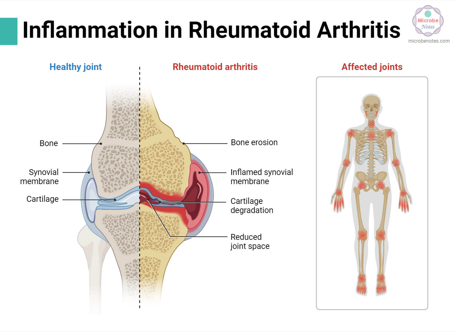
It is a condition where the joints connecting the bones get damaged. Usually, it happens naturally after the age of 50. Occasionally, it may also occur when the uric acid crystals are deposited in the joints. This type of arthritis is known as Gout arthritis.
Osteoporosis
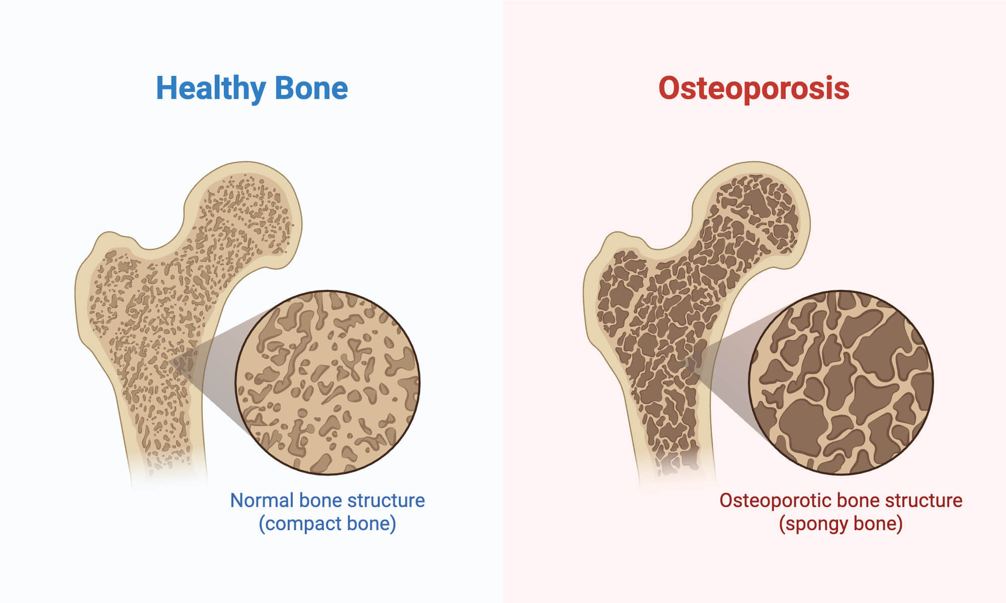
Bones are usually strong and dense structures. However, ageing slowly reduces the density of bones, causing them to be weaker than before. This type of condition, where bones become fragile and less dense, is called osteoporosis. Bone breaking is the most common symptom of osteoporosis.
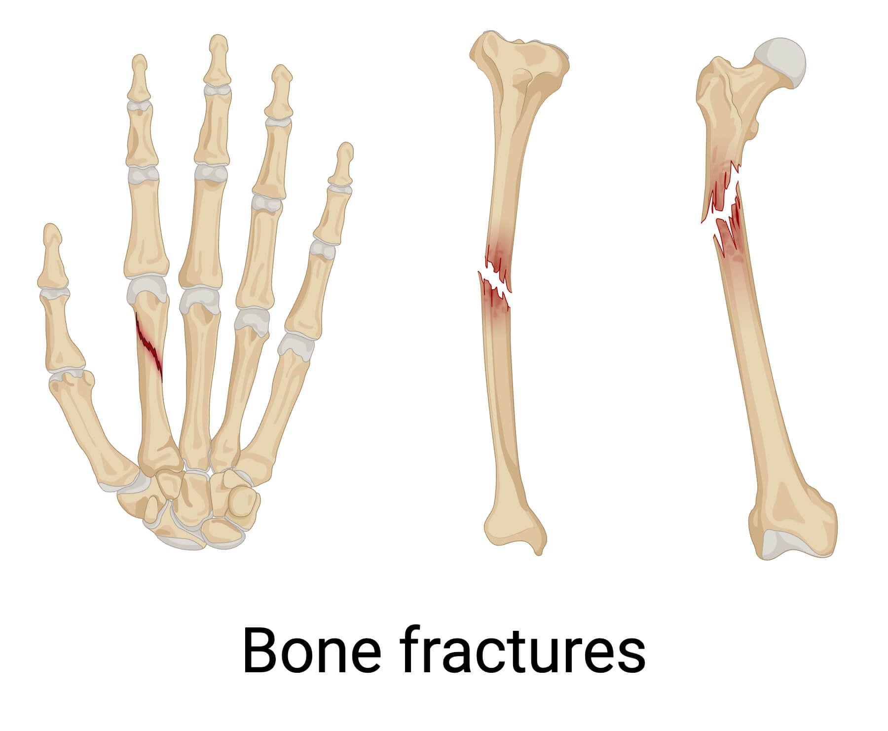
Bone fractures
Several accidents and injuries can break bones. This type of broken bone case is termed a bone fracture. There are several types of bone fractures depending on the pattern and severity of the bones broken, such as stress fractures, buckle fractures, oblique fractures, compression fractures, etc.
Muscle strains
When we use our muscles continuously for a long period of time, the myofibril strands stretch too much and tear apart. This condition is known as muscle strain.
Ligament tear (sprain)
Ligament tear, also known as a muscle sprain, is a condition where the connective tissue between the bones suddenly ruptures or overstresses, causing severe pain and swelling. It usually occurs in the knee and ankle, creating a characteristic popping sound.
How to keep the Human Musculoskeletal System healthy?
It is essential to take good care of our muscles and bones. Some of the ways to keep the human musculoskeletal system healthy include:
- Regular exercise
- Stretch and warm up before any physical activities
- Maintain a healthy, balanced diet
- Expose yourself to sunlight to absorb Vitamin D
- Regular health check-up
- Avoid smoking and drinking excess alcohol
- Drink water
Conclusion
The human musculoskeletal system is a complex biological system comprising bones, muscles, joints, and connective tissues, and it functions to provide foundational support and stability, protect vital organs, and facilitate controlled and uncontrolled movements in the body.
References
- Admin. (2022, August 3). Muscular and skeletal disorders. BYJUS. https://byjus.com/biology/disorders-muscular-and-skeletal-system/
- File:Anatomy and physiology of animals Structure of a muscle.jpg – Wikimedia Commons. (n.d.). https://commons.wikimedia.org/wiki/File:Anatomy_and_physiology_of_animals_Structure_of_a_muscle.jpg
- Healthdirect Australia. (n.d.). Bones, muscles and joints. Healthdirect. https://www.healthdirect.gov.au/bones-muscles-and-joints#:~:text=Skeletal%20muscle%20is%20a%20voluntary,the%20strength%20of%20your%20muscles.
- Malmquist, S., & Prescott, K. (n.d.). 4.7 musculoskeletal system. Human Biology. https://open.lib.umn.edu/humanbiology/chapter/4-7-musculoskeletal-system-2/
- Musculoskeletal system. (2023, November 3). Kenhub. https://www.kenhub.com/en/library/anatomy/the-musculoskeletal-system
- Musculoskeletal System | What is the Musculoskeletal System? | Skeletal Structure | Muscular System | Bones and Muscles | Orthopedic System | Bone and Muscle Health | Anatomy and Physiology | Body’s Support System | Movement System | Structural Framework | Musculoskeletal Health | Orthopedic Health | Biomechanical System | Physical Structure | Body’s Framework | Orthopedic Anatomy | Movement and Support | Structural Integrity | Physical Health | Bone and Muscle Function. (n.d.). Foundation Health Osteopathy. https://foundationhealth.com.au/musculoskeletal-system/#:~:text=What%20does%20the%20musculoskeletal%20system%20consist%20of?,important%20for%20preserving%20musculoskeletal%20function.
- Narayana Health. (n.d.). Narayana Health. https://www.narayanahealth.org/blog/skeletal-system-anatomy-location-function-and-conditions
- Professional, C. C. M. (2026, January 8). Skeletal system (Musculoskeletal system). Cleveland Clinic. https://my.clevelandclinic.org/health/body/21048-skeletal-system
- Ren, L., Qian, Z., & Ren, L. (2014). Biomechanics of Musculoskeletal System and its biomimetic Implications: A review. Journal of Bionic Engineering, 11(2), 159–175. https://doi.org/10.1016/s1672-6529(14)60033-0
- Shaffer, F., & Neblett, R. (2010). Practical Anatomy and Physiology: the skeletal muscle system. Biofeedback, 38(2), 47–51. https://doi.org/10.5298/1081-5937-38.2.47
- Villa-Forte, A. (2025, January 10). Introduction to the biology of the musculoskeletal system. MSD Manual Consumer Version. https://www.msdmanuals.com/home/bone-joint-and-muscle-disorders/biology-of-the-musculoskeletal-system/introduction-to-the-biology-of-the-musculoskeletal-system
- Visible Body, part of Cengage Learning. (n.d.). Muscle tissue types | Learn Muscular Anatomy. https://www.visiblebody.com/learn/muscular/muscle-types
- What is Musculoskeletal Health? | IU School of Medicine. (n.d.). https://medicine.iu.edu/expertise/musculoskeletal-health/what-is-musculoskeletal-health
- Wikipedia contributors. (2025, November 8). Human musculoskeletal system – Wikipedia. https://en.wikipedia.org/wiki/Human_musculoskeletal_system
