Special stains have been developed over time for identifying bacteria species, differentiating them morphologically, and even characterizing there very special features. The most common stain being Gram Staining, Acid-fast staining, endospore staining. Each of these stains aims at identifying and characterizing bacteria based on their morphologies.
Albert stain is no different. Its application aim at identifying bacteria that contain special structures known as metachromatic granules. Other staining techniques that are used to detect the presence of granules in the cytoplasmic membrane of bacteria are Nessers’s stain and Pugh’s stain.
Albert stain distinctly identifies metachromatic granules that are found in Corynebacterium diphtheriae.
Corynebacteria are gram-positive, non-spore forming, non-motile bacilli that contain metachromatic (Volutin) granules which are intracellular inclusion bodies, found in the cytoplasmic membrane of some bacterial cells for storage of complexed inorganic polyphosphate (poly-P) and enzymes. When these granules are subjected to stain with methylene blue dye, they appear reddish-purple color and not the blue dye.
The most common Corynebacterium is Corynebacterium diphtheriae, that caused diphtheria, a nasopharyngeal infection (affecting the nasal, throat) that can also affect the skin, after bacterial colonization and infection.
Basically, this bacteria is initially cultured in selective media either a Loeffler agar or Mueller-Miller tellurite agar, or Tinsdale tellurite agar, and its colonize isolated to prepare liquid culture that is then used for staining. Albert stain only acts as a confirmatory stain for the bacteria. Being a differential stain, it can only stain volutin granules, hence bacteria without these granules can not be stained nor identified with this technique.

Interesting Science Videos
Objective
To stain and observe metachromatic granules from a Corynebacterium diphtheriae culture.
Principle of Albert Staining
Albert staining technique aims at detecting the presence of metachromatic granulated bodies of Corynebacterium diphtheriae. Albert stain is made up of two staining solutions; designated as Albert Solution 1 and Albert Solution 2, their compositions being;
Albert Solution 1:
- toluidine blue, malachite green, glacial acetic acid, and alcohol
Albert solution 2:
- Iodine and Potassium iodide in water
To use Albert’s staining solutions, each of the two solutions must be prepared effectively with the right percentages of components in order to demonstrate the granules with the right color after staining.
Albert staining solution 1 acts as the staining solution while Albert solution 2 acts as the mordant, i.e an ion element that binds and holds a chemical dye, to make it stuck on the micro-organism.
How are these solutions prepared?
Albert Stain 1: Preparation of 100ml Albert stain 1
- Into 100ml of water, add 0.1ml of glacial acetic acid
- Add 2ml of 95% ethanol into the solution
- Then, dissolve 0.15g of toluidine blue into the solution
- Lastly, dissolve 0.2g of malachite green into the solution
Albert Stain 2: Preparation of 300ml of Albert stain 2
- Dissolve 2g of iodine in 50ml of distilled water
- Add 250ml of water to the solution
- Dissolve 3g of Potassium Iodide into the solution in the solution
Procedure of Albert Staining
Reagents:
Albert staining Solution 1
Albert staining solution 2
Distilled water
A. Staining:
- Aseptically, take a loopful culture of Corneybacterium diphtheriae
- Make a smear at the center of a clean sterile glass slide
- Heat fix the smear, gently
- On a staining rack, place the smeared glass slide.
- Add Albert staining Solution 1 into the smear and leave it for 3-5 minutes
- Wash the smeared slide with gently flowing tap water
B. Mordanting
- Add Albert staining solution 2 and leave it for 1 minute
- Wash the slide with gently flowing tap water.
- Blot to dry the smeared glass slide
- Add cedarwood oil on the smear
- Then observe under a microscope by oil immersion at 1000x
Result
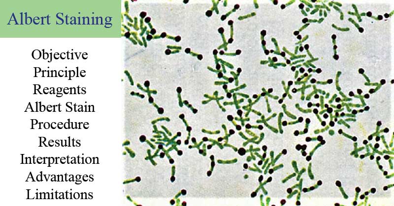
The metachromatic granules stain bluish black while the rest of the microbial cell stains green.
Interpretation of Albert Staining
Corynebacterium diphtheriae cytoplasmic membrane contains volutin granules, also known as metachromatic granules, which are a characteristic feature of this bacteria. The staining by Albert solutions, stains the granules making the appear as round-shaped blue-black dots at the bottom of L-shaped or V-shaped green Bacilli.
Applications of Albert Staining
- It is majorly used to identify the metachromatic granules found in disease-causing micro-organisms like Corynebacterium diphtheriae.
- Being a differential stain, it helps distinguish Corneybacterium diphtheriae from other nonpathogenic diphtheroid that lack the metachromatic granules.
Limitations of Albert Staining
- It can only be used to stain the metachromatic granular bodies and not any inclusions in the cytoplasmic membrane.
References
- Formation of volutin granules in Corynebacterium glutamicum Srinivas Reddy Pallerla, Sandra Knebel, Tino Polen, Peter Klauth, Juliane Hollender, Volker F. Wendisch, Siegfried M. Schoberth
- Bhumbla, Upasana. (2018). Albert’s Staining. 10.5005/jp/books/14206_10.
- Murphy JR. Corynebacterium Diphtheriae. In: Baron S, editor. Medical Microbiology. 4th edition. Galveston (TX): University of Texas Medical Branch at Galveston; 1996. Chapter 32. Available from: https://www.ncbi.nlm.nih.gov/books/NBK7971/
Internet Sources
- 1% – https://www.ncbi.nlm.nih.gov/books/NBK8477/
- 1% – https://www.ncbi.nlm.nih.gov/books/NBK7971/
- 1% – https://sciencing.com/advantages-stained-bacteria-12011291.html
- 1% – https://science.umd.edu/classroom/bsci424/PathogenDescriptions/Corynebacterium.htm
- 1% – https://quizlet.com/50988479/staining-methods-flash-cards/
- 1% – https://microbeonline.com/
- 1% – http://microrao.com/alberts_staining.htm
- <1% – https://www.slideshare.net/RESHMASOMAN3/staining-techniques
- <1% – https://www.chemistry.mcmaster.ca/adronov/resources/Stains_for_Developing_TLC_Plates.pdf

Your notes are really helpful
Dear Faith Mokobi, great to have found that particular description of an interesting stain – I’ve not known about for my 35 years of my active professional life… Thank you for your efforts
You may also adding viva questions and answers !!