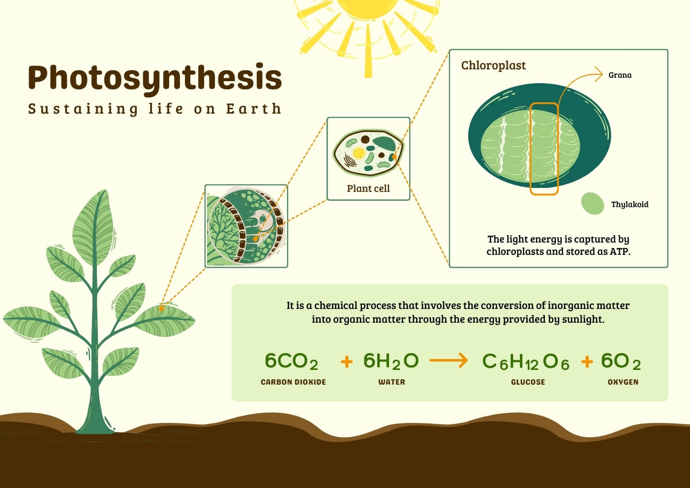
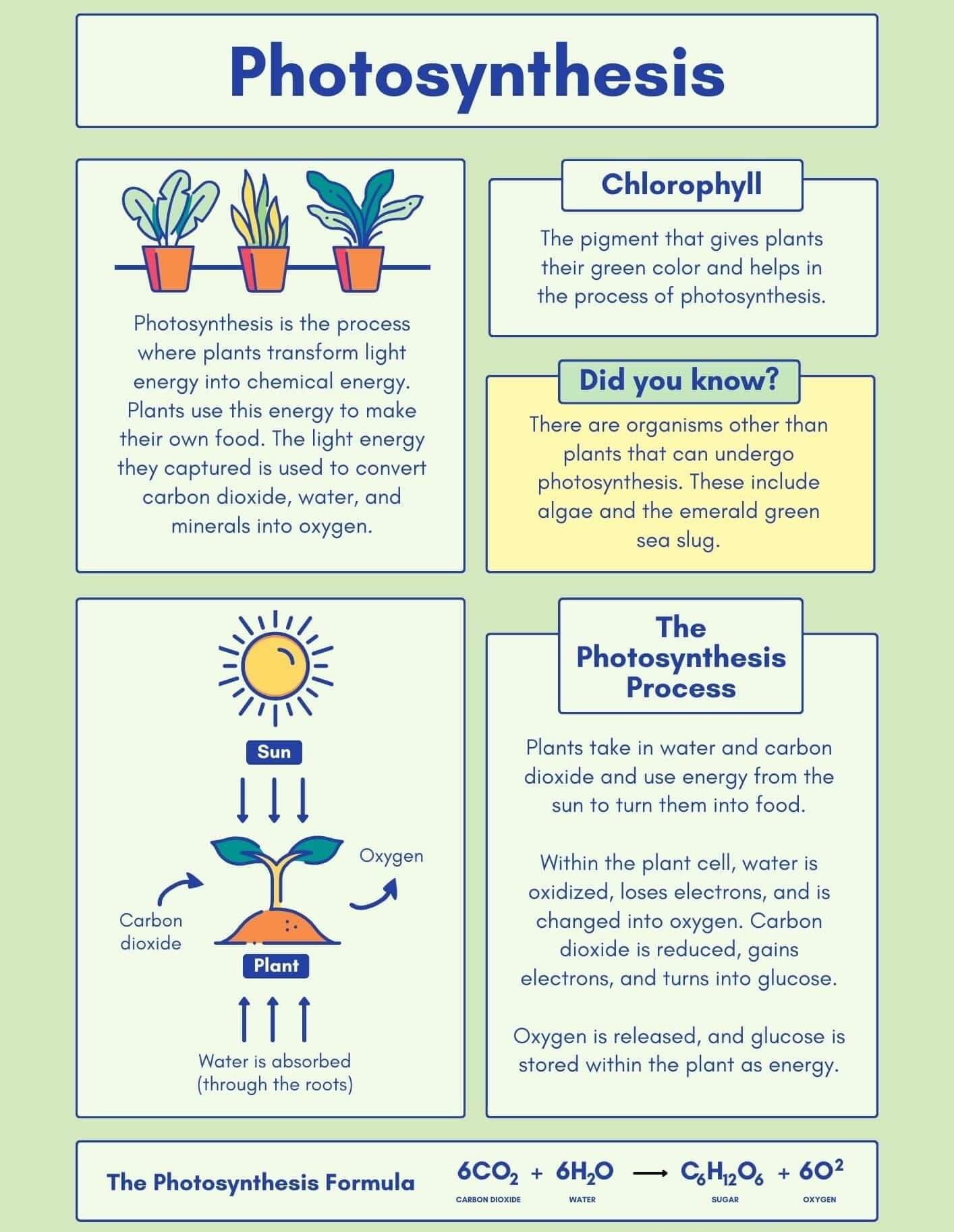
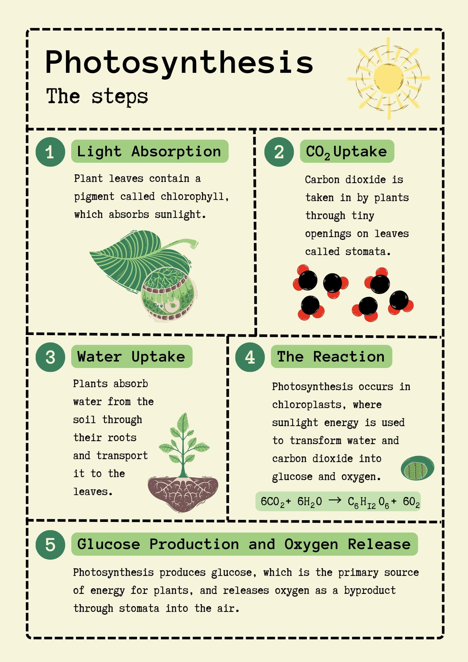
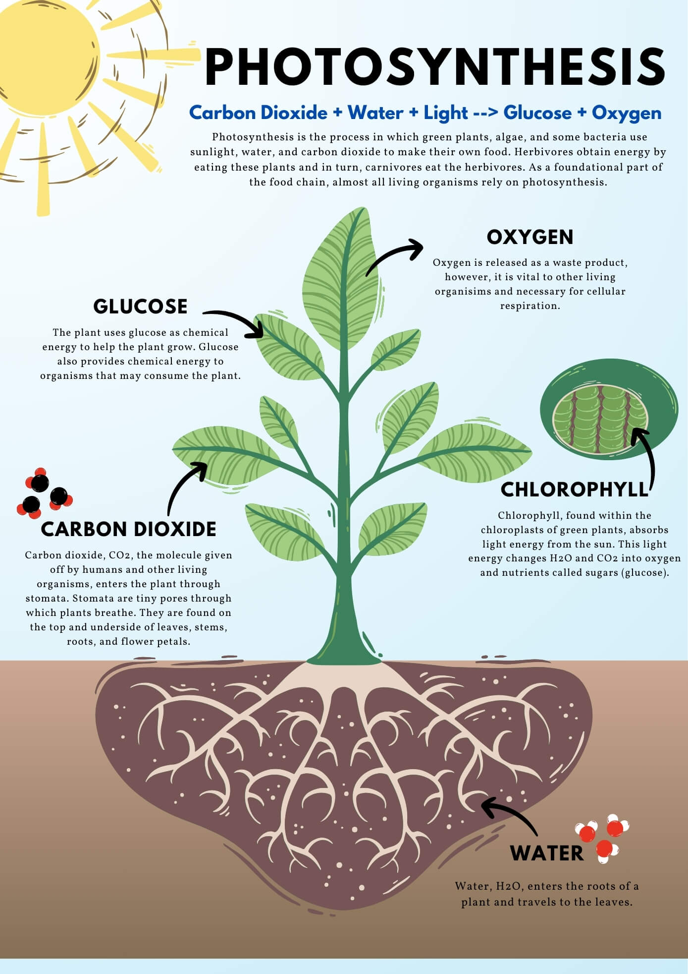
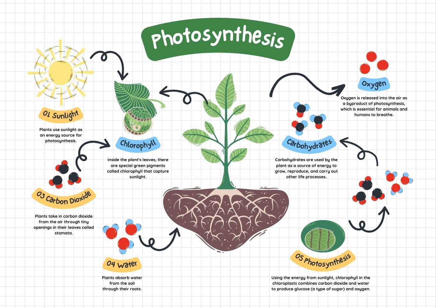
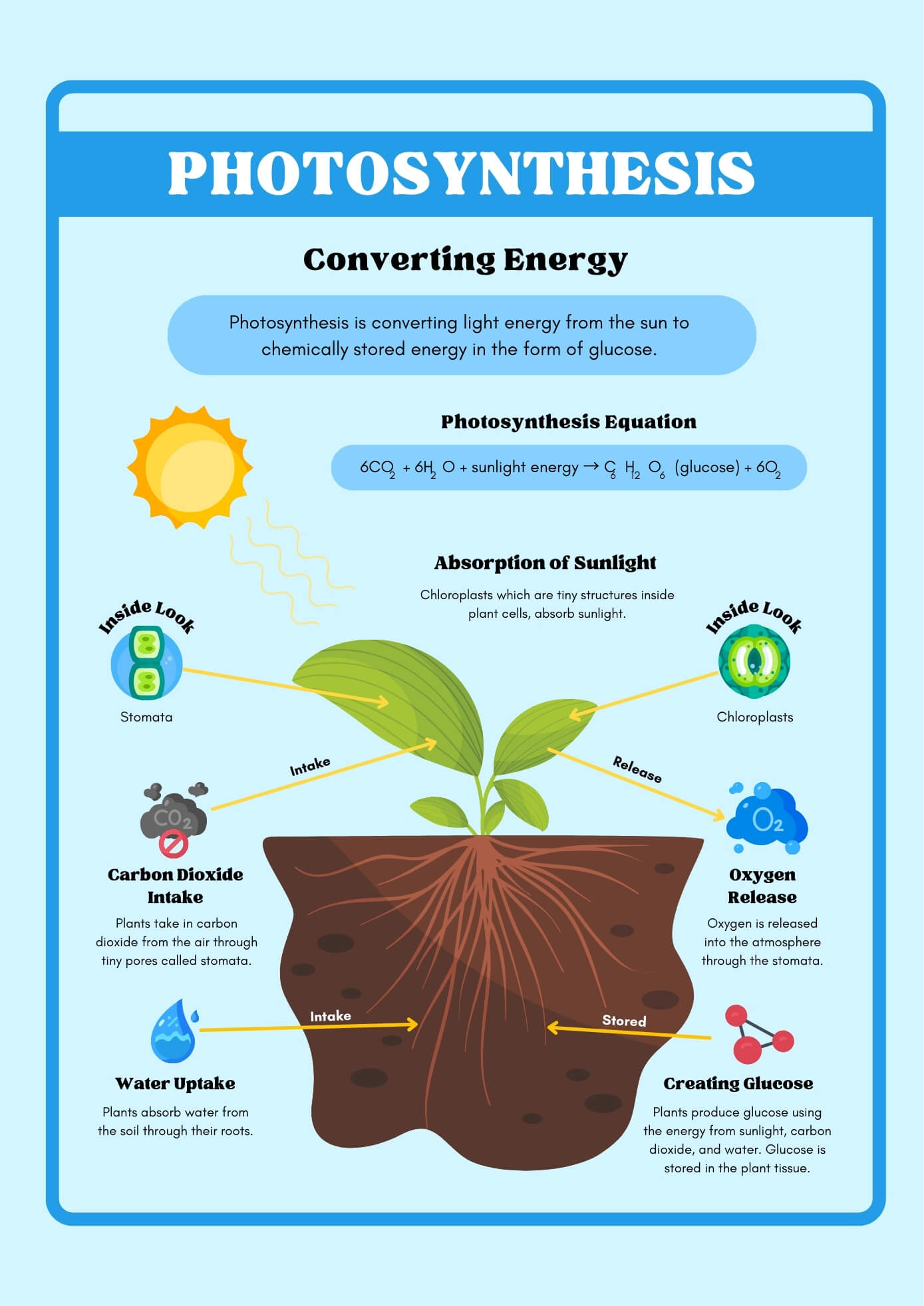
Photosynthesis is defined as the process of biosynthesis of simple carbohydrates like glucose by utilizing carbon dioxide and water in the presence of sunlight inside the chlorophyll-containing cells of green plants.
Photosynthesis Equation:
6CO2 (Carbon Dioxide) + 6H2O (Water) → C6H12O6 (Glucose) + 6O2↑ (Oxygen)
OR
6CO2 (Carbon Dioxide) + 12H2O (Water) → C6H12O6 (Glucose) + 6O2↑ (Oxygen) + 6H2O (Water)
Interesting Science Videos
Photosynthetic Unit
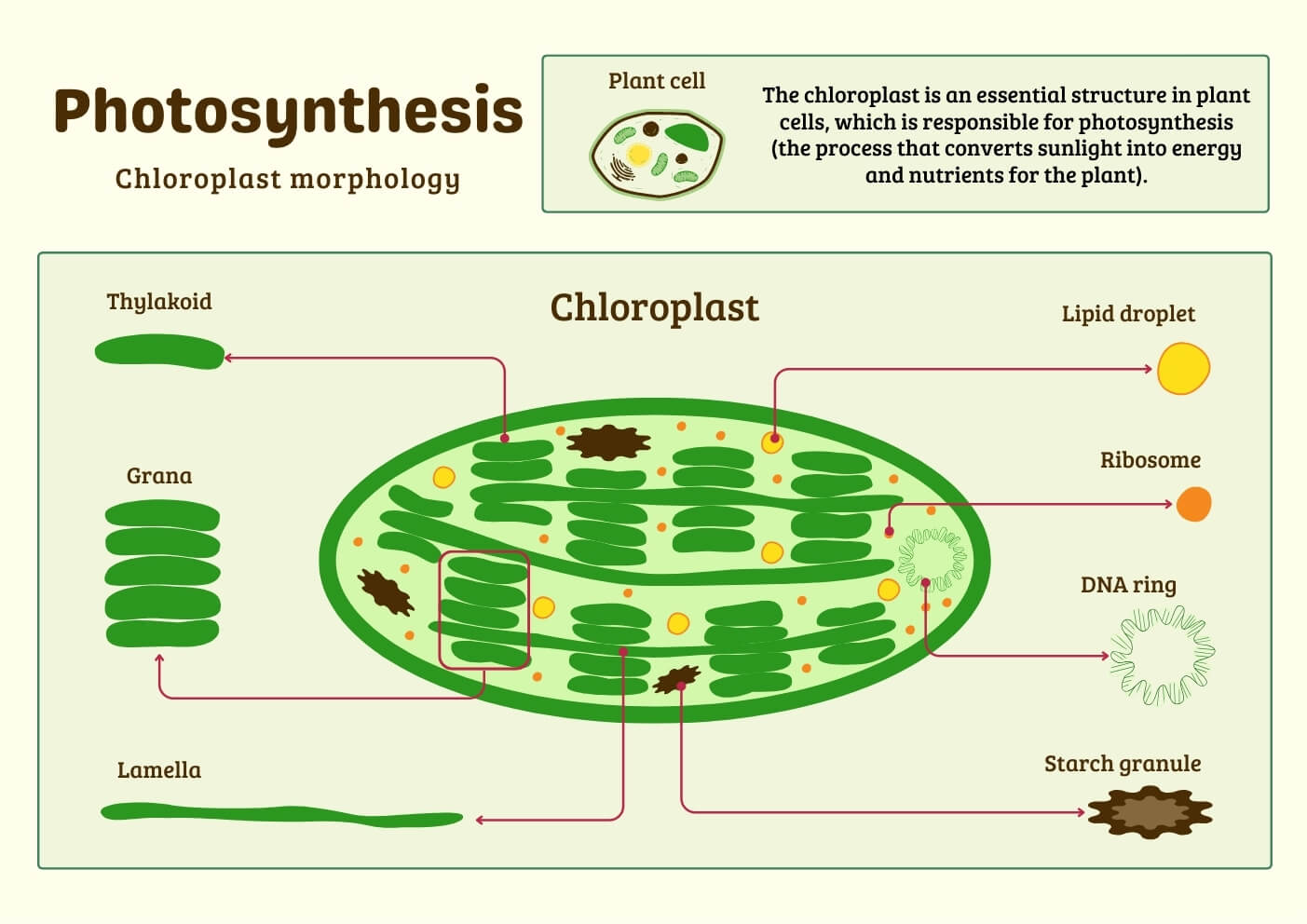
A photosynthetic unit is the smallest group of collaborating pigment molecules necessary to affect a photochemical act or conversion of light energy into chemical energy. Photosynthetic units are located in small particle units called quantasome.
A typical photosynthetic unit consists of a reaction center/photo center/trapping center which is fed by about 200-300 light-harvesting pigment molecules/ The reaction center is a special type of chlorophyll A molecule designated P700 in the case of Pigment System I or Photo System I (PSI) and P680 in the case of Pigment System II or Photo System II (PSII).
The light-harvesting pigment molecules are of two types viz: core molecules and antenna molecules. The light-harvesting pigment molecules consist of chlorophyll-b, carotene, xanthophyll, etc. The antenna molecules absorb light energy of wavelength lower than that absorbed by core molecules and transfer it to the core molecules by electron spin resonance.
The core molecules absorb light of wavelength lower than those absorbed by the reaction center. The light energy absorbed by the antenna molecules and core molecules is transferred to the reaction center.
Source of oxygen evolved during photosynthesis
Until 1930 AD, scientists believed photosynthesis was just the reverse of respiration, so the photosynthesis equation was given as follows.
In 1931 AD, Van Niel discovered that in the photosynthetic bacteria (green sulfur bacteria), photosynthesis occurred in the presence of Carbon dioxide but without the evolution of oxygen. If carbon dioxide was the source of oxygen, oxygen should’ve evolved. Those bacteria utilize H2S instead of water as a hydrogen donor so, elemental sulfur is produced instead of oxygen as follows:
6CO2 + 12H2S → C6H12O6 + 12S + 6H2O
So, Van Niel predicted that if elemental sulfur comes from H2S photosynthetic bacteria, oxygen should come from water in green plants. Finally, the experimental proof that oxygen evolved during photosynthesis comes from water was given by radio isotopic studies using heavy isotopes of oxygen (O18) by Samuel Ruben, Randal Kamen, and Hyde. When they grew the experimental plant in labeled CO218, the oxygen evolved was of normal type (O16). However, when the oxygen of water was labeled (H2O18), the evolved oxygen was off (O18).
6CO218 + 12H2O → C6H12O618 + 6O2 + 16H2O18
6CO218 + 12H2O18 → C6H12O6 + 6O218 + 16H2O
Hill Reaction
The phenomenon of the evolution of oxygen from water in the absence of carbon dioxide by the isolated chloroplast when illuminated in the presence of suitable hydrogen acceptors like ferric oxalic, quinone, etc.
2H2O + 2A → 2AH2 + O2↑
A : suitable hydrogen acceptor
AH2 : reduced form of A
Mechanism of Photosynthesis
The process of photosynthesis is a complicated oxidation-reduction process, ultimately resulting in the oxidation of water and the reduction of carbon dioxide. The mechanism of photosynthesis consists of two phases:
- Primary photochemical reaction / Light reaction / Photochemical decomposition of water / Light-dependent phase of photosynthesis.
- Dark reaction / Thermochemical reduction of Carbon dioxide / Light-independent phase of photosynthesis / Black man’s reaction / Calvin’s cycle/ C3 cycle.
Light Reaction of Photosynthesis
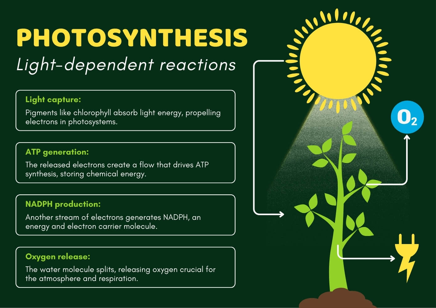
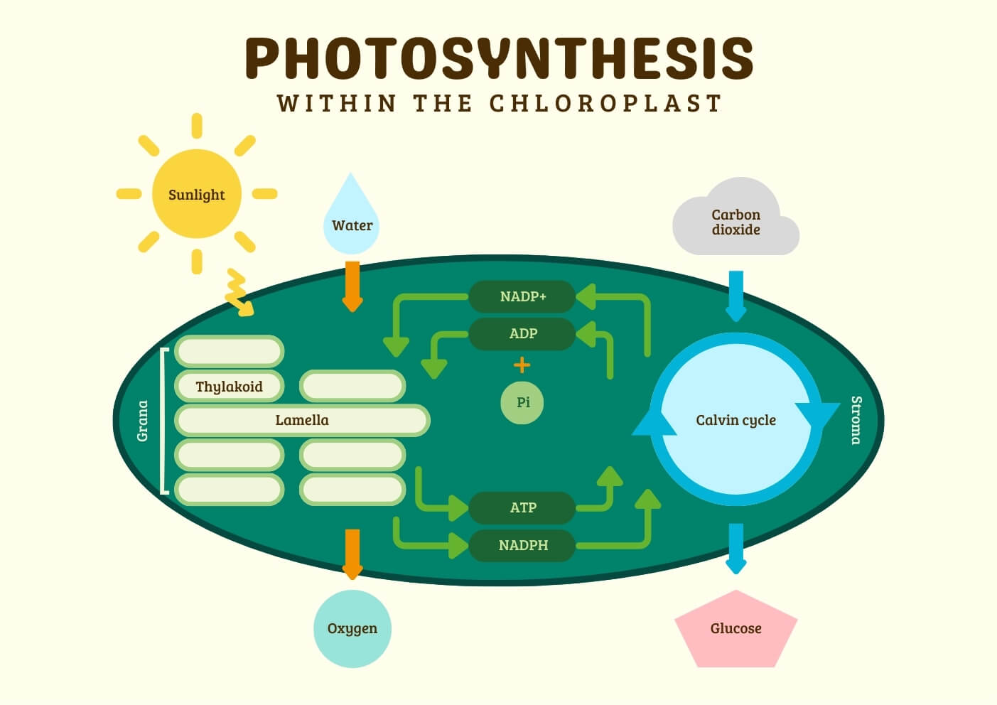
It is the first phase in photosynthesis during which the light energy of the sun is converted into chemical energy which is stored in the form of ATP and NADPH + H+. It occurs in the grana region of the chloroplast.
It consists of the following steps
a. Photoexcitation of chlorophyll-a
Light energy travels in the form of discrete energy packets called photons. Each photon contains 1 quantum of light energy. When a chlorophyll-a molecule (reaction center) absorbs a photon of light, it becomes excited. Sometimes, during excitation, an electron gets ejected from the chlorophyll-a molecule. The ejected electron contains an extra amount of energy received from the sun.
A part of this energy is used in the photolysis of water, and another part of the energy is used for photophosphorylation. Before the absorption of a photon of light, the chlorophyll molecule was in the ground state. But, after the ejection of an electron, an electron hole is formed in the chlorophyll-a molecule and the molecule is in the excited state.
b. Photolysis of water
It is the process of splitting water molecule into H+ and OH– ions during the light reaction of photosynthesis by utlising the light energy of sun. It required the presence of Mn2+ and Cl–.
2H2O → 2H+ + 2OH–
2OH– → 2e– + H2O + 1/2 O2
The H+ released is used for the reduction of NADP+ into NADPH + H+. The OH– ions after releasing their electrons recombine to form water and oxygen gas is released. The released electron is used for filling the electron-hole present in chlorophyll-a of PSII.
c. Photophosphorylation
The process of formation of ATP from ADP and inorganic phosphate (Pi) during the light reaction of photosynthesis by utilizing light energy is called photophosphorylation. It is of two types:
- Non-cyclic photophosphorylation
- Cyclic photophosphosphorylation
Non-cyclic photophosphorylation
It is the most common path of light reaction involving PSI and PSII. The PSI becomes activated after absorption of light of wavelength 700nm. When chlorophyll-a of PSI designated P700 absorbs a photon of light of wavelength 700nm it becomes excited. The excited chlorophyll-a ejects one of its electrons which is trapped by a Feredoxin-reducing substance (FRS). From FRS, the electron is transferred to Ferodoxin (FD). The FD transfers the electron NADP+ which gets reduced to form NADPH + H+. The proton used in this reduction is obtained from the photolysis of water. As the electron released by PSI is used for reducing NADP+ to NADPH + H+ an electron-hole is formed in the chlorophyll-a molecule.
Meanwhile, the PSII is activated after absorption of light of wavelength 680nm. When chlorophyll-a designated P680 absorbs a photon of light of wavelength in 680nm, it becomes excited. The excited chlorophyll-a ejects one of its electrons which is trapped by Quinone (Q). From Q, the electron is shuttled down to plastoquinone (PQ). From PQ, the electron is shuttled down to cytochrome (CYTF). During this electron shuttle, ATP is produced ADP and Pi. From CYTF, electrons make it to plastocyanin (PC) and then to chlorophyll-a of PSI.
Thus, the electron hole present in PSI is filled by the electron flowing from PSII. These electrons are released during the photolysis of water. Thus, we see that the movement of the electron donor and final electron acceptor are different. Hence, this type of photophosphorylation is called non-cyclic photophosphorylation.
Cyclic photophosphorylation
This type of photophosphorylation occurs when the plants are illuminated with light of wavelength greater than 680nm. Hence, cyclic phosphorylation involved only PSI. The PSI is activated after absorption of light of wavelength 700nm. When the chlorophyll-a of PSI designated P700 absorbs a photon of light of wavelength 700nm, it becomes excited.
The excited chlorophyll-a accepts one of its electrons which is trapped by FRS. From FRS, the electron is transferred to FD. On the availability of NADP+, FD transfers its electrons to cytochrome b6. During this electron shuttle, ATP is produced from ADP and Pi. From cytochrome b6, the electrons move downhill to PQ. From PQ, the electron is shuttled down to CYTF. During this electron shuttle, ATP is produced from ADP and Pi. From CYTF, electrons move downhill to PC and then back to chlorophyll-a of PSI.
Thus, we see that the movement of electrons is cyclic i.e. initial electron donor and final electron acceptor are the same. Hence, this type of photophosphorylation is called cyclic phosphorylation.
Dark Reaction of Photosynthesis
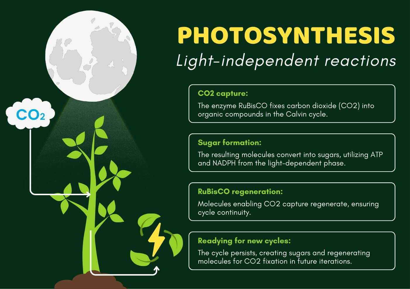
It is the second step in photosynthesis which is independent of light and during which the reducing power (ATP and NADPH + H+) formed during light reaction is utilized for the reduction of CO2 to glucose. It occurs in the stroma region of the chloroplast.
Scientists knew that the first stable compound formed after CO2 fixation is a three-carbon compound called Phosphoglyceric Acid (PGA). So, they were searching for a two-carbon compound that could act as an initial CO2 acceptor. But these two-carbon compounds weren’t found. The solution to these was given by Calvin and his co-workers by using the green algae Chlorella and the technique of autoradiography C14. They found that the initial CO2 acceptor wasn’t a two-carbon compound. Instead, it was a five-carbon compound called Ribulose-1,5-biphosphate (RuBP) which combined with the atmospheric CO2 to form an intermediate 6-carbon compound that immediately breaks down into two molecules of 3-PGA.
There are different pathways of CO2 fixation in different species of plants. These pathways are-
- Calvin cycle
- Hatch and Slack cycle
- CAM pathway
1. Calvin Cycle
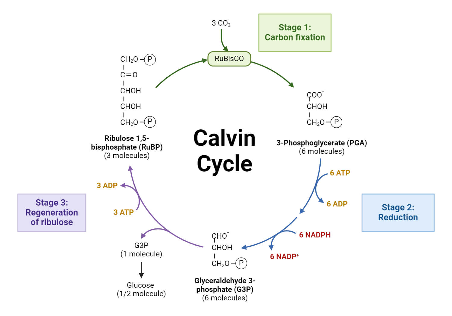
Calvin’s cycle consists of the following three stages: Carboxylation, Glycolytic reversal, and Regeneration of RuBP.
a. Carboxylation
The initial CO2 reversal acceptor is a five-carbon compound called ribulose-1,5-phosphate (RuBP) which combines with atmospheric CO2 in the presence of the enzymes ribulose-1,5-biphosphate carboxylase or oxygenase (RuBisCO) to form an intermediate 6 carbon compound which immediately breaks down to form 2 molecules of a 3-C compound called 3-phosphoglyceric acid (3PGA).
RuBP + CO2 → 3PGA
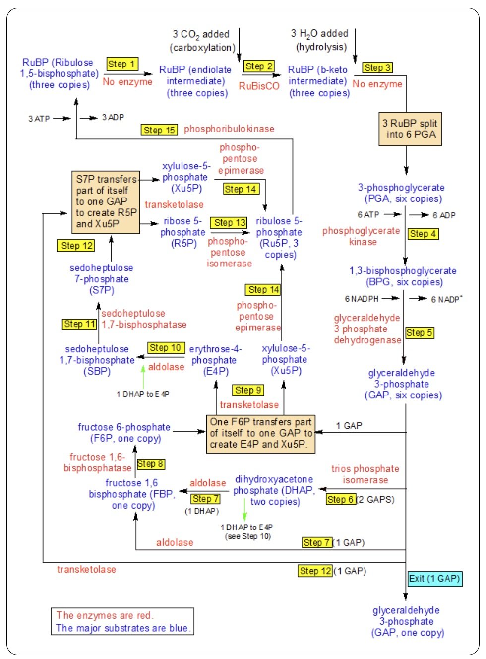
b. Glycolytic Reversal
It consists of the following steps:
- 3PGA combines with ATP in the presence of the enzyme Phosphoglycerokinase to form 1,3-diphosphoglyceric acid.
3PGA + ATP → 1,3-diphosphoglyceric acid - 1,3-diphosphoglyceric acid combines NADPH + H+ in the presence of the enzyme glyceraldehyde 3-phosphate dehydrogenase to form 3-phosphoglyceraldehyde (3-PGAld).
1,3-diphosphoglyceric acid + NADPH + H+ → 3-PGAld + NADP+ + H3PO4 - 3-PGAld isomerizes to form dihydroxyacetone phosphate (DHAP) in the presence of the enzyme Triose phosphate isomerase.
3-PGAld → Dihydroxyacetone phosphate (DHAP) - One molecule of DHAP combines with 1 molecule of 3-PGAld in the presence of the enzyme Aldolase to form Fructose-1,6-diphosphate.
DHAP + 3-PGAld → Fructose-1,6-diphosphate - Fructose-1,6-diphosphate reacts with water molecules to form Fructose-6-phosphate in the presence of the enzyme Phosphatase.
Fructose-1,6-diphosphate + H2O → Fructose-6-phosphate + H3PO4
Some of the fructose-6-phosphate molecules are utilized to produce hexose sugars.
c. Regeneration of RuBP
It consists of the following steps:
- One molecule of 3-PGAld combines with one molecule of Fructose-6-phosphate in the presence of the enzyme Transketone to form 1 molecule each of Erythrose-4-phosphate and Xylulose-5-phosphate.
3-PGAld + Fructose-6-phosphate → Erythrose-4-phosphate + Xylulose-5-phosphate - One molecule of Erythrose-4-phosphate combines with one molecule of DHAP in the presence of the enzyme Aldolase to form Sedoheptulose-1,7-diphosphate.
Erythrose-4-phosphate + DHAP → Sedoheptulose-1,7-diphosphate - Sedoheptulose-7-diphosphate combines with water molecules to form Sedoheptulose-7-phosphate in the presence of the enzyme Phosphatase,
Sedoheptulose-7-phosphate + H2O → Sedoheptulose-7-phosphate + H3PO4 - One molecule of Sedoheptulose-7-phosphate combines with a molecule of 3-PGAld in the presence of the enzyme Transketolase to form one molecule each of Ribose-5-phosphate and Xylulose-5-phosphate.
Sedoheptulose-7-phosphate + 3-PGAld → Ribose-5-phosphate + Xylulose-5-phosphate - Xylulose-5-phosphate is converted into Ribulose-5-phosphate with the help of the enzyme Phosphopentose epimerase.
Xylulose-5-phosphate → Ribulose-5-phosphate - Ribose-5-phosphate isomerizes into the Ribulose-5-phosphate with the help of the enzyme Phosphopentose isomerase.
Ribose-5-phosphate → Ribulose-5-phosphate - Ribulose-5-phosphate combines with ATP in the presence of the enzyme Phosphoribulokinase to form Ribulose-1,5-biphosphate.
Ribulose-5-phosphate + ATP → Ribulose-1,5-biphosphate (RuBP) + ADP
The RuBP so formed restarts the Calvin Cycle again.
2. Hatch and Slack Cycle
For a considerable period, the Calvin cycle as described earlier was 3rd to the only photosynthetic reaction sequence occurring in higher plants and algae. In 1965, Hartt and Burr reported that 4 carbon-containing di-carboxylic acids malate and aspartate were the major labeled product. When sugarcane leaves oil is allowed to photosynthesize for short periods in 14CO2. This finding was confirmed and greatly elaborated by Hatch and Slack who observed every labeling of oxaloacetate, malate, and aspartate only when sugarcane leaves oil exposed CO214 for 1 second. This and further study by these workers led to the establishment of another CO2 reduction pathway because C4 dicarboxylic acid pathway.
Besides sugarcane leaves, this pathway has been found to operate in many plant species of the family Graminea. Examples: maize, sorghum, Amaranthus, etc. These are all known as C4 plants distinguished by the absence of photorespiration and Krantz’s Anatomy of the Leaf. In the leaves of these plants, the vascular bundles are surrounded by a bundle sheath of large parenchymatous cells that in turn are surrounded by mesophyll cells. The chloroplast in cell of the bundle sheath is larger and usually lacks grana, the chloroplast in mesophyll cells is smaller and always contains grana.
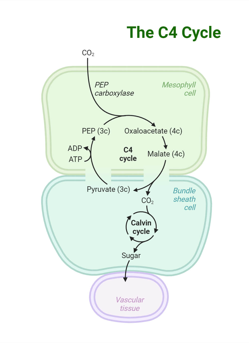
C4 cycle involves two carboxylation reactions; one taking place in the chloroplast of mesophyll cells and the other taking place in the chloroplast of bundle sheath cells.
- The first step involves the carboxylation of phosphoenol pyruvic acid (PEP) in the chloroplast of mesophyll cells to form C4 dicarboxylic acid, oxaloacetic acid. This reaction is a catalyst by phosphoenol pyruvate carboxylase.
- Oxaloacetic acid equilibrates with other C4 dicarboxylic acids, aspartic acid, and malic acid in the presence of the enzyme transaminase.
- From the chloroplast of mesophyll cells, the malic acid is transferred to the chloroplast of bundle sheath cell where it is decarboxylated form CO2 and pyruvate in the presence of enzyme malic.
- Second carboxylation occurs in the chloroplast of bundle sheath cells. Ribulose biphosphate is CO2 produced in step 3 in the presence of Rubisco and ultimately produces 3-PGA as in the case of the Calvin cycle.
- The pyruvic acid produced in step 3 is transferred to the chloroplast of the mesophyll cell where it is phosphorylated to regenerate PEP and this reaction is catalyzed by pyruvate kinase.
3. Crassulacean Acid Metabolism (CAM) pathway
During the night, large amounts of starch are consumed during acidification which indicates that carbohydrates are the source of malic acid synthesis. The distinctive diurnal fluctuation in acidity in plants showing the CAM cycle is predominantly due to the changes in the amount of vacuolar malic acid that is synthesized, utilizing CO2, which then accumulates in the vacuole and may account for 85% of the total acid content.
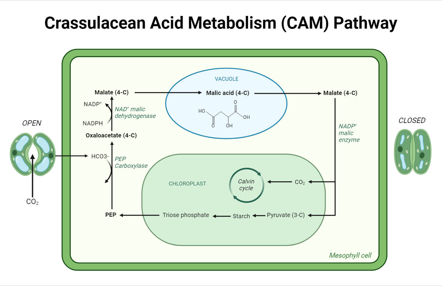
Malic acid is synthesized during the night in reaction. Pyruvate or PEP is carboxylated to produce malic acid either directly or first forming oxaloacetic acid which is then reduced to malate. At day time, during the following day, when acidified organs are exposed to light, rapid consumption of malic acid occurs resulting in deacidification due to decarboxylation of malic acid.
Photosynthesis Infographics
References
- Cooper GM. The Cell: A Molecular Approach. 2nd edition. Sunderland (MA): Sinauer Associates; 2000. Photosynthesis. Available from: https://www.ncbi.nlm.nih.gov/books/NBK9861/
- Stirbet A, Lazár D, Guo Y, Govindjee G. Photosynthesis: basics, history and modelling. Ann Bot. 2020;126(4):511-537. doi:10.1093/aob/mcz171
- Intro to photosynthesis (article) | Khan Academy. (n.d.). Retrieved from https://www.khanacademy.org/science/ap-biology/cellular-energetics/photosynthesis/a/intro-to-photosynthesis
- Lambers, H., & Bassham, J. A. (2024, March 1). Photosynthesis | Definition, Formula, Process, Diagram, reactants, Products, & Facts. Retrieved from https://www.britannica.com/science/photosynthesis
- Photosynthesis. (n.d.). Retrieved from https://education.nationalgeographic.org/resource/photosynthesis/
- https://www.eagri.org/eagri50/PPHY261/lec11.pdf
- https://www.uv.es/symbiosis/pdfs/2011_Calvin-Benson_cycle.pdf
- https://ncert.nic.in/textbook/pdf/kebo111.pdf
- Libretexts. (2022, December 24). 5.12C: the Calvin Cycle. Retrieved from https://bio.libretexts.org/Bookshelves/Microbiology/Microbiology_(Boundless)/05%3A_Microbial_Metabolism/5.12%3A_Biosynthesis/5.12C%3A_The_Calvin_Cycle
- Hatch-Slack Pathway (C4 cycle). (2020, April 27). [Slide show]. Retrieved from https://www.slideshare.net/EmaSushan/hatchslack-pathway-c4-cycle
