
Messenger RNA (mRNA) isolation is a targeted RNA purification approach designed to selectively isolate polyadenylated RNA molecules from a complex pool of total cellular RNA.
In eukaryotic cells, mature mRNA molecules possess a characteristic poly(A) tail at their 3′ end, which distinguishes them from ribosomal RNA (rRNA), transfer RNA (tRNA), and most non-coding RNAs. Modern mRNA extraction methods exploit this structural feature to achieve selective enrichment of mRNA using immobilized oligo(dT) molecules (Macherey-Nagel, 2014; Rio et al., 2010).
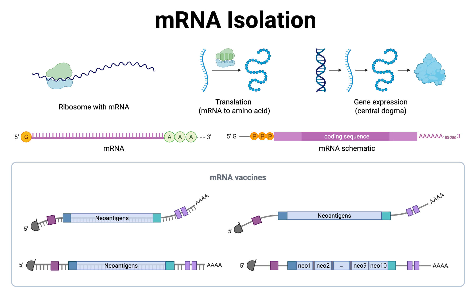
Unlike total RNA extraction, which recovers all RNA species present in a sample, mRNA extraction focuses on enriching biologically relevant transcripts that directly reflect gene expression. This enrichment is particularly important because mRNA typically represents only 1–5% of total RNA in eukaryotic cells (Thermo Fisher Scientific, 2024). Therefore, isolating mRNA improves sensitivity and efficiency in downstream applications such as RNA sequencing (RNA-seq), cDNA library construction, reverse transcription-PCR (RT-PCR), and quantitative gene expression analysis.
Most contemporary protocols employ either magnetic oligo(dT) beads or oligo(dT) immobilized on solid matrices such as silica or cellulose supports. These approaches offer high specificity, scalability, and compatibility with automation (NEB, 2023; Promega, n.d.). Despite differences in commercial implementations, the fundamental principle underlying all mRNA extraction methods remains consistent: hybridization between the poly(A) tail of mRNA and complementary oligo(dT) sequences under optimized ionic conditions.
Key Reagents of mRNA Isolation
The successful isolation of mRNA depends on carefully formulated reagents that promote specific binding, efficient washing, and gentle elution of poly(A)+ RNA. Although reagent compositions may vary slightly between manufacturers, the core components remain largely consistent across protocols (Macherey-Nagel, 2014; Promega, n.d.; NEB, 2023).
| Reagent | Typical Concentration / Amount | Purpose |
| Oligo(dT) magnetic beads or matrix | 20–100 µL slurry | Selective binding of poly(A)+ mRNA |
| Binding buffer (high salt) | Provided ready-to-use | Promotes stable hybridization between poly(A) tails and oligo(dT) |
| Wash buffer | Provided ready-to-use | Removes unbound RNA, DNA, and proteins |
| Elution buffer or RNase-free water | 10–50 µL | Releases mRNA by disrupting hybridization |
| RNase inhibitor | As recommended | Protects RNA from enzymatic degradation |
| Total RNA sample | 1–100 µg | Starting material containing mRNA |
| Magnetic stand (for bead-based methods) | 1 unit | Enables separation of beads from solution |
Principle of mRNA Isolation
The principle of mRNA extraction is based on sequence-specific hybridization between the polyadenylated tails of eukaryotic mRNA and complementary oligo(dT) sequences immobilized on a solid support. When total RNA is incubated with oligo(dT) under high-salt conditions, stable hydrogen bonding occurs between adenine residues in the poly(A) tail and thymine residues of the oligo(dT) probe (Rio et al., 2010).
High ionic strength buffers stabilize this interaction by shielding negative charges on the RNA backbone, thereby enhancing binding efficiency. Non-polyadenylated RNA species lack the complementary sequence and therefore remain unbound. Subsequent washing steps remove these contaminants, resulting in selective retention of mRNA on the solid phase (Pertea, 2019).
Elution is achieved by reducing ionic strength or increasing temperature, which destabilizes the hydrogen bonds between poly(A) tails and oligo(dT). This releases intact mRNA molecules into solution while leaving the capture matrix behind (Macherey-Nagel, 2014). The method is gentle and preserves RNA integrity, making it suitable for sensitive downstream applications.
Protocol and Steps of mRNA (Poly(A)+ RNA) Isolation
Oligo(dT) Latex Bead-Based Isolation of Poly(A)+ RNA (NucleoTrap® Method)
Materials and Reagents
- Total RNA (100-1000 µg)
- Buffer RM0 (binding adjustment buffer)
- Buffer RM1 (RNA solubilization buffer)
- Buffer RM2 (wash buffer 1)
- Buffer RM3 (wash buffer 2)
- Oligo(dT) Latex Beads suspension
- NucleoTrap® Microfilter and collection tubes
- RNase-free water (pre-warmed to 68 °C)
- Microcentrifuge capable of ≥11,000 g
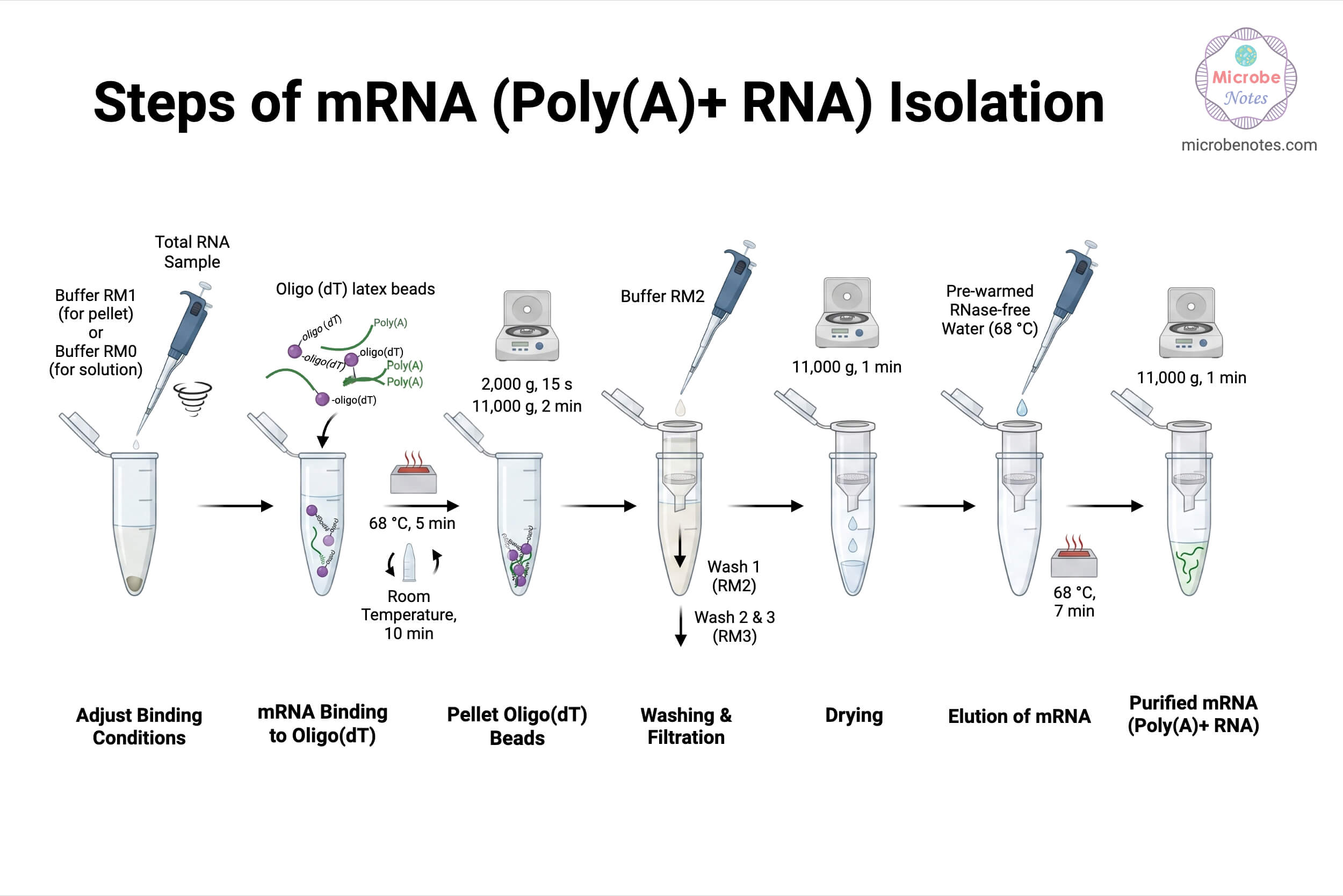
Step 1: Adjustment of Binding Conditions:
For RNA pellets:
-Add 500 µL Buffer RM1 to pellets containing 100–500 µg total RNA
-Add 1000 µL Buffer RM1 to pellets containing up to 1000 µg total RNA
-Resuspend completely by pipetting and vortexing.
For RNA in solution (200–500 µL):
-Add 1 volume of Buffer RM0
-Mix thoroughly to ensure homogeneous binding conditions.
Step 2: Binding of Poly(A)+ RNA to Oligo(dT) Latex Beads:
-Vortex the Oligo(dT) Latex Beads to fully resuspend.
-Add 15 µL bead suspension per 100 µg total RNA.
-Mix thoroughly.
-Incubate at 68 °C for 5 minutes to denature RNA secondary structures.
-Incubate at room temperature for 10 minutes, inverting the tube every 2 minutes.
Centrifugation:
–2,000 g for 15 seconds
–Followed by 11,000 g for 2 minutes
Note: This step pellets the oligo(dT) beads with bound poly(A)+ RNA.
Step 3: Washing of Bound Poly(A)+ RNA:
First Wash (RM2):
-Discard the supernatant.
-Resuspend bead pellet in 600 µL Buffer RM2 until the suspension becomes uniformly “milky.”
-Transfer to a NucleoTrap® Microfilter.
Centrifuge:
–2,000 g for 15 seconds
–11,000 g for 2 minutes
-Discard flow-through and place the Microfilter in a fresh tube.
Note: If more than 500 µg total RNA was used, perform an additional wash using 400 µL Buffer RM2 under identical conditions.
Second and Third Washes (RM3):
Add 500 µL Buffer RM3, resuspend completely, and centrifuge:
–2,000 g for 15 seconds
–11,000 g for 2 minutes
Repeat once more with 500 µL Buffer RM3.
Note: These washes remove ribosomal RNA and residual contaminants.
Step 4: Drying of Oligo(dT) Latex Beads:
-Centrifuge the Microfilter at 11,000 g for 1 minute to remove residual wash buffer.
-Transfer the Microfilter to a clean, RNase-free 1.5 mL tube.
Step 5: Elution of Poly(A)+ RNA:
-Add 20 µL pre-warmed (68 °C) RNase-free water per 10 µL beads.
-Resuspend completely until the solution appears “milky.”
-Incubate at 68 °C for 7 minutes.
-Centrifuge at 11,000 g for 1 minute.
-Collect the eluate containing purified poly(A)+ RNA.
Note: A second elution may be performed to increase yield by 10–20%.
Magnetic Bead-Based mRNA Isolation Using Biotinylated Oligo(dT) (PolyATtract® Method)
Materials and Reagents:
- Total RNA (50 µg – 5 mg)
- Biotinylated oligo(dT) probe
- 20× SSC
- Streptavidin MagneSphere® paramagnetic particles
- 0.5× SSC and 0.1× SSC wash buffers
- RNase-free water
- Magnetic stand
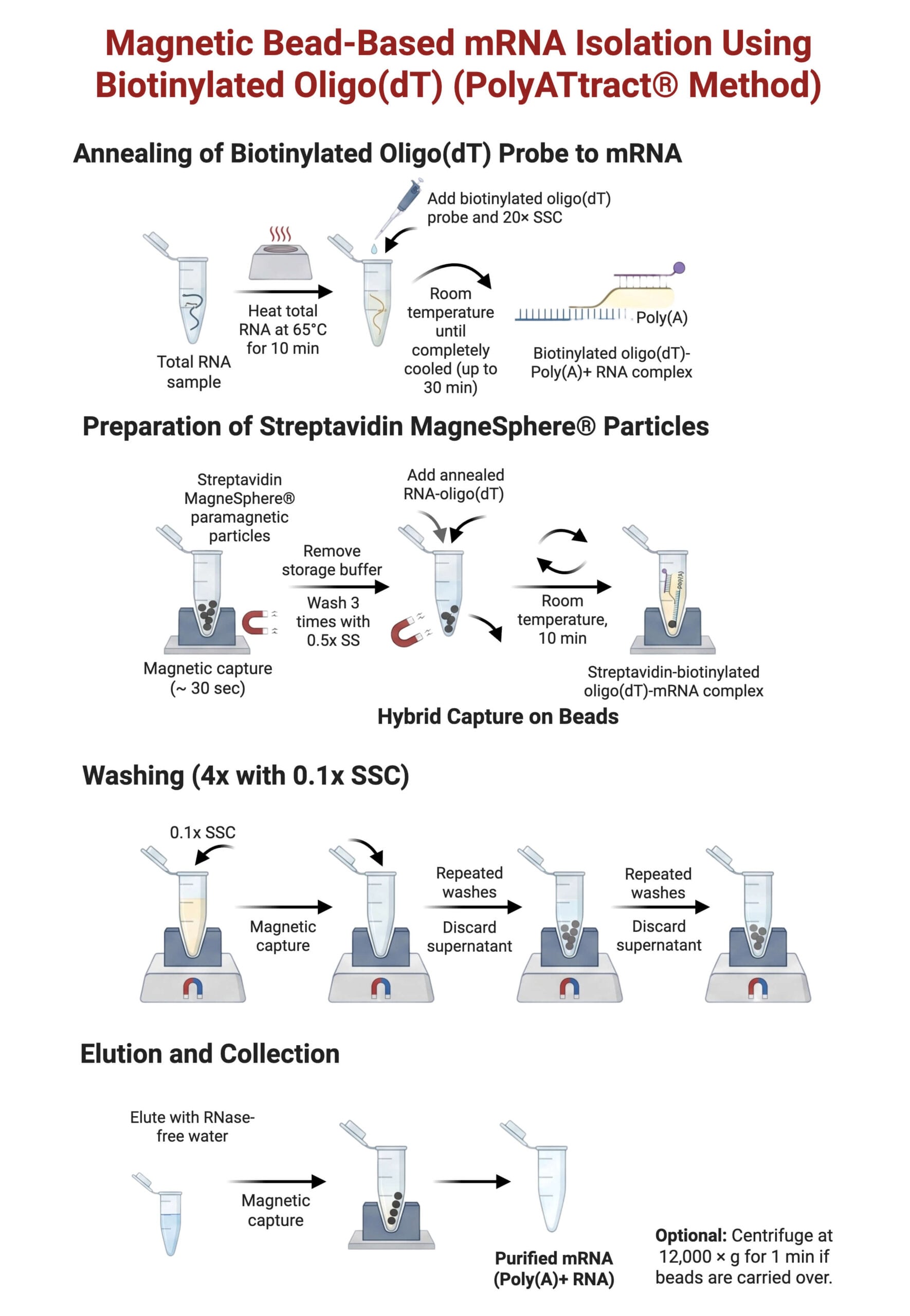
Step 1: Annealing of Biotinylated Oligo(dT) Probe to mRNA:
Large-scale (1-5 mg RNA):
–Adjust RNA volume to 2.43 mL with RNase-free water.
–Heat at 65 °C for 10 minutes.
–Add 10 µL biotinylated oligo(dT) and 60 µL 20× SSC.
–Incubate at room temperature until fully cooled (≤30 minutes).
Small-scale (0.1-1 mg RNA):
–Adjust RNA to 500 µL.
–Heat at 65 °C for 10 minutes.
–Add 3 µL biotinylated oligo(dT) and 13 µL 20× SSC.
–Cool at room temperature for ≤10 minutes.
Step 2: Preparation of Streptavidin Magnetic Beads:
-Resuspend beads by gentle inversion.
-Capture beads on a magnetic stand (~30 seconds).
-Remove storage buffer without centrifugation.
Wash beads three times with 0.5× SSC:
–1.5 mL per wash (large-scale)
–300 µL per wash (small-scale)
Resuspend beads in:
–0.5 mL 0.5× SSC (large-scale)
–100 µL 0.5× SSC (small-scale)
Step 3: Capture of Oligo(dT)–mRNA Hybrids:
-Add annealed RNA-oligo(dT) mixture to prepared beads.
-Incubate at room temperature for 10 minutes, gently inverting every 1–2 minutes.
-Capture beads magnetically and remove supernatant.
Step 4: Washing of Magnetic Beads:
-Wash beads four times with 0.1× SSC:
–1.5 mL per wash (large-scale)
–300 µL per wash (small-scale)
-Fully resuspend beads during each wash to ensure removal of non-specific RNA.
Step 5: Elution of Poly(A)+ RNA:
Large-scale:
–Resuspend beads in 1.0 mL RNase-free water
–Capture beads magnetically and collect eluate
Small-scale:
–Elute first with 100 µL RNase-free water
–Perform a second elution with 150 µL
–Pool eluates (total 250 µL)
If beads are carried over, clarify the eluate by centrifugation at 12,000 g for 1 minute.
Observations and Results of extracted mRNA
Following successful mRNA extraction, the recovered RNA typically exhibits a substantial reduction in rRNA bands when analyzed by gel electrophoresis or electropherograms. The mRNA yield is lower than total RNA but demonstrates significantly higher transcript specificity (Promega, n.d.).
High-quality preparations show intact mRNA suitable for reverse transcription and sequencing. The absence of degradation products indicates effective RNase control throughout the procedure (NEB, 2023).
Modifications of mRNA Isolation
- Double-round purification: Performing two successive binding steps increases mRNA purity by further reducing residual rRNA contamination (Thermo Fisher Scientific, 2024).
- Reduced elution volume: Smaller elution volumes produce more concentrated mRNA for low-input downstream applications (Macherey-Nagel, 2014).
- Temperature-controlled elution: Mild heating improves elution efficiency without compromising RNA integrity (Rio et al., 2010).
Troubleshooting of mRNA Isolation
| Problem | Likely Cause | Solution |
| Low mRNA yield | Incomplete binding | Increase incubation time or ensure correct salt concentration |
| RNA degradation | RNase contamination | Use RNase-free reagents and consumables |
| rRNA contamination | Insufficient washing | Increase wash steps or perform second purification |
| Poor downstream performance | Impure RNA | Repeat purification or improve washing stringency |
Quality Assessment of the Isolated mRNA
- Spectrophotometric analysis: A260/A280 ratios around 2.0 indicate high RNA purity (Thermo Fisher Scientific, 2024).
- Agarose gel electrophoresis: Reduced or absent rRNA peaks confirm successful mRNA enrichment (Promega, n.d.).
- Functional testing: Efficient cDNA synthesis validates mRNA integrity and usability (NEB, 2023).
Safety Tips and Precautions of mRNA Isolation
- Use RNase-free consumables: Prevents RNA degradation during processing.
- Wear gloves at all times: Minimizes RNase contamination from skin.
- Avoid repeated freeze–thaw cycles: Preserves RNA integrity (Macherey-Nagel, 2014).
Storage and Long‑Term Stability of Isolated mRNA
- Short-term storage at −20 °C: Suitable for immediate downstream use, such as reverse transcription or short-term expression analysis, provided the mRNA is stored in RNase-free conditions and used within a limited timeframe to avoid gradual degradation.
- Long-term storage at −80 °C: Maintains RNA stability for extended periods by significantly reducing enzymatic activity and chemical degradation, making it appropriate for archival storage of purified mRNA intended for future sequencing or expression studies.
- Aliquoting samples: Prevents degradation caused by repeated freeze–thaw cycles by minimizing physical stress and RNase exposure, thereby preserving mRNA integrity, concentration consistency, and performance reliability in repeated downstream applications (Promega, n.d.).
Applications of mRNA Isolation
- RNA sequencing (RNA-seq): Enables comprehensive transcriptome profiling by enriching poly(A)+ RNA, thereby reducing ribosomal RNA background and improving sequencing depth, read alignment efficiency, and accurate quantification of gene expression levels.
- cDNA library construction: Provides high-quality, mRNA-derived templates for reverse transcription, ensuring that constructed cDNA libraries predominantly represent protein-coding transcripts and improving the reliability of downstream sequencing and cloning experiments.
- Gene expression analysis: Improves sensitivity and specificity in RT-PCR and qPCR assays by minimizing competing RNA species, allowing more accurate detection and quantification of low-abundance transcripts across different biological conditions.
- Functional genomics studies: Facilitates the analysis of actively expressed genes by selectively isolating mature mRNA, enabling investigations into transcriptional regulation, differential gene expression, and cellular responses to physiological or experimental stimuli (Thermo Fisher Scientific, 2024).
Advantages of mRNA Isolation
- High specificity for mRNA: Selectively enriches poly(A)+ transcripts by exploiting the unique polyadenylated tail of eukaryotic mRNA, thereby efficiently separating coding RNA from abundant ribosomal and transfer RNA species.
- Improved downstream sensitivity: Reduces interference from highly abundant rRNA, enhancing signal-to-noise ratios in downstream molecular analyses and enabling reliable detection of low-expression genes in complex biological samples.
- Compatibility with automation: Magnetic bead-based formats support high-throughput and automated workflows, making the method suitable for large-scale studies, standardized processing, and reproducible results in research and diagnostic laboratories.
- Preservation of RNA integrity: The gentle hybridization-based approach minimizes mechanical and chemical stress on RNA molecules, preserving transcript integrity and suitability for sensitive applications such as sequencing and reverse transcription (Rio et al., 2010).
Limitations of mRNA Isolation
- Inapplicability to prokaryotic RNA: Most bacterial mRNA lacks stable poly(A) tails, making oligo(dT)-based enrichment ineffective and limiting the applicability of this method to eukaryotic samples only.
- Loss of non-polyadenylated transcripts: Certain regulatory and non-coding RNAs that lack poly(A) tails are excluded during purification, potentially resulting in incomplete representation of the cellular transcriptome.
- Higher cost than total RNA extraction: The requirement for specialized reagents such as oligo(dT) probes, magnetic beads, and proprietary buffers increases overall cost compared to conventional total RNA extraction methods (Thermo Fisher Scientific, 2024).
Conclusion
The method of mRNA extraction based on poly(A) selection represents a cornerstone technique in modern molecular biology. By exploiting the unique structural feature of eukaryotic mRNA, this approach enables selective enrichment of biologically meaningful transcripts while minimizing background RNA species. The protocols described across Macherey-Nagel, NEB, Promega, and Cold Spring Harbor resources demonstrate that oligo(dT)-based purification is robust, reproducible, and adaptable to diverse experimental needs.
Despite certain limitations, particularly regarding non-polyadenylated RNAs, mRNA extraction remains indispensable for transcriptomic and gene expression studies. When performed with proper precautions and quality control, it yields high-integrity mRNA suitable for a wide range of downstream applications in research and biotechnology.
References
- Macherey-Nagel. (2014). NucleoTrap Poly(A) mRNA purification user manual. https://www.takarabio.com/documents/User%20Manual/NucleoTrap%20Poly(A)%20mRNA%20Purification%20User%20Manual%20(PT4020/NucleoTrap%20Poly(A)%20mRNA%20Purification%20User%20Manual%20(PT4020-1)_Rev_05.pdf
- New England Biolabs (NEB). (2023). Protocol for NEBNext Poly(A) mRNA magnetic isolation module (E7490). https://www.neb.com/en/products/e7490-nebnext-polya-mrna-magnetic-isolation-module
- Pertea, M. (2019). Isolation of poly(A)+ messenger RNA using magnetic oligo(dT) beads. Cold Spring Harbor Protocols, 2019(10). https://doi.org/10.1101/pdb.prot097410
- Promega Corporation. (n.d.). PolyATtract® mRNA isolation systems protocol. https://www.promega.com/-/media/files/resources/protocols/technical-manuals/0/polyattract-mrna-isolation-systems-protocol.pdf
- Rio, D. C., Ares, M., Jr., Hannon, G. J., & Nilsen, T. W. (2010). Enrichment of poly(A)+ mRNA using immobilized oligo(dT). Cold Spring Harbor Protocols, 2010(9), pdb.prot5435. https://doi.org/10.1101/pdb.prot5435
- Thermo Fisher Scientific. (2024). mRNA extraction and enrichment. https://www.thermofisher.com/us/en/home/life-science/dna-rna-purification-analysis/rna-extraction/rna-types/mrna-extraction.html
- Hamid, S. (n.d.). mRNA isolation [PowerPoint slides]. SlideShare. https://www.slideshare.net/slideshow/mrna-isolation/251126951
