
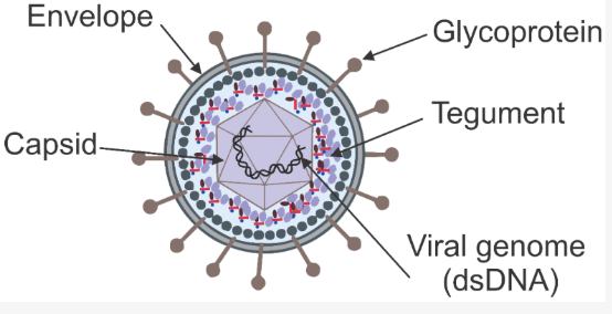
Human herpesviruses (HHV) are enveloped, large double-stranded DNA viruses capable of lifelong persistence and latency in their hosts. These viruses have a common virion structure but vary in terms of cell specificity, genomic arrangement, and pathogenesis.
| Component | Key Features in HHVs |
| Envelope | Lipid bilayer, >10 glycoproteins, polymorphic |
| Tegument | Amorphous protein layer, 9–20+ proteins, regulatory and structural roles |
| Capsid | T=16 icosahedral, ~125–130 nm, 150 hexons + 11 pentons + portal vertex |
| Genome | Linear dsDNA, ~125–241 kbp, 70–170 genes, complex UL/US + repeats |
What are Human Herpesviruses?
Nature of Human Herpesviruses
- Large, spherical/enveloped virions with linear dsDNA genomes.
- Virion: icosahedral nucleocapsid, proteinaceous tegument, and lipid envelope containing multiple glycoproteins.
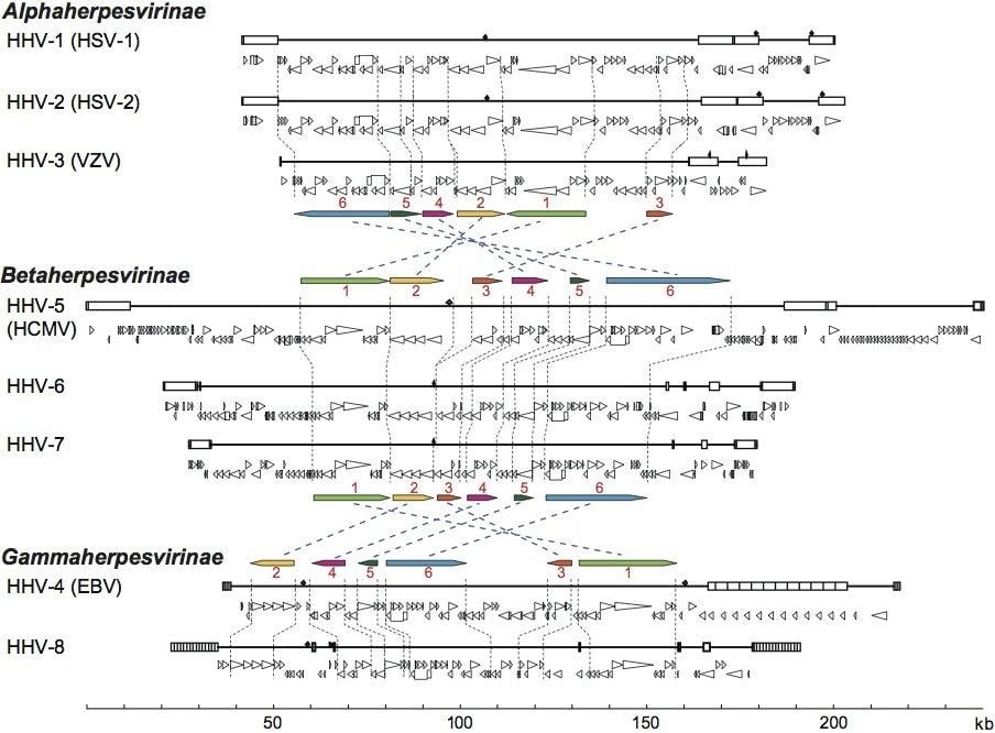
- Genome sizes ~125–241 kbp; 70–170 ORFs, with a core set (~43 genes) conserved from an ancestral herpesvirus.
Biological hallmarks
- Establish lifelong latency after primary infection, with periodic reactivation.
- Highly adapted to their hosts; severe disease mainly in fetuses, neonates, immunocompromised, or aberrant host species.
Human herpesvirus complement
- Nine human herpesviruses described in recent systematics:
Taxonomy and Classification
Higher Taxa
Order and families
- Order Herpesvirales, three families: Orthoherpesviridae (mammals, birds), Alloherpesviridae (fish, amphibians), Malacoherpesviridae (molluscs).
- All human herpesviruses lie within the family Herpesviridae.
Subfamilies Within Herpesviridae
Alphaherpesvirinae
- Features: short replication cycle, efficient cell lysis, latency in sensory ganglia.
- Human members: HHV-1 (HSV-1), HHV-2 (HSV-2), HHV-3 (VZV).
- Genomic: often high G+C content; characteristic UL/US architectures, but with subfamily-specific gene complements.
Betaherpesvirinae
- Features: long replicative cycle, cytomegaly, latency in mononuclear cells (e.g., myeloid lineage).
- Human members: HHV-5 (HCMV), HHV-6A, HHV-6B, HHV-7.
- Genomes: large, complex, often with extensive repeats and immune-modulatory genes.
Gammaherpesvirinae
- Features: latency in lymphocytes (B or T cells), frequent association with malignancy (EBV, KSHV).
- Human members: HHV-4 (EBV) and HHV-8 (KSHV).
- Genomes: encode multiple latency and transformation-associated proteins and non-coding RNAs.
Taxonomic Criteria
Modern classification emphasizes:
- Conserved genes and gene clusters (DNA polymerase, major capsid protein, glycoprotein B, ssDNA-binding protein).
- Genome organization: arrangement of UL/US and repeat regions, composition/placement of packaging termini.
- Host range and cell tropism, latency sites, and disease patterns.
Structure and Morphology of Human Herpesviruses
Overall Virion Architecture
Size and shape
- Spherical to pleomorphic, typically 150–200 nm in diameter; envelope spikes extend the overall diameter to ~225 nm in HSV-1.
Capsid
- T=16 icosahedral capsid (~125–130 nm) with:
- 150 hexons and 11 pentons composed of major capsid protein (e.g., VP5 in HSV-1).
- One vertex replaced by a portal complex (dodecamer of UL6 in HSV-1) for DNA entry/exit.
Tegument
- Partially ordered protein layer between the capsid and envelope.
- Contains 9–20+ viral proteins, plus recruited host proteins; total up to ~71 structural proteins per virion in some mammalian herpesviruses.
- Subsets remain capsid-associated to mediate nuclear targeting; others are released to modulate early infection and host responses.
Envelope and glycoproteins
- Lipid bilayer derived from host membranes, populated by >10 viral glycoproteins essential for attachment, fusion, and egress.
- Gammaherpesviruses (EBV, KSHV) show less dense glycoprotein arrays than HSV-1 or HCMV and distinctive arrangements of fusion protein gB trimers.
Subfamily- and Species-Specific Structural Variation
Capsid cores
- All Human Herpesviruses share a conserved capsid scaffold, but cryo-EM reveals subfamily-specific differences in triplex structure, vertex organization, and capsid protein domain insertions.
- Capsids function as pressurized DNA containers, and structural variations relate to differences in genome size and packaging pressures.
Tegument organization
- EBV and KSHV: eccentric capsid positioning and polarized tegument distribution, with tegument “spanning” from capsid to envelope and likely scaffolding virion assembly.
- HSV-1: well-defined capsid-associated tegument complex (CATC; pUL17, pUL25, pUL36) at each penton-adjacent triplex, essential for axonal transport and nuclear egress.
Genome Organization and Proteins of Human Herpesviruses
Global Genome Features
Genome size and complexity
- Linear dsDNA, ~125–241 kbp, among the largest of human viruses.
- Encode 70–170 proteins, plus extensive non-coding RNAs (miRNAs, long noncoding RNAs, EBV EBERs).
- ~43 genes conserved across Herpesviridae, comprising a core gene set for replication, capsid assembly, DNA packaging, and virion egress.
Segmented unique and repeat regions
- Canonical architecture: unique long (UL) and unique short (US) segments flanked by terminal and internal inverted repeats (TR/IR), generating genome isomers via inversion.
- HCMV is classified as an E-class genome (UL and US, each bounded by inverted repeats).
- Such isomerization may impact gene regulation and life-cycle transitions.
Functional Classes of Genes
Core replication and structural genes
- DNA replication: DNA polymerase, helicase-primase complex, single-stranded DNA-binding protein, origin-binding proteins.
- Capsid proteins: major capsid protein (e.g., VP5), triplex proteins (VP19C, VP23 in HSV-1), small capsid proteins (VP26), portal protein (UL6).
- DNA packaging apparatus: products of UL6, UL15, UL17, UL25, UL28, UL32, UL33 in HSV-1; homologs in other HHVs.
- Envelope glycoproteins: gB, gH/gL, gC, gD (alphaherpesviruses), and homologous entry complexes in beta- and gamma herpesviruses.
Subfamily-specific and accessory genes
- Betaherpesviruses (HCMV, HHV-6/7): expand gene families for immune evasion, chemokine/chemokine receptor homologs, and modulation of antigen presentation.
- Gammaherpesviruses (EBV, KSHV): encode oncogenic latency proteins (EBNA, LMP1/2; KSHV LANA and others) and multiple miRNAs, supporting transformation and immune escape.
Representative Examples
HSV-1 capsid and packaging proteins
Capsid
- VP5: major capsid protein forming hexons/pentons.
- VP19C + VP23: triplex heterotrimers linking capsomers.
- VP26: small capsid protein atop hexons (~900 copies per capsid).
- UL6: dodecameric portal replaces one penton vertex.
- Capsid-associated tegument complex (CATC): pUL17, pUL25, pUL36 assemble as heterotetrameric complexes at vertices, coupling DNA-filled capsids to transport machinery.
Packaging
- UL6, UL15, UL17, UL25, UL28, UL32, UL33 coordinate cleavage of concatemers and genome encapsidation.
HCMV genome organization
- E-class UL/US–TR/IR arrangement; >70 virion proteins identified, ~50 essential for replication.
- Large fraction of ORFs involved in host immunomodulation and manipulation of cellular signaling, plus multiple polyadenylated RNAs and ≥26 miRNAs.
EBV genome and tegument
- 100 proteins encoded; ~30 well characterized, including latent proteins (EBNAs, LMP1/2), lytic-cycle transactivators, and structural proteins.
- Tegument proteins include highly conserved structural regulators and gammaherpesvirus-specific factors with roles in oncomodulation, immune evasion, and reprogramming of infected cells.
Replication Cycle of Human Herpesviruses
Human herpesviruses have lytic and latent/persistent phases.
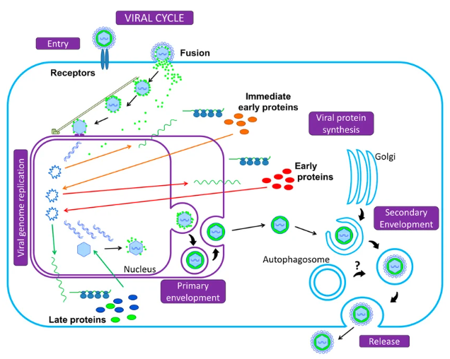
- Lytic cycle: Following uptake by the cell, the linear DNA is transported to the nucleus. The expression of viral genes hierarchically takes place – immediate early genes (IE) (transcription factors), early genes (E) (replication), and late genes (L) (structural proteins). DNA replication occurs by rolling circle replication to produce large concatemers, which are subsequently cleaved and packaged within capsids.
- Capsids assemble in the nucleus, acquire tegument and envelope, and exit the cell to infect new cells.
- Latency/persistence: Replication of the virus is inhibited, or is done so on a “low” level. Gene expression is limited to a specific set of genes (LATs), and no virions are produced. Subfamilies have varying preferences for either neurons (herpes simplex viruses, varicella zoster viruses) or leukocytes/lymphocytes (CMV, EBV, HHV 6/7/8).
| Phase | Main Events | Typical Sites |
| Lytic | IE–E–L gene cascade, DNA replication, virion production | Epithelia, many tissues |
| Latent/persistent | Minimal genes, no or low replication | Neurons, leukocytes |
Pathogenesis and Host Immune Response of Human Herpesviruses
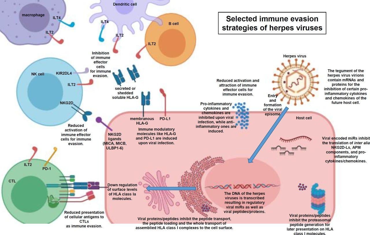
Human herpesviruses cause disease by direct cell damage and by dysregulated immune responses. Symptoms can be virus-driven (e.g., lesions that aid transmission) or caused by collateral immune damage.
Immune control and evasion
- Innate and adaptive immunity normally restrain replication and reactivation.
- Human herpesviruses encode numerous proteins that block antigen presentation (interfering with MHC I/II pathways), modulate interferon signaling, and alter cytokine responses, enabling latency and chronic infection.
- Betaherpesviruses (CMV, HHV-6/7) have especially elaborate immune evasion systems, hampering T-cell recognition and delaying effective adaptive responses.
Disease mechanisms
- HSV: lytic infection of mucosal and corneal tissue, with latency in neuronal cells; pathogenesis associated with stromal keratitis, encephalitis, and genital infection, with host-mediated inflammation playing an important role in pathology.
- EBV/KSHV: latency of infection in B and other leukocyte cells; expression of oncogenic latent proteins, such as LMP1, LANA, and vIL6, leads to increased cell survival and growth, resulting in lymphomas, Kaposi’s sarcoma, and other cancers.
- HHV 6/7: common activation in immunocompromised individuals, leading to encephalitis, bone marrow, and pulmonary disorders, and other systemic inflammatory conditions.
Epidemiology and Transmission of Human Herpesviruses
Human herpesviruses are ubiquitous, with >90% of adults infected with at least one type.
Transmission routes
- Saliva and mucosal secretions: major route for EBV, CMV, HHV-6/7, KSHV, and also for HSV-1.
- Sexual contact: important for HSV-2, some HSV-1, and KSHV.
- Vertical (mother-to-child): CMV and HHV-6/7 can cross the placenta or be transmitted perinatally, causing congenital or early-life infection.
- Blood products/organ transplantation: CMV, EBV, HHV-6/7, and KSHV can be transmitted via transfusion or grafts and then reactivate under immunosuppression.
Primary infection with HHV 6/7 and usually EBV generally takes place during early childhood, with primary infection with HHV 6B occurring at around 6-12 months of age, usually presenting as roseola. Seroprevalence of CMV may be up to 60-100% in developing nations.
Reactivation is frequent and generally asymptomatic; however, in certain clinical conditions like chemotherapy and HIV, reactivation leads to considerable morbidity.
Clinical Manifestations of Human Herpesviruses
Clinical outcomes range from benign self-limited illness to severe organ disease and cancer. Many infections or reactivations are silent.
Typical primary infections
- HHV-6B: roseola infantum (sudden high fever followed by rash) and undifferentiated febrile illnesses in infants; infectious mononucleosis–like illness can also occur.
- EBV: infectious mononucleosis with fever, lymphadenopathy, and fatigue; also chronic active infection.
- HSV-1/2, VZV: mucocutaneous lesions, cold sores, genital herpes, chickenpox.
Reactivation and opportunistic disease
- HSV: recurrent orolabial/genital lesions, stromal keratitis, encephalitis.
- HHV-6/7: encephalitis, pneumonitis, graft rejection, severe rash, and systemic illness in transplant and other immunocompromised patients; many reactivations remain asymptomatic.
- EBV/KSHV: multiple lymphomas, nasopharyngeal carcinoma, Kaposi’s sarcoma, multicentric Castleman disease, and primary effusion lymphoma.
- Co-infection (e.g., CMV + HHV-6) in leukemia patients on chemotherapy is associated with severe lymphopenia, rash, pneumonia, bacteremia, and invasive fungal infections, suggesting synergistic pathogenic effects.
Detection of viral DNA in neural ganglia post-mortem (notably HSV-1, VZV, EBV, CMV, HHV-6) confirms widespread lifelong latency in sensory and cranial ganglia.
Laboratory Diagnosis of Human Herpesviruses
Targets and specimen types
- Direct detection of viral DNA (or occasionally antigen) from blood, CSF, tissue, or other fluids is central for CMV, HHV-6/7, EBV, KSHV, and for latent-site studies.
- Serology (IgG/IgM) is useful to determine past exposure and, in selected settings, primary infection, but is limited for ubiquitous viruses with early childhood infection (HHV-6/7, CMV, EBV).
Real-time PCR
- qPCR for CMV, HHV-6, HHV-7, and EBV is the common method employed for diagnosing, monitoring viral load, and pre-emptive treatment.
- Multiplex PCR allows simultaneous detection of several HHVs in one sample (e.g., multiplex PCR for HSV-1/2, VZV, EBV, CMV, HHV-6 in ganglia or plasma).
- Qualitative PCR is used to define DNAemia, recurrent vs new episodes, and co-infection patterns, but lacks quantitative thresholds for disease in many settings.
- Antigen detection (e.g., pp65 for CMV) is now largely supplanted by DNA quantification in many centers but can still complement PCR in transplant medicine.
Serology
- High IgG seroprevalence (>80–90%) for many HHVs limits specificity for acute disease; IgM and avidity assays can help for primary CMV/EBV in selected populations.
Diagnostic Challenges and Pitfalls
Latency and asymptomatic shedding
- HHVs often reactivate without symptoms, with DNA detected in mucosa, saliva, or blood, complicating causal attribution.
- EBV and CMV can be shed asymptomatically in the oral mucosa; detection alone does not prove disease.
Tissue vs plasma detection
- End-organ disease (e.g., CMV pneumonia, HHV-6 encephalitis) may require PCR from local samples (BAL, CSF, tissue biopsy) in addition to plasma.
Chromosomally integrated HHV-6 (ciHHV-6)
- Approximately 1% of the population harbors HHV-6 DNA integrated into telomeres, and such individuals possess extremely high HHV-6 DNA concentrations in blood and tissues regardless of viral replication.
- Distinguishing ciHHV-6 from active infection requires high-level viral loads, testing hair/nail DNA, and clinical correlation.
Co-infections and viral interactions
- In leukemia patients on chemotherapy, at least one HHV was detectable in 63% of patients; CMV was most frequent, followed by HHV-6, HHV-7, and EBV.
- Co-infection with CMV and HHV-6 was common and associated with severe lymphopenia, rash, pneumonia, and opportunistic infections, implying synergistic pathogenicity.
- These findings argue for systematic multi-HHV PCR panels in high-risk immunocompromised hosts.
Selected Virus-Specific Diagnosis
HHV-6/HHV-7
- Diagnosis: mainly qPCR on blood and CSF; serology and antigen detection have substantial limitations.
- HHV-6: wide tissue tropism; primary infection (exanthem subitum), later reactivation in HSCT with encephalitis.
- HHV-7: diagnosis and disease associations are less well defined; evidence base dominated by case reports, with few systematic pathogenesis studies.
CMV
- qPCR of plasma is central for diagnosis, viral load monitoring, and pre-emptive therapy in transplant patients.
- Serology (IgG) is used for risk stratification (donor/recipient serostatus) before transplantation.
EBV and KSHV
- EBV: combination of serology and qPCR (blood, tissues) for infectious mononucleosis, chronic active infection, and EBV-associated malignancies.
- KSHV: PCR, serology, and immunohistochemistry for tumor tissues; underdiagnosed, particularly in HIV-endemic areas.
What treatments and antiviral therapies are available for human herpesviruses?
General Therapeutic Principles
No curative therapy: all current antivirals are virostatic, targeting lytic replication (viral DNA polymerase) but not eliminating latent episomes or integrated genomes.
Indications focus on:
Severe primary infection (e.g., neonatal HSV, disseminated VZV).
- Reactivation or primary infection in immunocompromised individuals (transplant recipients, advanced HIV, leukemia).
- HHV-associated malignancies (EBV, KSHV), where treatment targets both the lytic virus and transformed cells.
Available Antivirals and Activity Spectrum
Nucleoside analog DNA polymerase inhibitors
- Ganciclovir/valganciclovir
- Active against beta-herpesviruses (CMV, HHV-6/7) and some gamma-herpesviruses.
- First-line for tissue-invasive CMV disease and for many HHV-6 diseases in high-risk hosts.
- Acyclovir/valacyclovir
- Strong activity for HSV-1/2 and VZV; much weaker against CMV, HHV-6/7.
- Example: successful treatment of severe periodontitis with high EBV levels using valacyclovir, with viral DNA clearance and clinical improvement.
- Cidofovir, foscarnet
- Broad-spectrum DNA polymerase inhibitors, used for CMV and HHV-6 in cases of ganciclovir resistance or intolerance; significant toxicity (renal) limits use.
Specific notes for HHV-6/7
- Ganciclovir, valganciclovir, foscarnet, and cidofovir display in vitro and clinical efficacy against HHV-6 and have been used for HHV-6-induced encephalitis in HSCT patients; no indication or dose threshold exists officially yet.
- HHV 7: Few data exist on this virus and its treatment, with no indication of efficacy for antiviral drugs for CMV infections.
KSHV and EBV-associated disease
- Kaposi’s sarcoma/Primary effusion lymphomas: The treatment is a combination of chemotherapy, immune reconstitution (through cART in patients suffering from HIV), and sometimes antiviral drugs like ganciclovir and foscarnet, which primarily act on lytic virus replication.
- EBV: In lymphomas caused by EBV, the treatment includes chemotherapy, rituximab, and immunomodulation therapies since the viral effect cannot be controlled by antivirals.
Antiviral Strategies in Transplant Medicine
CMV: prophylaxis vs pre-emptive therapy
- For a clinical trial done in liver transplant patients, whose donors were positive but patients seronegative for CMV, universal valganciclovir prophylaxis was found to have equal efficacy with pre-emptive therapy depending on viral load detection.
HHV-6 in HSCT
- No consensus thresholds; centers use variable qPCR cut-offs and clinical criteria to initiate ganciclovir or foscarnet.
- Guidelines emphasize individualized risk assessment (graft type, GVHD, immunosuppression intensity).
| Claim | Reasoning |
| qPCR is the primary diagnostic for active HHV-6/7 and CMV | Multiple reviews emphasize DNA quantification in blood/tissue as the mainstay, superior to serology/antigen tests |
| Ganciclovir/valganciclovir, foscarnet, and cidofovir are active against HHV-6 | Consistent in vitro data and clinical experience, but indications and dosing criteria are not standardized. |
| HHV co-infections (CMV + HHV-6) worsen outcomes in leukemia patients | One prospective cohort shows a strong association with lymphopenia, pneumonia, and opportunistic infections |
How to Prevent and Control Human Herpesviruses?
General Measures
Basic infection control
- Transmission routes include saliva, genital secretions, blood products, organ transplantation, and vertical transmission for several HHVs.
- Standard precautions (hand hygiene, safe sex practices, blood screening, careful donor selection) reduce spread, particularly for CMV and KSHV.
Transplant setting
- Serostatus-based risk stratification (CMV D+/R− highest risk) guides antiviral prophylaxis or pre-emptive monitoring strategies.
- Regular qPCR monitoring for CMV and often HHV-6/7 enables early intervention before end-organ disease develops.
Vaccines and Immune-Based Prevention
EBV and CMV
- Both have strong rationale for vaccination due to oncogenic or congenital disease burden, yet no licensed vaccines exist; multiple candidates are under evaluation.
HSV and VZV
- Vaccines against VZV include live attenuated vaccines and subunit vaccines; no abstract is available on these. Vaccines against HSV infections are now at the phase 3 testing stage, but are not commercially available.
Betaherpesviruses (HCMV, HHV-6/7)
- Vaccine development is hampered by extensive immune evasion and complex antigenic repertoires.
- Recent advances in vaccine technology, including novel platforms and adjuvants, are being explored, particularly for CMV.
Control of KSHV and HHV-Associated Malignancies
KSHV
- Public health measures: HIV prevention and treatment (cART) to reduce KSHV-driven Kaposi’s sarcoma.
- Research directions: vaccines targeting KSHV structural or latency proteins, monoclonal antibodies against entry receptors, and immune checkpoint inhibitors (PD-1/PD-L1) to target latent infection.
Conclusion
Laboratory testing for human herpesviruses mainly focuses on nucleic acid testing, particularly real-time PCR, along with proper interpretation in the light of latency, ciHHV 6, co-infections, and tissue compartmentalization.
The antivirals mainly used for human herpesviruses include nucleoside and pyrophosphate analogues that are quite efficient in inhibiting lytic replication of CMV, HHV 6/7, HSV, and VZV, but fail to eliminate latency or recurrent episodes of the diseases, although no established guidelines for treating these viruses exist for many betaherpesviruses.
Preventing the disease depends on infection-control procedures and risk-adapted antiviral treatment in vulnerable patients. Developing a suitable vaccine for CMV, EBV, HSV, and KSHV is an important area, but it has been unsuccessful so far.
References
- Grinde, B. (2013). Herpesviruses: latency and reactivation – viral strategies and host response. Journal of Oral Microbiology, 5, 22766. https://doi.org/10.3402/jom.v5i0.22766
- Lan, K., & Luo, M. H. (2017). Herpesviruses: epidemiology, pathogenesis, and interventions. Virologica Sinica, 32(5), 347–349. https://doi.org/10.1007/s12250-017-4069-2
- Gatherer, D., Depledge, D. P., Hartley, C. A., Szpara, M. L., Vaz, P. K., Benkő, M., Brandt, C. R., Bryant, N. A., Dastjerdi, A., Doszpoly, A., Gompels, U. A., Inoue, N., Jarosinski, K. W., Kaul, R., Lacoste, V., Norberg, P., Origgi, F. C., Orton, R. J., Pellett, P. E., … Davison, A. J. (2021). ICTV Virus Taxonomy Profile: Herpesviridae 2021. Journal of General Virology, 102(10), 001673. https://doi.org/10.1099/jgv.0.001673
- Strausbaugh, L. J., Caserta, M. T., Mock, D. J., & Dewhurst, S. (2020). Human herpesvirus 6. Clinical Infectious Diseases, 71(8), 2042–2049. https://doi.org/10.1093/cid/ciz1182
- Dotto-Maurel, A., Arzul, I., Morga, B., & Chevignon, G. (2025). Herpesviruses: overview of systematics, genomic complexity and life cycle. Virology Journal, 22, 123. https://doi.org/10.1186/s12985-025-02203-4
- Agut, H., Bonnafous, P., & Gautheret-Dejean, A. (2016). Human herpesviruses 6A, 6B, and 7. Microbiology Spectrum, 4(3), DMIH2-0007-2015. https://doi.org/10.1128/microbiolspec.DMIH2-0007-2015
- Agut, H., Bonnafous, P., & Gautheret-Dejean, A. (2015). Laboratory and clinical aspects of human herpesvirus 6 infections. Clinical Microbiology Reviews, 28(2), 313–335. https://doi.org/10.1128/CMR.00122-14
- Lan, K., & Luo, M. H. (2017). Herpesviruses: epidemiology, pathogenesis, and interventions. Virologica Sinica, 32(5), 347–349. https://doi.org/10.1007/s12250-017-4069-2
- Grinde, B. (2013). Herpesviruses: latency and reactivation – viral strategies and host response. Journal of Oral Microbiology, 5, 22766. https://doi.org/10.3402/jom.v5i0.22766
- Sausen, D. G., Reed, K. M., Bhutta, M. S., Gallo, E. S., & Borenstein, R. (2021). Evasion of the host immune response by betaherpesviruses. International Journal of Molecular Sciences, 22(14), 7447. https://doi.org/10.3390/ijms22147447
- Ptaszyńska-Sarosiek, I., Dunaj, J., Zajkowska, A., Niemcunowicz-Janica, A., Król, M. E., Pancewicz, S. A., & Zajkowska, J. (2019). Post-mortem detection of six human herpesviruses (HSV-1, HSV-2, VZV, EBV, CMV, HHV-6) in trigeminal and facial nerve ganglia by PCR. PeerJ, 7, e6351. https://doi.org/10.7717/peerj.6351
- Jassim, M., Mahmood, M., & Hussein, M. (2021). Human herpetic viruses and immune profiles. In Innate immunity in health and disease (pp. 287–323). Springer. https://doi.org/10.1007/978-3-030-83697-8_14
- Handous, I., Achour, B., Marzouk, M., Rouis, S., Hazgui, O., Brini, I., Khelif, A., Hannachi, N., & Boukadida, J. (2020). Co-infections of human herpesviruses (CMV, HHV-6, HHV-7, and EBV) in non-transplant acute leukemia patients undergoing chemotherapy. Virology Journal, 17, 42. https://doi.org/10.1186/s12985-020-01315-9
- Zhu, S., & Viejo-Borbolla, A. (2021). Pathogenesis and virulence of herpes simplex virus. Virulence, 12(1), 2670–2702. https://doi.org/10.1080/21505594.2021.1962460
- Heming, J. D., Conway, J. F., & Homa, F. L. (2017). Herpesvirus capsid assembly and DNA packaging. Advances in Anatomy, Embryology and Cell Biology, 223, 119–142. https://doi.org/10.1007/978-3-319-53168-7_6
- Verzosa, A. L., McGeever, L. A., Bhark, S., Delgado, T., Salazar, N., & Sanchez, E. L. (2021). Herpes simplex virus 1 infection of neuronal and non-neuronal cells elicits specific innate immune responses and immune evasion mechanisms. Frontiers in Immunology, 12, 644664. https://doi.org/10.3389/fimmu.2021.644664
- Bai, L., Xu, J., Zeng, L., Zhang, L., & Zhou, F. (2024). A review of HSV pathogenesis, vaccine development, and advanced applications. Molecular Biomedicine, 5, 31. https://doi.org/10.1186/s43556-024-00168-7
- Dotto-Maurel, A., Arzul, I., Morga, B., & Chevignon, G. (2025). Herpesviruses: overview of systematics, genomic complexity and life cycle. Virology Journal, 22, 123. https://doi.org/10.1186/s12985-025-02203-4
- Verbeek, R., Vandekerckhove, L., & Van Cleemput, J. (2024). Update on human herpesvirus 7 pathogenesis and clinical aspects as a roadmap for future research. Journal of Virology, 98(6), e01552-23. https://doi.org/10.1128/jvi.01552-23
- Nasir, M. W., & Gao, Y. (2025). Advances in KSHV research: Molecular pathogenesis, immune evasion, and evolving therapeutic horizon. Journal of Medical Virology, 97(11), e12345. https://doi.org/10.1002/jmv.29340
- Kamel, M. S., Munds, R. A., & Verma, M. S. (2023). The Quest for Immunity: Exploring human herpesviruses as vaccine vectors. International Journal of Molecular Sciences, 24(22), 16112. https://doi.org/10.3390/ijms242216112
- Wan, J., Wang, M., Cheng, A., Zhang, W., Yang, Q., Tian, B., Ou, X., Sun, D., He, Y., Zhao, X., Wu, Y., Zhang, S., Huang, J., Wu, Z., Yu, Y., Zhang, L., Zhu, D., Liu, M., Chen, S., & Jia, R. (2024). Multiple functions of the herpesvirus UL14 gene product in viral infection. Frontiers in Microbiology, 15, 1483022. https://doi.org/10.3389/fmicb.2024.1483022
- Lussignol, M., & Esclatine, A. (2017). Herpesvirus and autophagy: “All right, everybody be cool, this is a robbery!” Viruses, 9(12), 372. https://doi.org/10.3390/v9120372
- Jasinski-Bergner S, Mandelboim O, Seliger B. Molecular mechanisms of human herpes viruses interfering with host immune surveillance. Journal for ImmunoTherapy of Cancer. 2020;8: e000841. https://doi.org/10.1136/jitc-2020-000841
- https://ictv.global/report_9th/dsDNA/Herpesvirales
