
Down syndrome or Down’s syndrome (DS) is a genetic disorder resulting from an extra copy of chromosome 21, also named trisomy 21. According to the World Health Organization (WHO), about 1 in 1000 babies are born annually with this condition.
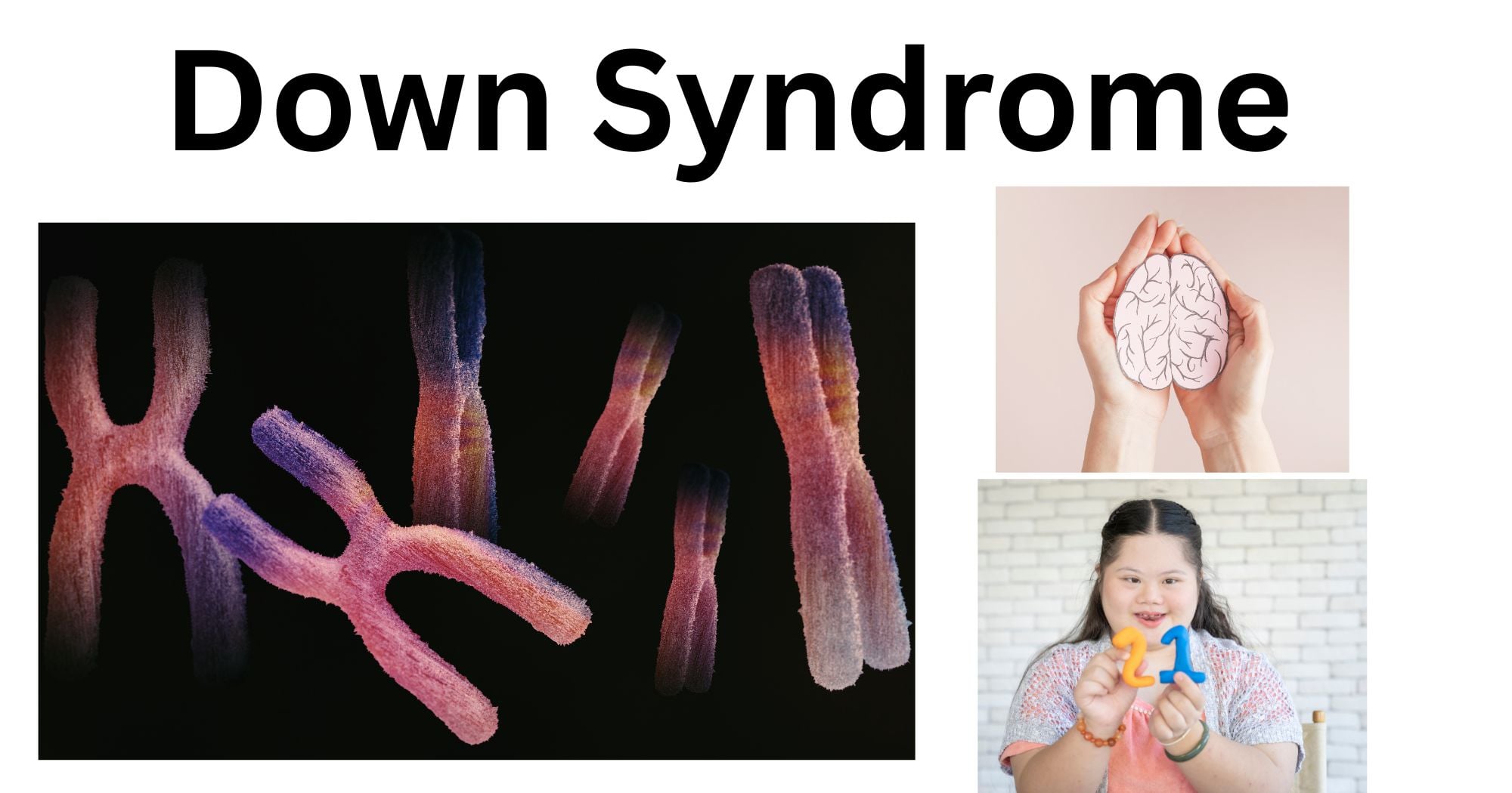
The general features of Down syndrome are weak muscle tone (hypotonia) in infants, intellectual disability, and a characteristic facial appearance. People with DS may have a shorter life expectancy; however, they can live a long and fulfilling life as well.
Down syndrome has been around for thousands of years, but its first full description was recorded by John Langdon Down in 1866, where it got its name. In 2015, DS was prevalent in 5.4 million individuals globally and resulted in 27,000 deaths.
What causes Down syndrome?
The cause of Down syndrome is the addition of an extra chromosome in the smallest human autosome, chromosome 21. This extra chromosome can either be of paternal or maternal origin. The chromosomal abnormality can be caused in three different ways, which are as follows:
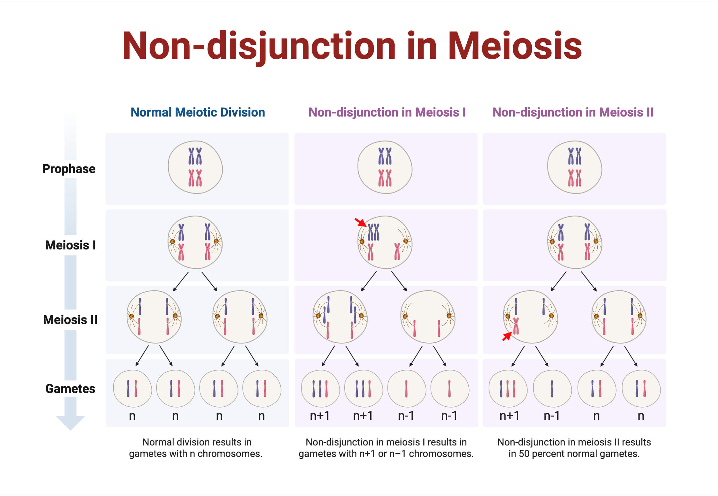
Non-disjunction in meiosis
About 95% of trisomy 21 cases are the result of non-disjunction in meiosis.
- In the normal meiotic division, the homologous chromosomes with their sister chromatids are separated into four haploid cells. However, in non-disjunction, the chromosomes are not separated uniformly, resulting in an abnormal number of chromosomes (n + 1 or n – 1).
- Non-disjunction can occur in anaphase I or in anaphase II. In anaphase I, the homologous chromosomes fail to separate, resulting in four abnormal daughter cells.
- In anaphase II, a haploid chromosome fails to separate its sister chromatids, resulting in two daughter cells being affected.
- When these gametes (n + 1) get fertilized, an abnormal trisomy zygote is formed, resulting in Down syndrome.
Robertsonian Translocation
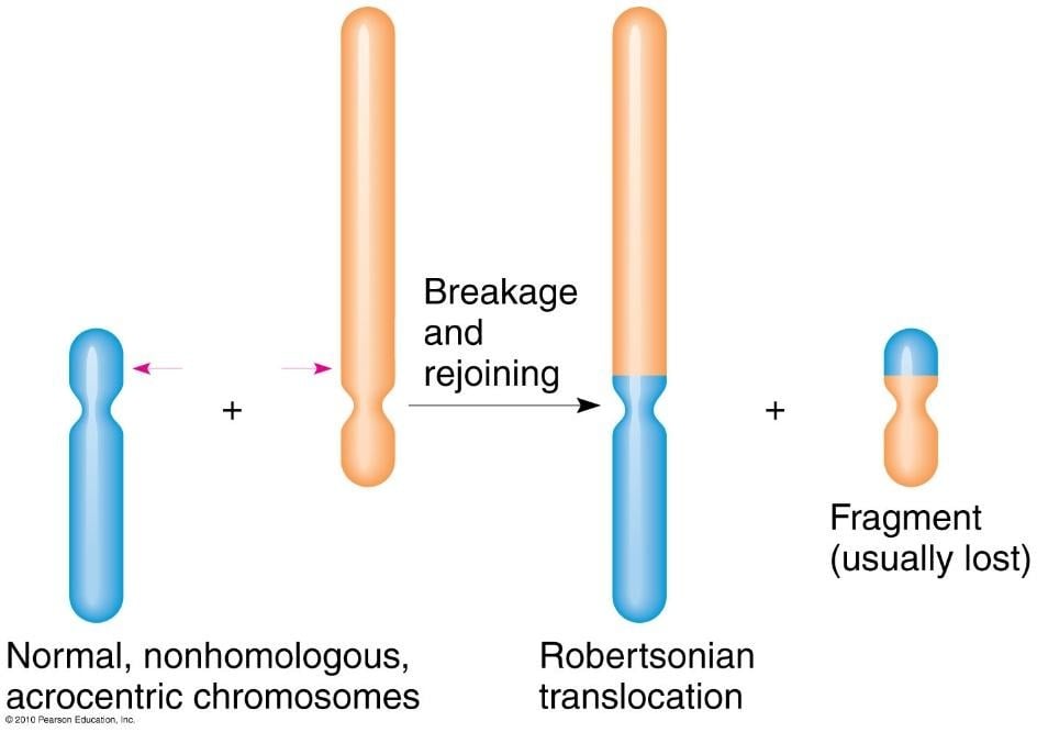
Robertsonian translocation accounts for about 4% of trisomy cases.
- The short arm of the acrocentric chromosome 14 gets translocated with the long arm of chromosome 21, producing two hybrid chromosomes.
- Chromosome 21 has two long arms, whereas chromosome 14 has two short ones. The latter one carries only a little bit of genetic information and generally gets lost in meiosis.
- When this kind of hybrid chromosome is inherited, there is replication, which results in two of these in one cell and none in the other (as the smaller one gets lost).
- The fertilization of this cell results in trisomy 21, and the other one gives monosomy 14.
Mosaic Down syndrome
Lastly, 1% of the patients are mosaic—they have mixed cells, having both trisomy 21 and normal cells.
- This is the result of non-disjunction in the mitotic division. If trisomy happens in this case, one cell will have 47 chromosomes, whereas the other one will lack one chromosome (i.e., 45 chromosomes).
- The 45 chromosome cells do not survive, but the trisomy one does and will produce more cells with its karyotype.
- The earlier this event happens, the more fetal it will be, as there will be more cells with the abnormal chromosomal number.
Symptoms of Down Syndrome
The symptoms of Down syndrome can be mild, moderate, or even severe. Some patients may exhibit extreme health problems, while others may appear relatively healthy. A few common symptoms include:
- Flattened face and short neck
- Almond-shaped eyes that slant up, with tiny white parts on the iris
- Small ears, hands, and feet
- Shorter height in children and adults
- A single line across the palm (palmar crease)
- Small pinky fingers that curve inwards
- The gap between the first two toes
- Poor muscle and joint connection
People with Down syndrome have slower speech patterns than most children and a mild-to-moderately low range IQ.
Having an extra chromosome 21 results in a lot of complications for every organ in the body. This is because of the overexpression of genes in the cells and tissues, showing phenotypic abnormalities. The extrachromosomal copy may affect the development of the organs listed below:
- Hearing and vision problems, including crossed eyes or even cataracts
- Heart problems: About half of the babies born with DS have septal defects in the heart chambers (usually between the two atria) and may require surgery.
- Presence of gastrointestinal (GI) atresia, which is the narrowing or closure of the intestine
- Blood: Increased risk of leukemia
- Brain: Mental retardation and sleep apnea with increased risk of Alzheimer’s disease
- Reproductive: Sterility in males
- Abnormal immune system: People with DS are at higher risk of autoimmune diseases, certain cancers, and infectious diseases such as pneumonia.
Major Risk Factor of Down Syndrome
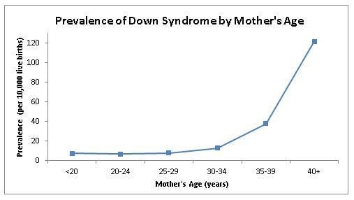
Down syndrome shows an increased probability with ascending maternal age. Older mothers tend to have a higher probability of having a child with this syndrome. The graph below shows an exponential increment as the mother’s age passes 35 years of age. There is approximately 1 in 350 chances of conceiving a child with Down syndrome at this age, which increases gradually to 1 in 100 by age 40 and approximately 1 in 30 by age 45. It is suggested that older eggs have a higher chance of improper division, but more research is still being conducted on the topic.
Diagnosis of Down Syndrome
The doctor may diagnose a baby with Down syndrome based on their appearance. To diagnose pregnant women, there are two distinct techniques: diagnostic tests and screening tests. They can be done in different periods of pregnancy, which are as follows:
- Blood tests: Blood tests involve monitoring of markers, which are proteins and hormones. In the first trimester, a protein called PAPP-P and a hormone called hCG are measured. Triple or quad-screen tests are done in the second trimester to measure protein AFP and hormone estriol. If any of these deviates from the normal levels, then the baby is suspected to have DS.
- Ultrasound: The doctor will examine the ultrasound of the baby and look for characteristic DS features.
Other kinds of prenatal tests include:
- Chorionic villus sampling (CVS): Chorionic villi are cells near the placenta that attach to the walls of the uterus. They are made from fertilized eggs, so they consist of the same genes as the baby. The examination of these cells is used to elucidate DS.
- Amniocentesis: Amniotic fluid consists of cells shed by the fetus with genetic information that can be used to diagnose DS.
- Percutaneous umbilical blood sampling (PUBS): Blood is removed from the umbilical cord during the third trimester and tested. It is the most accurate diagnostic method out of the three.
Treatment of Down Syndrome
Down syndrome has no treatment; however, a wide range of physical and mental therapies exists to help patients flourish to their utmost potential. These therapies have proven to be better if started earlier. Some of them are:
- Physical, occupational, and speech therapy
- Social and recreational activities
- Specialized education services
- Programs offering job training and self-care skills
Current Interventions for Down Syndrome
Due to the advancements in medical technologies, individuals with Down syndrome are living longer and healthier lives. In the 1960s, the average lifespan of people with this disorder was only about 10 years. This has significantly increased over the years. Today, with corrective heart surgeries, as many as 80% of affected people reach 50 to 60 years of age. Early intervention strategies mentioned previously have been helping children with DS to better adapt to the situation.
- Diagnosis of Down syndrome with invasive prenatal screening such as CVS, amniocentesis, etc. have the risk of miscarriage. With innovations in genomics and related technologies, a better way known as non-invasive prenatal screening (NIPS) test has been developed, which offers low false-positive rates and an extremely low risk of fetal death. However, a negative result from this test would not mean that there are no abnormalities.
- Another method, known as the cell-free fetal DNA (cffDNA) test, provides more accurate results but comes with a hefty cost.
- New genomic techniques have brought about many studies to identify the correlation between the genotype and phenotype of this condition. Scientists have identified some genes for certain phenotypic traits.
- With the onset of different experiments in model organisms such as mice, the study of the brain and other organs is in different stages of development as well. With accelerating cytogenetic investigation, some insights into the abnormal developmental pathways and gene expressions of DS will surely be provided.
References
- Akhtar, F., & Bokhari, S. R. A. (2025). Down Syndrome. In StatPearls. StatPearls Publishing. http://www.ncbi.nlm.nih.gov/books/NBK526016/
- Antonarakis, S. E., Skotko, B. G., Rafii, M. S., Strydom, A., Pape, S. E., Bianchi, D. W., Sherman, S. L., & Reeves, R. H. (2020). Down syndrome. Nature Reviews. Disease Primers, 6(1), 9. https://doi.org/10.1038/s41572-019-0143-7
- Bull, M. J. (2020). Down Syndrome. New England Journal of Medicine, 382(24), 2344–2352. https://doi.org/10.1056/NEJMra1706537
- Down Syndrome: Facts, Statistics, and You. (2018, October 31). Healthline. https://www.healthline.com/health/down-syndrome/down-syndrome-facts
- Down Syndrome: Symptoms & Causes. (n.d.). Cleveland Clinic. Retrieved April 27, 2025, from https://my.clevelandclinic.org/health/diseases/17818-down-syndrome
- Down syndrome—Symptoms and causes—Mayo Clinic. (n.d.). Retrieved April 27, 2025, from https://www.mayoclinic.org/diseases-conditions/down-syndrome/symptoms-causes/syc-20355977
- Mai, C. T., Kucik, J. E., Isenburg, J., Feldkamp, M. L., Marengo, L. K., Bugenske, E. M., Thorpe, P. G., Jackson, J. M., Correa, A., Rickard, R., Alverson, C. J., Kirby, R. S., & National Birth Defects Prevention Network. (2013). Selected birth defects data from population-based birth defects surveillance programs in the United States, 2006 to 2010: Featuring trisomy conditions. Birth Defects Research. Part A, Clinical and Molecular Teratology, 97(11), 709–725. https://doi.org/10.1002/bdra.23198
- Non-Disjunction | BioNinja. (n.d.). Retrieved April 27, 2025, from https://old-ib.bioninja.com.au/standard-level/topic-3-genetics/33-meiosis/non-disjunction.html
- PLAIASU, V. (2017). Down Syndrome – Genetics and Cardiogenetics. Mædica, 12(3), 208–213.
- Robertsonian Translocation. (n.d.). Retrieved April 27, 2025, from https://www.mun.ca/biology/scarr/Robertsonian_fusion.html
- Russell, P. J. (2014). IGenetics: A molecular approach (Third edition. Pearson new international edition). Pearson.
