
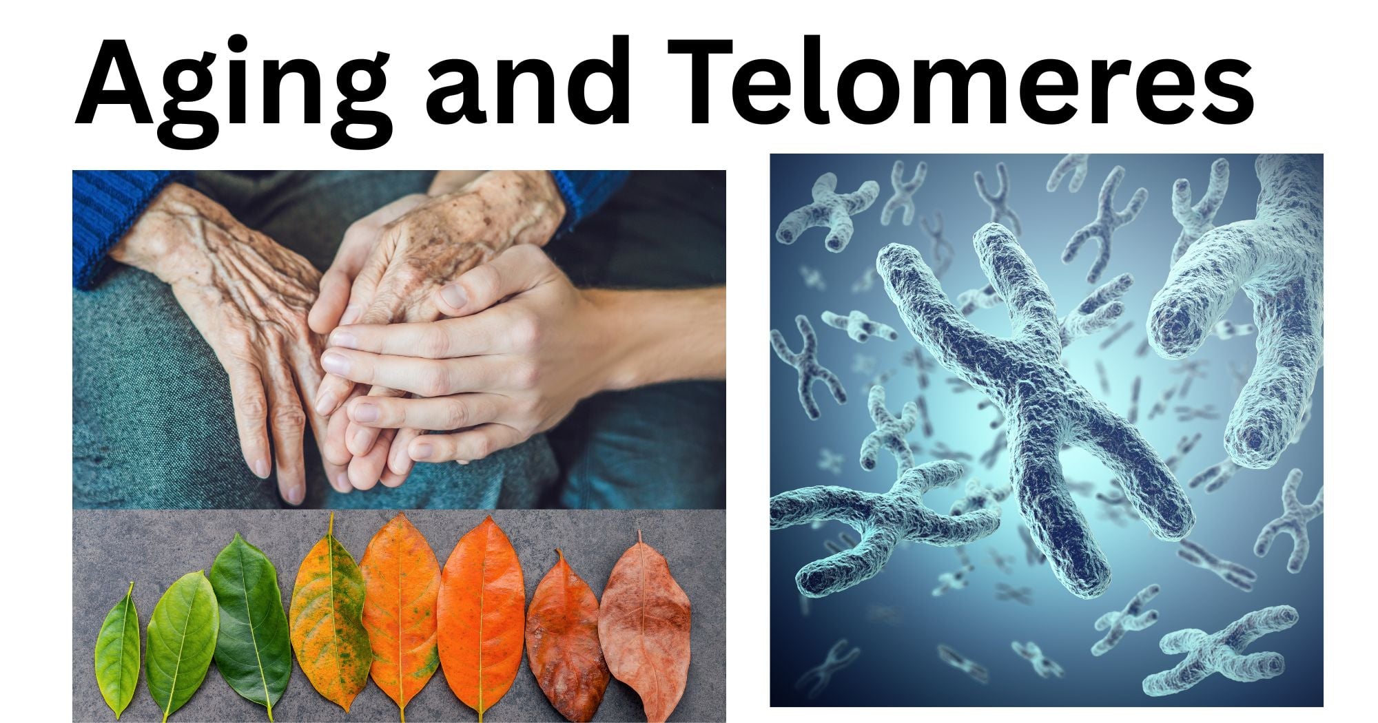
Aging, or senescence, is the gradual deterioration of the cellular and physiological functions of the body necessary for survival and fertility over time. The process is a culmination of the loss of specific regenerative and bioprotective mechanisms that occur in an organism over time.
Aging is affected by genetic and environmental components. One of such factors is the shortening of the telomeres, a repetitive sequence of DNA that protects the ends of the chromosome.
What are Telomeres?
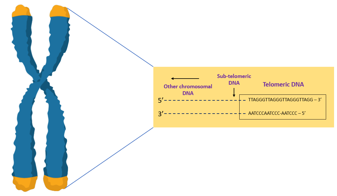
The DNA sequence in the cells is arranged in a closely wound compact structure known as a chromosome. Humans comprise 46 total chromosomes (22 pairs of autosomes and 1 pair of sex chromosomes). Among them, telomeres exist as repeating sequences of 6 base pairs, TTAGGG.
The length of telomeres is about 8000 base pairs in newborns and gradually decreases to about 3000 base pairs in adults, and as low as 1500 base pairs in elderly people. An entire chromosome has about 150 million bp, with each mitotic division costing approximately 30 to 200 bp from the ends of telomeres. Normal cells can divide only about 50 to 70 times in a lifetime, with telomeres progressively getting shorter. The cells eventually become senescent or die. In 2009, Elizabeth Blackburn, Jack Szostak, and Carol Greider were awarded the Nobel Prize in Physiology or Medicine for this discovery.
Significance of Telomeres
- Telomeres act as a protective barrier for the chromosome. It gets shorter each time a cell divides; therefore, the repeated palindromic region protects the actual genes from being degraded.
- The absence of telomeres could also lead to chromosome fusion, which might corrupt the DNA. The malfunction can detrimentally impact cellular health, which can lead to cell death or cancer.
- In normal cells, once the DNA is damaged, the cellular mechanisms can recognize and repair the damage. This happens at the ends of the chromosomes, in telomeres. Once this region is absent, DNA repair mechanisms such as non-homologous end-joining (NHEJ) can be faulty.
- Telomeres do not shorten in cells that do not continuously divide, such as in heart muscle cells.
- Shortened telomeres are associated with senescence, apoptosis, or even cancer, affecting an individual’s lifespan. Telomere shortening has been linked to increased disease incidence and poorer survival.
The End Replication Problem
DNA replication involves an enzyme known as DNA polymerase to synthesize an identical strand of DNA. The machinery works by annealing an RNA primer to the template strands and adding complementary nucleotide bases from the 5’ to the 3’ end. The leading strand can be continuously synthesized till the end of the template strand. However, synthesis of the lagging strand occurs in a discontinuous fashion, known as the Okazaki fragments.
The end replication problem only arises at the lagging strand of DNA replication, i.e., for replicating the ends of the Okazaki fragments. As the replication machinery reaches the end of the chromosome, the RNA primase no longer has sufficient space to anneal the RNA primer. This results in incomplete replication with a short single-stranded DNA (ssDNA) region at the 3’ end of the lagging-strand DNA. When this product is replicated in the next division, one of the two products gets shortened and lacks the region that was fully duplicated in the previous round.
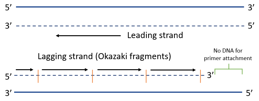
This means that each round of DNA replication would lead to the shortening of one of the two daughter DNA molecules. This gradual change would slowly hinder the transfer of genetic material from one generation to the next.
How do Cells Counter the End Replication Problem?
Organisms solve the end replication problem in varying ways. Most eukaryotic cells use telomeres, rich in the TG DNA sequence, to mitigate the problem. To elaborate, human telomeres comprise numerous repeats of the sequence 5’-TTAGGG-3’. Many of these repeats are double-stranded, with the 3’ end of each chromosome extending beyond the 5’ end as single-stranded DNA (ssDNA). This structure acts as a novel origin of replication that compensates for the end replication problem. The origin cannot interact with the same proteins as other eukaryotic origins and recruits a specialized DNA polymerase known as telomerase.
Mechanism of Telomerase
Telomerase is a remarkable enzyme that replicates the telomeric sequence of DNA at the end of the chromosome. It is made up of multiple protein subunits and an RNA component, called telomerase RNA (TER). TER varies in size from 150 to 1300 bp, depending on the organism. It includes a short region of DNA that encodes about 1.5 copies of the complement of the telomere sequence. In humans, the TER sequence is 5’-AAUCCCAAUC-3’.
Unlike DNA polymerase, telomerase does not require an exogenous DNA template to direct the addition of new dNTPs. It uses the RNA component as a template for adding sequence to the 3’ end of the chromosome. The RNA primer can anneal to the ssDNA at the 3’ end of the telomere, in such a way that the RNA template remains single-stranded, creating a primer: template junction which can be acted on by telomerase.
Telomerase also consists of reverse transcriptase, known as telomerase reverse transcriptase (TERT), which can transcribe RNA into DNA. TERT can synthesize DNA to the end of the TER template region, but cannot continue to copy the RNA beyond this point. The RNA template eventually disengages from the DNA product and reattaches to the last four nucleotides of the telomere, repeating the process.
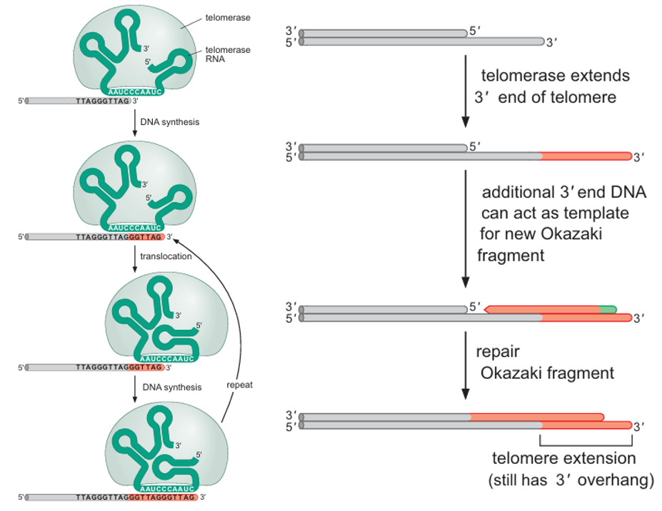
The enzyme specifically elongates the 3’-OH of telomeric ssDNA sequences using its RNA as a template. Because of this reason, the newly synthesized DNA is single-stranded. This 3’ overhang may serve to protect the functioning of the telomere. Such tiny, repetitive, and non-coding telomeric variation can be easily tolerated by the cell.
Hayflick’s Limit and Maximum Lifespan
Before the 1960s, somatic cells grown in vitro were thought to grow endlessly. This paradigm changed when Leonard Hayflick discovered that isolated cells would become senescent after 40 to 60 divisions. This limit of cell division is known as Hayflick’s limit.
Maximum lifespan, on the other hand, is the maximum number of years an organism is known to survive. Despite the increasing maximum aging, humans have shorter telomeres (5 to 15 kb) than mice (50 kb). However, the telomeres of humans shorten at a slower pace than those of mice.
| Organism | Maximum Aging |
| Human | 121 years |
| Dogs | 20 years |
| Drosophila | 3 months |
| Mouse | 4.5 years |
Telomerase and Cancer
If the telomere shortens with each cell division, a fascinating question arises regarding cancer cells, which are immortal and divide rapidly without any sign of cellular senescence. Telomeres are notably short in many cancer cells, such as those in the bone, prostate, bladder, lung, and kidney. These cells enhance the production of the telomerase enzyme to prevent these repetitive sequences from degrading, thus protecting the integrity of the genome.
Consequently, inhibiting telomerase activity could eliminate cancer cells and lead to their death. However, blocking telomere function could impede fertility, regeneration, and even the production of blood and immune cells.
References
- Are Telomeres the Key to Aging and Cancer. (n.d.). Retrieved May 20, 2025, from https://learn.genetics.utah.edu/content/basics/telomeres/
- Frontiers | Telomere Length as a Marker of Biological Age: State-of-the-Art, Open Issues, and Future Perspectives. (n.d.). Retrieved May 20, 2025, from https://www.frontiersin.org/journals/genetics/articles/10.3389/fgene.2020.630186/full
- Gilbert, S. F. (2000). Aging: The Biology of Senescence. In Developmental Biology. 6th edition. Sinauer Associates. https://www.ncbi.nlm.nih.gov/books/NBK10041/
- Shammas, M. A. (2011). Telomeres, lifestyle, cancer, and aging. Current Opinion in Clinical Nutrition and Metabolic Care, 14(1), 28–34. https://doi.org/10.1097/MCO.0b013e32834121b1
- The Hayflick Limit | Embryo Project Encyclopedia. (n.d.). Retrieved May 22, 2025, from https://embryo.asu.edu/pages/hayflick-limit
- Pederson, T. (2015). Molecular Biology of the Gene: By James D. Watson: W. A. Benjamin (1965): New York, New York. FASEB Journal: Official Publication of the Federation of American Societies for Experimental Biology, 29(11), 4399–4401. https://doi.org/10.1096/fj.15-1101ufm
