
The billions of neurons in the brain are connected with one another, and these connections are regulated by precise timing of electrical and chemical signals that influence behavior, memory and physiology. The ability to control specific cells with light, as is made possible with optogenetics, has revolutionized the way that scientists have studied these connections.
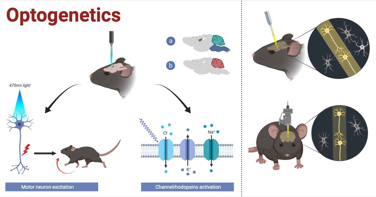
This method involves engineering of the cells and activating them with light, by inserting light sensitive proteins called opsins into specific cells. These proteins were first identified in microorganisms like Chlamydomonas reinhardtii, and can modulate cell function when stimulated by certain wavelengths of light (Boyden, 2011; Fenno et al., 2011; Emiliani et al., 2022).
Optogenetics and the Nobel Prize in Physiology or Medicine 2026
The importance of optogenetics was formally recognized with the 2026 Nobel Prize in Physiology or Medicine, awarded jointly to Peter Hegemann, Georg Nagel, and Karl Deisseroth for their discoveries of light-gated ion channels and optogenetics. Their work transformed an unusual light-sensing mechanism found in microorganisms into one of the most powerful tools for studying how the brain works.

The story began with a seemingly simple question: how can a microorganism such as Chlamydomonas detect light and respond to it? Unlike the human eye, which relies on a complex network of proteins to detect light and ultimately generate signals in neurons, Chlamydomonas uses specialized light-sensitive proteins that can directly influence the movement of ions across its cell membrane.
Peter Hegemann investigated these light-sensing mechanisms in microorganisms, while Georg Nagel helped establish the properties of channelrhodopsins (light-gated ion channels that can act both as light sensors and molecular gates). When exposed to light, these proteins open and allow ions to pass through the cell membrane, changing the cell’s electrical state. This unusual combination of light detection and ion movement provided researchers with an idea: what if such a protein could be placed inside a neuron?
Karl Deisseroth and his colleagues helped turn that idea into reality. They introduced microbial opsins into neurons, demonstrating that light could be used to control the electrical activity of these cells. This laid the foundation for optogenetics, in which genes encoding light-sensitive proteins are introduced into selected neurons, allowing researchers to switch specific populations of nerve cells on or off with light.
The real power of optogenetics is that it helps scientists move from simply observing the brain to testing cause and effect. Researchers can activate or inhibit a particular group of neurons and immediately observe what happens. This has revealed neural circuits involved in pain, hunger, sleep, learning, and memory, while also helping scientists understand disorders such as Parkinson’s disease and schizophrenia.
Principles and Mechanism of Optogenetics
The process can be understood in several steps:
- Selection of target cells: Researchers first identify the cell population or neural circuit they want to study.
- Genetic targeting: A gene encoding an appropriate opsin is delivered to the target cells using methods such as viral vectors or genetically modified animals.
- Opsin expression: The target cells produce the light-sensitive protein, which becomes incorporated into their cell membranes.
- Light delivery: Light of the appropriate wavelength is delivered to the cells using an optical fiber, LED, laser, or other optical system.
- Ion movement: Activation of the opsin changes ion permeability across the cell membrane.
- Change in cellular activity: Depending on the opsin, the cell becomes activated or inhibited.
- Physiological or behavioral response: Researchers observe how manipulating the selected cells affects neural circuits, behavior, or other biological functions.
An important feature of optogenetics is that genetic specificity and optical precision work together. Genetics determines which cells respond to light, while optics determines when and where those cells are activated (Fenno et al., 2011; Emiliani et al., 2022).
Components and Tools Used in Optogenetics
An optogenetic experiment requires both biological and optical components.
| Component/Tool | Function |
| Opsin gene | Encodes the light-sensitive protein responsible for controlling cellular activity. |
| Opsin protein | Responds to a specific wavelength of light and changes ion movement or cellular signaling. |
| Genetic delivery system | Introduces the opsin gene into selected cells, commonly using viral vectors or transgenic approaches. |
| Promoter/enhancer | Helps restrict opsin expression to particular cell types or populations. |
| Optical fiber | Delivers light directly to targeted tissues, especially deep regions of the brain. |
| LED or laser | Provides controlled illumination at the wavelength required to activate the selected opsin. |
| Light source controller | Controls light intensity, timing, pulse duration, and stimulation frequency. |
| Optical cannula/implant | Provides stable access for light delivery during experiments in living animals. |
| Electrophysiological recording system | Measures changes in neuronal electrical activity during optical stimulation. |
| Fluorescent reporter | Helps identify cells expressing the opsin or monitor cellular activity. |
| Microscope/imaging system | Allows visualization of cells, circuits, and optical stimulation. |
| Optrode | Combines optical stimulation with electrophysiological recording to manipulate and measure neural activity simultaneously. |
(Kushibiki et al., 2014; Emiliani et al., 2022).
Opsins and Light-Sensitive Proteins in Optogenetics
Opsins are a group of light-sensitive proteins that form the central molecular component of optogenetic systems. Many of which originate from microorganisms belonging to the microbial rhodopsin family.
They operate by means of a light-sensitive chromophore, typically retinal, that is linked to the protein opsin. When the correct wavelength of light is absorbed, retinal changes shape resulting in a change in the shape of the opsin protein. This finally results in either an ion-conducting or ion-pumping reaction (Piatkevich & Boyden, 2023).
Major examples of opsins include:
- Channelrhodopsins: Light-gated ion channels generally used for neuronal excitation.
- Halorhodopsins: Light-driven chloride pumps that can inhibit neuronal activity.
- Archaerhodopsins: Light-driven proton pumps that can reduce neuronal excitability.
- Engineered chloride-conducting opsins: Designed to produce inhibitory currents through chloride conductance.
- Red-shifted opsins: Engineered or naturally occurring variants activated by longer wavelengths of light, which can provide advantages for deeper tissue applications (Lin, 2010; Klapoetke et al., 2014).
Types of Optogenetic Tools
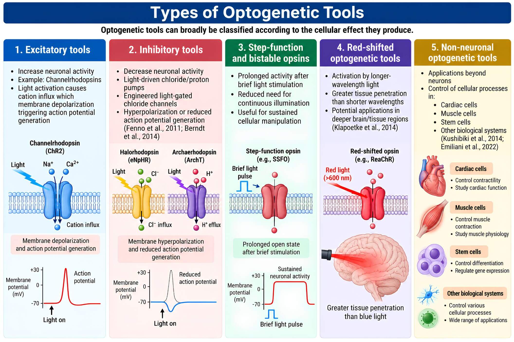
Optogenetic tools can broadly be classified according to the cellular effect they produce.
1. Excitatory tools:
- Increase neuronal activity
- Example: Channelrhodopsins
- Light activation causes cation influx which membrane depolarization triggering action potential generation
2. Inhibitory tools:
- Decrease neuronal activity
- Light-driven chloride/proton pumps
- Engineered light-gated chloride channels
- Hyperpolarization or reduced action potential generation (Fenno et al., 2011; Berndt et al., 2014)
3. Step-function and bistable opsins:
- Prolonged activity after brief light stimulation
- Reduced need for continuous illumination
- Useful for sustained cellular manipulation
4. Red-shifted optogenetic tools:
- Activation by longer-wavelength light
- Greater tissue penetration than shorter wavelengths
- Potential applications in deeper brain/tissue regions (Klapoetke et al., 2014)
5. Non-neuronal optogenetic tools:
- Applications beyond neurons
- Control of cellular processes in:
- Cardiac cells
- Muscle cells
- Stem cells
- Other biological systems (Kushibiki et al., 2014; Emiliani et al., 2022)
Optogenetic Stimulation and Inhibition of Neurons
Bidirectional control is the major advantage of optogenetics in neuroscience.
Neuronal stimulation
Excitatory opsins function as light-gated cation channels. When illuminated, they allow positively charged ions to cross the neuronal membrane. This causes depolarization, increasing the probability that the neuron will generate an action potential.
The timing of the light pulse can also be precisely controlled. Researchers can therefore reproduce specific patterns of neuronal activity and examine how those patterns influence downstream circuits and behavior.
Neuronal inhibition
Inhibitory opsins suppress neuronal activity by altering ion movement across the membrane. An example being halorhodopsin, while other approaches use proton pumps or engineered chloride-conducting channels.
An important development was the engineering of light-activated chloride channels, which demonstrated that opsins could be structurally modified to produce new functional properties (Berndt et al., 2014).
This allows researchers to control circuits by switching them on and off.
Applications of Optogenetics in Neuroscience
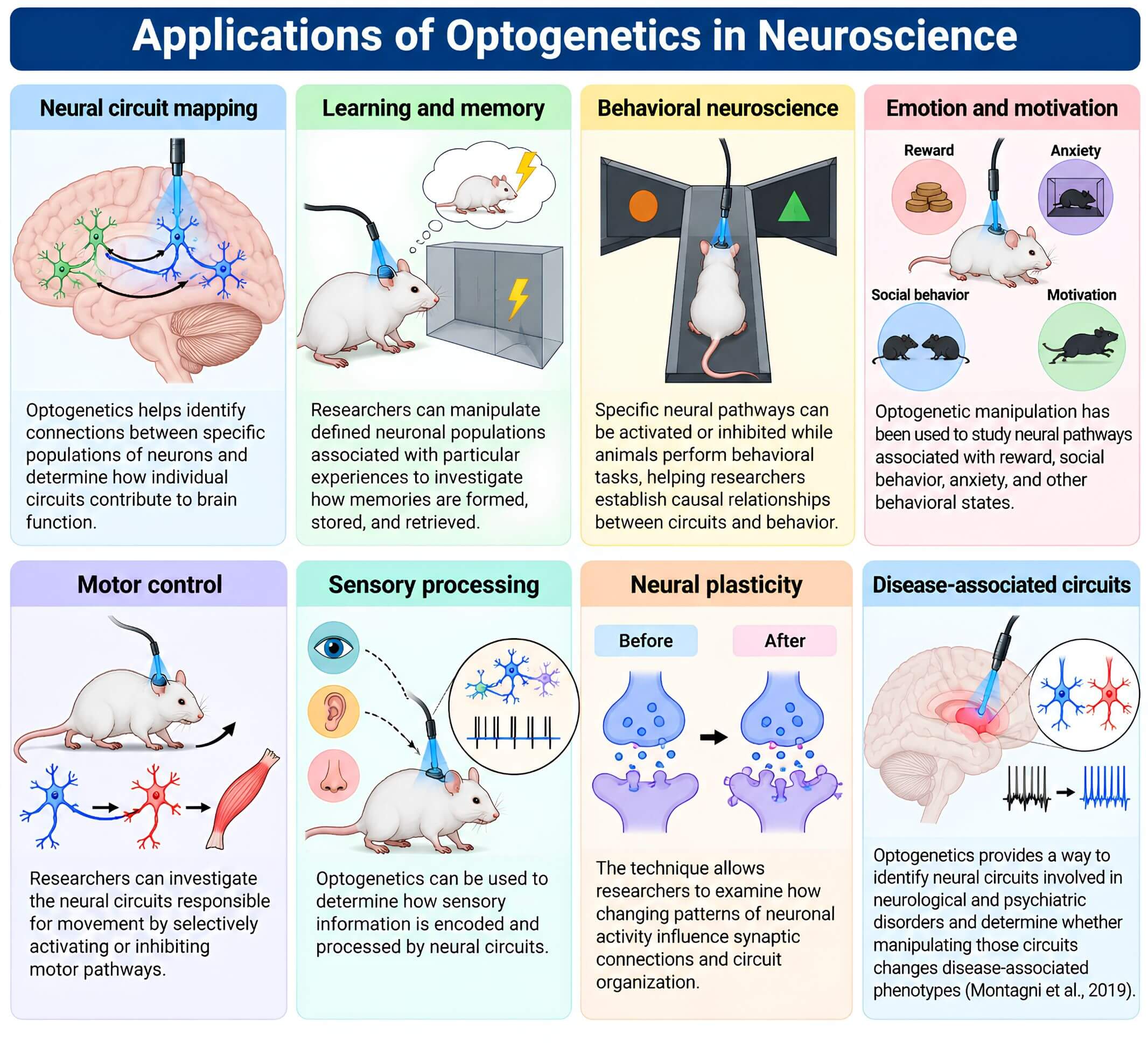
- Neural circuit mapping: Optogenetics helps identify connections between specific populations of neurons and determine how individual circuits contribute to brain function.
- Learning and memory: Researchers can manipulate defined neuronal populations associated with particular experiences to investigate how memories are formed, stored, and retrieved.
- Behavioral neuroscience: Specific neural pathways can be activated or inhibited while animals perform behavioral tasks, helping researchers establish causal relationships between circuits and behavior.
- Emotion and motivation: Optogenetic manipulation has been used to study neural pathways associated with reward, social behavior, anxiety, and other behavioral states.
- Motor control: Researchers can investigate the neural circuits responsible for movement by selectively activating or inhibiting motor pathways.
- Sensory processing: Optogenetics can be used to determine how sensory information is encoded and processed by neural circuits.
- Neural plasticity: The technique allows researchers to examine how changing patterns of neuronal activity influence synaptic connections and circuit organization.
- Disease-associated circuits: Optogenetics provides a way to identify neural circuits involved in neurological and psychiatric disorders and determine whether manipulating those circuits changes disease-associated phenotypes (Montagni et al., 2019).
Optogenetics in Research and Disease Studies
Optogenetics has become an important tool for understanding neurological and psychiatric disorders, allowing researchers to manipulate specific neural pathways rather than simply observing which brain regions are active.
In Parkinson’s disease, optogenetic studies have revealed how particular circuits contribute to abnormal motor control, while studies of depression have explored pathways involved in reward, motivation, mood, and social behavior. Similar approaches have helped investigate epilepsy, Alzheimer’s disease, addiction, and other conditions by linking abnormal circuit activity with disease-related behaviors.
Its potential also extends beyond understanding disease. Light-controlled cellular activity is being explored in neural regeneration and tissue repair, where researchers hope to influence cellular behavior to support recovery (Davletshin et al., 2025). Another promising application is vision restoration, with light-sensitive proteins and gene-delivery strategies being developed to restore light responsiveness in damaged retinal cells (Poboży et al., 2025).
Advantages of Optogenetics
- High temporal precision: Light can be delivered in precisely timed pulses, allowing researchers to control neuronal activity on a millisecond timescale.
- High spatial precision: Optical stimulation can be directed to specific cells, regions, or neural projections.
- Cell-type specificity: Genetic targeting allows opsins to be expressed in selected populations of cells rather than indiscriminately stimulating an entire tissue.
- Bidirectional control: Different opsins allow researchers to either activate or inhibit neuronal activity.
- Causal investigation: Optogenetics allows researchers to test whether activity in a particular circuit is sufficient or necessary for a physiological or behavioral response.
- Compatibility with other techniques: Optogenetics can be combined with electrophysiology, fluorescence imaging, anatomical tracing, and behavioral experiments.
- Broad biological applications: Although initially developed primarily for neuroscience, optogenetic approaches can be adapted to other cell types and biological systems (Emiliani et al., 2022).
Limitations and Challenges of Optogenetics
- Requirement for genetic modification: Opsin expression generally requires delivery of genetic material, which introduces additional experimental complexity.
- Light delivery to deep tissues: Light scattering and absorption limit penetration into biological tissues. Deep brain structures often require implanted optical fibers or specialized optical systems.
- Potential tissue damage: Repeated implantation and illumination can produce tissue damage or inflammatory responses if experimental conditions are not carefully controlled.
- Variable opsin expression: Differences in gene delivery and protein expression can affect the strength and consistency of cellular responses.
- Heating and phototoxicity: High-intensity or prolonged illumination can potentially heat tissue or cause unwanted biological effects.
- Limited clinical translation: Therapeutic use in humans requires careful consideration of gene delivery, immune responses, long-term expression, light delivery, safety, and ethical issues (Shen et al., 2020).
- Opsin kinetics and limitations: Different opsins have different activation and deactivation speeds, ion selectivities, and light sensitivities, meaning that no single opsin is ideal for every experiment (Lin, 2010; Piatkevich & Boyden, 2023).
- Experimental models vs Humans: Results obtained from animal models may not directly translate into human physiology or disease.
Future Applications of Optogenetics
The future of optogenetics is likely to involve the development of tools that are less invasive, more selective, and better suited for clinical applications.
One major direction is the development of improved opsins with greater light sensitivity, longer-wavelength activation, faster kinetics, and improved expression in target cells. Red-shifted proteins are particularly interesting because longer wavelengths can provide improved tissue penetration (Klapoetke et al., 2014; Poboży et al., 2025).
The development of wireless and minimally invasive light-delivery systems may also expand the potential applications of optogenetics. Reducing dependence on implanted optical fibers would be particularly important for long-term therapeutic use.
Future systems could combine optogenetic control with real-time recording. Such closed-loop systems could detect neural activity and automatically adjust optical stimulation according to the physiological state of the subject providing more adaptive control than predetermined stimulation patterns.
Research is exploring applications in cardiac biology, tissue regeneration, cell signaling, and other areas where controlling cellular activity with light could provide experimental or therapeutic advantages (Emiliani et al., 2022; Davletshin et al., 2025).
Conclusion
Optogenetics has transformed neuroscience by enabling precise control of selected cells using light. From microbial opsins to advanced neural applications, it has revealed how specific circuits shape physiology and behavior. With the Nobel Prize in Physiology or Medicine in 2026, continued advances may expand its potential to understand disease and develop future therapies.
References
- Berndt, A., Lee, S. Y., Ramakrishnan, C., & Deisseroth, K. (2014). Structure-guided transformation of channelrhodopsin into a light-activated chloride channel. Science, 344. https://doi.org/10.1126/science.1252367
- Boyden, E. S. (2011). A history of optogenetics: The development of tools for controlling brain circuits with light. F1000 Biology Reports, 3, 11. https://doi.org/10.3410/b3-11
- Davletshin, E., Albert, S., Tatyana, A., Galina, S., Albert, R., & Yana, M. (2025). Optogenetic approaches for neural tissue regeneration: A review of basic optogenetic principles and target cells for therapy. Neural Regeneration Research. https://doi.org/10.4103/nrr.nrr-d-24-00685
- Emiliani, V., Entcheva, E., Hedrich, R., Hegemann, P., Konrad, K., Lüscher, C., Mahn, M., Pan, Z., Sims, R. R., Vierock, J., & Yizhar, O. (2022). Optogenetics for light control of biological systems. Nature Reviews Methods Primers, 2. https://doi.org/10.1038/s43586-022-00136-4
- Fenno, L. E., Yizhar, O., & Deisseroth, K. (2011). The development and application of optogenetics. Annual Review of Neuroscience, 34. https://doi.org/10.1146/annurev-neuro-061010-113817
- Klapoetke, N. C., Murata, Y., Kim, S. S., Pulver, S. R., Birdsey-Benson, A., Cho, Y. K., … Boyden, E. S. (2014). Independent optical excitation of distinct neural populations. Nature Methods, 11. https://doi.org/10.1038/nmeth.2836
- Kushibiki, T., Okawa, S., Hirasawa, T., & Ishihara, M. (2014). Optogenetics: Novel tools for controlling mammalian cell functions with light. International Journal of Photoenergy, 2014, 895039. https://doi.org/10.1155/2014/895039
- Lin, J. Y. (2010). A user’s guide to channelrhodopsin variants: Features, limitations and future developments. Experimental Physiology. https://doi.org/10.1113/expphysiol.2009.051961
- Montagni, E., Resta, F., Allegra Mascaro, A. L., & Pavone, F. S. (2019). Optogenetics in brain research: From a strategy to investigate physiological function to a therapeutic tool. Photonics, 6(3), 92. https://doi.org/10.3390/photonics6030092
- Piatkevich, K. D., & Boyden, E. S. (2023). Optogenetic control of neural activity: The biophysics of microbial rhodopsins in neuroscience. Quarterly Reviews of Biophysics, 56. https://doi.org/10.1017/S0033583523000033
- Poboży, K., Poboży, T., Domański, P., Derczyński, M., Konarski, W., & Domańska-Poboża, J. (2025). Evolution of light-sensitive proteins in optogenetic approaches for vision restoration: A comprehensive review. Biomedicines, 13(2), 429. https://doi.org/10.3390/biomedicines13020429
- Shen, Y., Campbell, R. E., Côté, D. C., & Paquet, M. E. (2020). Challenges for therapeutic applications of opsin-based optogenetic tools in humans. Frontiers in Neural Circuits, 14, 41. https://doi.org/10.3389/fncir.2020.00041
- Harz, H., Nonnengasser, C., Hegemann, P. The photoreceptor current of the green alga Chlamydomonas. Philos. Trans. R. Soc. Lond. Ser. B: Biol. Sci. 1992:338:39–52.
- Nagel G, Ollig D, Fuhrmann M, Kateriya S, Musti AM, Bamberg E, Hegemann P. Channelrhodopsin-1: a light-gated proton channel in green algae. Science. 2002:296:2395–2398.
- Nagel G, Szellas T, Huhn W, Kateriya S, Adeishvili N, Berthold P, Ollig D, Hegemann P, Bamberg E. Channelrhodopsin-2, a directly light-gated cation selective membrane channel. Proc. Natl. Acad. Sci. U. S. A. 2003:100:13940–13945.
- Boyden ES, Zhang F, Bamberg E, Nagel G, Deisseroth K. Millisecond-timescale, genetically targeted optical control of neural activity. Nat. Neurosci. 2005:8:1263–1268.
- Aravanis AM, Wang LP, Zhang F, Meltzer LA, Mogri MZ, Schneider MB, Deisseroth K. An optical neural interface: in vivo control of rodent motor cortex with integrated fiberoptic and optogenetic technology. J Neural Eng. 2007:4:S143–156.
