
The genus Nocardia is an aerobic, catalase-positive, Gram-positive, rod-shaped actinomycete with a branching filamentous form, first described in 1888 by Edmond Nocard. Nocardia sp. is found worldwide in a myriad of environments. Nocardia infection causes a variety of symptoms depending on the site of infection. Nocardia asteroides is the most common isolate in nonhuman primates.
Typically, Nocardia is an opportunistic pathogen that primarily affects immunosuppressed patients. Infection occurs following contact with skin wounds, inhalation or ingestion. Nocardiosis is an uncommon bacterial infection with a wide variety of clinical manifestations in immunocompetent and immunocompromised patients. Clinical manifestations of nocardiosis range from cutaneous infections caused by traumatic inoculation in normal hosts to severe pulmonary and central nervous system diseases in immunocompromised hosts.
Taxonomy and Classification of Nocardia asteroides
Domain: Bacteria
Kingdom: Bacillati
Phylum: Actinomycetota
Class: Actinomycetes
Order: Mycobacteriales
Family: Nocardiaceae
Genus: Nocardia
Species: Nocardia asteroides
Prior to the molecular era, the taxonomy of Nocardia species and the identification of Nocardia isolates from patient specimens were relatively uncomplicated. With the advent of molecular methodologies, specifically, gene sequencing, a huge number of new Nocardia species have been identified.
Morphology of Nocardia asteroides
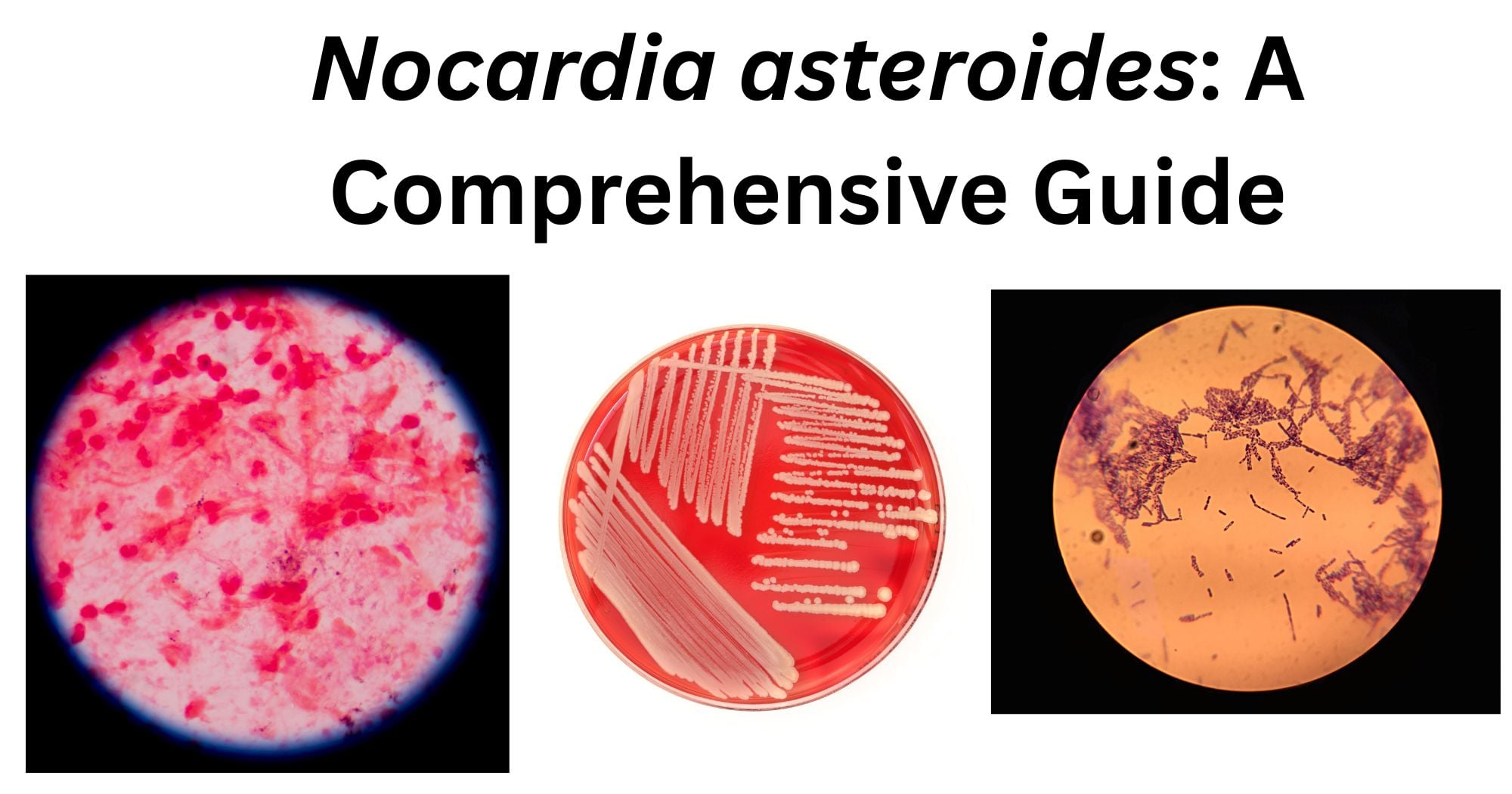
Nocardia asteroides is an aerobic, gram-positive, partially acid-fast actinomycete. This organism exhibits a characteristic branching filamentous structure. It belongs to the genus Nocardia which are commonly found in soil, water, dust and decaying organic matters. When Nocardia asteroides is cultured in a routine non-selective media such as blood agar, N. asteroides grow slowly, around 48-72 hours though some strains may require longer incubation period. The colonies that grow after adequate incubation, are dry, chalky wrinkled or velvety and may appear to be white, cream, yellowish or light brown varying according to the strains of the bacteria and medium used. Many colonies produce aerial hyphae, giving them a fuzzy or powdery appearance similar to fungi. The growing colonies are adherent to the agar surface and crumble when manipulated with a loop, making hem difficult to emulsify in saline. Earthy or musty odor are characteristic for many Nocardia species. They contain tuberculostearic acids but differ from the mycobacteria by possession of short chained mycolic acids. They have a Type IV cell wall, characterized by a peptidoglycan made up of meso-diaminopimelic acid, arabinose, and galactose.
How does Nocardia asteroides look under a microscope?
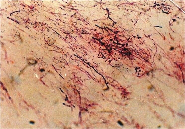
Nocardiae are gram positive bacillary, branching bacteria whose hyphae often fragment to coccobacillary forms. The characteristic morphology is branching, filamentous, gram-positive rods when observed under microscope. They can resemble Actinomycetes species on gram stain. However, Nocardia is an obligate aerobe that stains weakly positive on acid-fast staining. This partial/weak acid-fastness is caused by the presence of intermediate-length mycolic acids within the cell walls. Hence why, modified acid-fast stains like the Kinyoun stain are commonly used for laboratory identification.
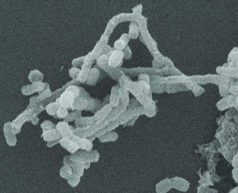
When observed under the electron microscope, N. asteroides is seen to possess a complex cell envelope with an irregularly thick outer layer. Ribosomes like granules, nuclear fibrils and polyphosphate granules are present in the cytoplasm.
In a light microscope, N. asteroides appear as delicate, thin and branching filaments resembling a fungal hypha. The filaments are around 0.5-1.0µm in diameter and may fragment into bacillary or coccoid forms.
Cultural and Growth Characteristics of Nocardia asteroides
Macroscopically, cultures of Nocardia present as glabrous, either orange or white chalky, “gypsoides” colonies. Microscopically, when stained with gram stain and Kinyoun stain, these colonies show delicate, branching, filamentous coccobacilli that are variably gram-positive and weakly acid-fast positive.
N. asteroides is a strict aerobe and can grow well at temperatures ranging from 25°C to 37°C, optimum being around 37°C. it can however thrive on a wide variety of non-selective laboratory media. N. asteroides is observed to grow readily on 5% sheep blood agar, chocolate agar and Sabouraud dextrose agar. Since N. asteroides are typically non-hemolytic organisms, the blood agar plates should be carefully examined. Nocardia species can also grown in media like Lowenstein-Jensen medium and Middlebrook 7H10/7H11 agar, which are designed for mycobacteria. It is because these Nocardia are partially acid-fast.
N. asteroides is a slow grower. It takes around 5 days for N. asteroides to show some colony growth at ⁓37°C in SDA plates. However, the grown colonies are still quite small at this stage. They may take about 2-3 weeks to grow in medium like LJ media.
The young colonies forming in medium agars are usually smooth and butyrous in nature but are quick to become dry, chalky and turn matte like appearance. This is dues to the production of abundant aerial hyphae. Mature colonies are often wrinkled and convoluted making them look like brains. Many strains of Nocardia asteroides is strong and distinctive earthy or musty odor, similar to other Actinomycetes. The isolates from this colonial growth are seen to be white, cream or chalky-white on initial isolation. On further incubation, the colonies may develop pigments that are bound to the colonies themselves turning yellow, tan, orange or even pinkish. The pigments do not diffuse into surrounding medium.
Epidemiology of Nocardia asteroides
Nocardia is a gram positive, facultative aerobic actinomycete that is a widespread pathogen in the environment and can cause serious infections of the lings, skin, brain and central nervous system. Nocardiosis is primarily an opportunistic infection in immunocompromised individuals but immunocompetent people are also at risk. In recent years, with the global population growth and the increase in the number of immunosuppressed people, the number of Nocardia infection cse reports and detections has increased significantly. This change is not only attributed to the expansion of the susceptible population but also related to the increase in detection rate brought about by advances in diagnostic technology.
Nocardia infection carries a high risk of mortality, with a reported one-year mortality rate of approximately 25%. Risk factors for mortality include immunosuppression, use of glucocorticoids, disseminated infection, cavitary or pleural involvement and infection with specific species.
Nocardiosis affects all ages and races but is more common in males than females in a 3 to 1 ratio. Nocardia typically manifests as an opportunistic infection in immunocompromised patients, with 60% of patients with nocardiosis having immunosuppressive conditions. however, up to 33% of patients are immunocompetent, this is especially true of cutaneous infections where immunocompetent individuals are the majority of cases. Dissemination of both cutaneous and pulmonary nocardiosis is more likely in immunocompromised hosts.
Immunocompromised conditions associated with Nocardia infections include:
- HIV/AIDS
- Cancer
- Chemotherapy treatment
- Corticosteroid treatment
- Solid organ or bone marrow transplant recipients
- Allogenic hematopoietic stem cell transplant recipients
- Diabetes
- Autoimmune diseases
- Chronic lung diseases such as COPD
- Any condition that results in deficient cell-mediated immunity
- Connective tissue disorder
Pathogenesis of Nocardia asteroides
Nocardia species, such as N. asteroides and N. brasiliensis are primarily responsible for human infections. N. asteroides usually leads to pulmonary and disseminated infections. Infections occur through inhalation of dust containing the bacteria or direct inoculation via cuts or scrapes when contaminated soil or water enters the body. These infections are not necessarily associated with immunocompromised host states. But the dissemination from these sites of inoculation is more likely in immunocompromised hosts, particularly those with impaired cell-mediated immunity. Nocardia asteroides is an opportunistic bacterial pathogen causing nocardiosis, a chronic suppurative and granulomatous infectious disease affecting the lungs, skin, CNS and other organs.
As soon as the bacteria is inhaled through respiratory tract, they reach alveoli. As N. asteroides reach the alveoli, they are phagocytosed by macrophages and neutrophils. But the bacteria consist of several virulence factors that aids it in survival intracellular and evade the host immune defenses. This explains why Nocardia can produce a spectrum of disease ranging from localized skin infections to life-threatening abscesses. As the bacteria reaches alveoli, they can establish a primary pulmonary infection. Another route can be traumatic inoculation through breaks in skin leading to cutaneous nocardiosis.
Once the bacteria reach the tissues, N. asteroides must survive an immediate attack by alveolar macrophages and neutrophils which can be neutralized by the wide array of virulence factors possessed by N. asteroides.
The host systems attempt to contain the infection by forming granulomas. But, Nocardia granulomas tend to be suppurative, and these appear as an abscess surrounded by a wall of epithelioid histocytes and chronic inflammatory cells. Both the bacterial products and host’s own inflammatory response lead to the tissue damage. However, if the pulmonary or cutaneous infection is not contained, usually in an immunocompromised host, N. asteroides can spread via bloodstream to almost any organ. The bacterium exhibits a significant preference for the CNS. The mechanisms behind the neurotropism are not fully defined but may involve specific adhesion ligands for brain endothelial cells and the ability to traverse blood-brain barrier within circulating macrophages.
The pathogenesis is heavily dependent on host defenses. Cell-mediated immunity is the most important for controlling Nocardia infection. In the immunocompromised individuals, the disease is frequently progressive and disseminated. On the other hand, immunocompetent hosts may develop a self-limited, chronic and indolent pulmonary infection that mimics tuberculosis or fungal pneumonia or a localized skin pustule that progresses slowly along lymphatics.
What are the Virulence Factors of Nocardia asteroides?
- Inhibition of Phagosome-Lysosome Fusion: After engulfment by macrophages, N. asteroides can persist intracellularly by preventing the fusion of phagosomes with lysosomes. This makes N. asteroides able to avoid the destruction and replicate within the cells designed to kill it.
- Neutralization of Reactive Oxygen Species: phagocytic cells employ reactive oxygen species, such as superoxide radicals and hydrogen peroxide as a part of the oxidative burst to eliminate invading microorganisms. N. asteroides has a surface-associated superoxide dismutase (SOD) that converts superoxide to hydrogen peroxide and a periplasmic catalase which then breaks hydrogen peroxide down into water and oxygen. This entire process makes the bacterium highly resistance to the oxygen-dependent killing mechanisms of neutrophils and macrophages.
- Trehalose Dimycolate: N. asteroides’ cell wall contains trehalose 6,6’-dimycolate, a glycolipid which is structurally similar to the cord factor in Mycobacterium tuberculosis. This molecule is toxic to mammalian cells, inhibits leukocyte migration and damaging the mitochondrial membranes. All of this directly contribute to the formation of necrotic, granulomatous lesions.
Clinical Manifestations of Nocardia asteroides
Pulmonary Nocardiosis
Pulmonary nocardiosis is the most common manifestation of nocardial infection. Involvement of lung occurs in 70% of all cases. Nocardia asteroides are the most common organism through the nine species have been associated with human disease. Pulmonary nocardiosis can occasionally occur through gastrointestinal tract especially through appendix rarely pulmonary infection follows dental procedures or peri odontal infection. Symptom include cough, fever, dyspnea, chest pain, weight loss and hemoptysis. It can present as an acute, sub-acute or chronic disease with remissions and exacerbations. Lung involvement can be a part of disseminated nocardiosis. Other findings include bronchiectasis, pleural thickening, ground glass opacity, mass-like consolidation, intrathoracic lymphadenopathy, pleural effusion, reticular infiltration and pericardial effusion.
Nocardiosis of CNS
The most common extra pulmonary site for Nocardiosis is CNS. It is uncommon but clinically important disease as it is associated with high mortality. It accounts for 15-50% of nocardiosis and 2% brain abscesses. It can affect the brain or spinal cord. It occurs along the pulmonary disease or as a isolated disease. It usually occurs in immunosuppressed individuals. The presentation is usually with headache, nausea, vomiting, focal neurological deficit and seizures or as cord compression.
Nocardiosis of Skin and Subcutaneous Tissues
Cutaneous nocardiosis usually occurs 9in immunocompetent individuals. The infection is most common in individuals from rural areas in developing countries, attributed to minor localized trauma due to walking barefoot. Foot is the most common site of infection, followed by hand, neck and face. The disease maybe subdivided into four clinical types: mycetoma, lymphocutaneous infection, superficial skin infection and secondary cutaneous involvement with disseminated disease. Mycetoma is a chronic, localized, slowly progressive, painless infection with tumefaction, subcutaneous nodules, destructive granulomata and formation of intermittent fistulas with pus. In lymphocutaneous form, he spread is beyond the cutaneous focus to involve the regional lymphatics and results in regional lymphadenopathy. Superficial skin infection is characterized by cellulitis with erythema, pain and swelling. There may be ulceration and abscess formation. Cutaneous nocardiosis secondary to disseminated disease occurs in immunocompromised individuals and associated with high mortality.
Disseminated Nocardiosis
It is a life-threatening infection, occurring in severely immunocompromised individuals and associated with high mortality. It is an endogenous infection with a primary focus usually in the lung and rarely in the skin. CNS involvement is the most common and serious complication, though any organ may be involved.
Laboratory Diagnosis of Nocardia asteroides
The clinical diagnosis of nocardiosis is difficult. Signs, symptoms and radiologic studies may suggest the diagnosis but are not pathognomonic.
Specimen
When nocardiosis is clinically suspected, multiple specimens should be submitted for culture, as smears and cultures are simultaneously positive only in a third of the cases. Depending on the clinical presentation, samples include sputum, pus from an abscess, and granules.
Direct Microscopy
- Presence of gram-positive branching or partially branching, filamentous bacilli often with beaded appearance suggest the presence
- Appear as branching and filamentous red-coloured acid-fast bacilli when stained by a modified acid-fast staining method
- H and E staining (for histopathology) of the granules show multilobulated with sunray appearance
Culture
They do not have complex growth requirement and can grow on various media such as SDA, BHI agar, sheep blood agar, etc. colonies will be visible after 2-3 days of incubation at 37°C. to overcome the problem of contaminating flora (since N. asteroides are slow growers), various selective media have been in use for their isolation like, buffered yeast extract containing polymyxin and vancomycin, SDA with chloramphenicol, BHI agar with chloramphenicol and cycloheximide, LJ medium, etc.
The colonies are creamy, wrinkled, pigmented and adhere firmly to the medium. Some colonies possess abundant aerial growth and have a cotton wool ball appearance.
Differential Diagnosis
Nocardia needs to be differentiated from actinomycetes and mycobacteria. Actinomycetes are also Gram-positive but are not acid-fast. Actinomycetes grow under anaerobic conditions in culture, whereas Nocardia grow under aerobic conditions. Weak acid fastness and a beaded appearance on microscopy differentiate Nocardia from mycobacteria.
Molecular Methods
Conventional methods based on morphology, staining characteristics, and biochemical tests are time-consuming and may not be reliable for accurate identification of Nocardia asteroides. Molecular methods provide faster, more accurate identification of the bacterium.
The 16S rRNA gene contains conserved regions that allow universal amplification and variable regions that enable differentiation among bacterial species. After DNA extraction, the 16S rRNA gene is amplified by PCR and sequenced. The resulting sequence from the PCR is then subjected to comparison against public databases. This method identifies N. asteroides and many other Nocardia species in a reliable manner and is often regarded as a reference method in clinical microbiology laboratories.
- PCR-based Identification
PCR enables direct detection of Nocardia DNA from either cultured isolates or clinical specimens. For confirmation of the presence of N. asteroides, species-specific primers that target conserved genetic regions are used. The most common genetic targets include the 16S rRNA gene, the hsp65 gene, the secA1 gene, the gyrB gene, etc. The use of PCR methods significantly reduces the time required for identification of the organism in comparison with the traditional culture-based techniques.
WGS provides the highest level of taxonomic resolution by analyzing the entire genome of the organism. Its applications range from identification and strain-level typing to the detection of antimicrobial resistance determinants and phylogenetic reconstruction. Even though this technique is highly informative, it remains one of the more expensive methods and is primarily used in research and specialized reference laboratories only.
- MALDI-TOF Mass Spectrometry
Although it is not a molecular genetic method, Matrix Associated Laser Desorption/Ionization-Time of Flight (MALDI-TOF) mass spectrometry is used as a modern identification technique along with other molecular methods. It identifies Nocardia isolates based on unique protein species-level spectra and can provide species level identification when supported by comprehensive reference databases.
What are the treatments for Nocardia asteroides Infections?
The mainstay treatment for Nocardia infections has been sulfonamides. Primary agents that have been used successfully are minocycline, amikacin, imipenem, and linezolid. Combination therapy with a sulfa-containing agent and one 0of the primary agents has been recommended for serious, systemic disease. Other potentially efficacious choices include the extended-spectrum cephalosporins, amoxicillin-clavulanate, newer macrolides, other aminoglycosides, and the fluoroquinolones. The duration of therapy is uncertain, but it should be prolonged because of the occurrence of considerable numbers of relapses after shorter courses of therapy.
The most common regimen includes TMP-SMX, amikacin and imipenem. However, if the patient has CNS involvement, it is important to pick agents with good CNS penetration such as TMP-SMX and ceftriaxone.
Other agents to consider include:
- Carbapenems: imipenem, meropenem or ertapenem with imipenem having the best efficacy
- Quinolones: especially moxifloxacin
- Tetracycline: minocycline
How to prevent Nocardia asteroides Infections?
Risk of nocardiosis can be reduced by:
- Wearing gloves and mask when working with dirt or dust
- Wearing clothing that covers skin to prevent cuts or wounds
- Bandaging any cuts or open wounds in the skin
- Washing hands frequently, especially after being outdoors or working with dust/dirt
- Treating injuries right away by washing them with soap and water and covering them
- Carefully monitoring for early signs of infection in risk group
- For certain high-risk patients, particularly solid-organ transplant recipients and individuals receiving prolonged immunosuppressive therapy, prophylaxis with trimethoprim-sulfamethoxazole (TMP-SMX) may reduce the risk of nocardiosis
References
- Brown-Elliott, B. A., Brown, J. M., Conville, P. S., & Wallace, R. J., Jr. (2006). Clinical and laboratory features of Nocardia spp. based on current molecular taxonomy. Clinical Microbiology Reviews, 19(2), 259–282. https://doi.org/10.1128/CMR.19.2.259-282.2006
- Cleveland Clinic. (n.d.). Nocardiosis. https://my.clevelandclinic.org/health/diseases/nocardiosis
- Conville, P. S., Brown-Elliott, B. A., Smith, T., & Zelazny, A. M. (2018). The complexities of Nocardia taxonomy and identification. Journal of Clinical Microbiology, 56(1), e01419-17. https://doi.org/10.1128/JCM.01419-17
- Global Biodiversity Information Facility. (n.d.). Nocardia asteroides. https://www.gbif.org/species/102536645
- IntechOpen. (2014). Nocardia: Virulence and pathogenicity. https://www.intechopen.com/chapters/41359
- McNeil, M. M., Brown, J. M., & the Nocardia Working Group. (2003). The medically important aerobic actinomycetes: Epidemiology and microbiology of Nocardia species. Annals of Clinical and Laboratory Science, 33(3), 329–337. https://www.annclinlabsci.org/content/33/3/329.full
- Microbe Online. (n.d.). Nocardia: Properties, pathogenesis, and diagnosis. https://microbeonline.com/nocardia-properties-pathogenesis-diagnosis/
- National Center for Biotechnology Information. (2024). Nocardia. In StatPearls. StatPearls Publishing. https://www.ncbi.nlm.nih.gov/books/NBK560872/
- Ramos, F., et al. (2014). Nocardiosis: Review of clinical and diagnostic aspects. Clinical Microbiology and Infection, 20(6), 47–53. https://www.sciencedirect.com/science/article/pii/S1198743X14617596
- ResearchGate. (n.d.). Nocardia asteroides: Filamentous gram-positive organism in sulfur granule [Figure]. https://www.researchgate.net/figure/Nocardia-asteroides-Filamentous-gram-positive-organism-in-sulfur-granule_fig1_221728284
- ResearchGate. (n.d.). Scanning electron micrograph showing detail of coccobacillary to filamentous aspect of Nocardia [Figure]. https://www.researchgate.net/figure/Scanning-electron-micrograph-showing-detail-of-coccobacillary-to-filamentous-aspect-of_fig3_5335497
- ScienceDirect. (n.d.). Nocardia asteroides. In ScienceDirect Topics. Elsevier. https://www.sciencedirect.com/topics/medicine-and-dentistry/nocardia-asteroides
- Scientific Reports. (2025). Article on Nocardia species and related research. Scientific Reports. https://doi.org/10.1038/s41598-025-26244-1
- Uhde, K. B., Pathak, S., McCullum, I., Jannat-Khah, D. P., Shadomy, S. V., Dykewicz, C. A., Clark, T. A., Smith, T. L., Brown, J. M., & others. (2007). Antimicrobial-resistant nocardia isolates, United States, 1995–2004. Clinical Infectious Diseases, 44(11), 1445–1448. https://pmc.ncbi.nlm.nih.gov/articles/PMC1471991/
- Wilson, J. W. (2012). Nocardiosis: Updates and clinical overview. Mayo Clinic Proceedings, 87(4), 403–407. https://pmc.ncbi.nlm.nih.gov/articles/PMC5744224/
- Yassin, A. F., Rainey, F. A., Steiner, U., et al. (2003). Molecular identification and classification of Nocardia species. Journal of Clinical Microbiology, 41(10), 4497–4501. https://doi.org/10.1128/JCM.41.10.4497-4501.2003
- Zitelli, K. B., & Bronson, M. J. (2015). Nocardia infection of the foot and ankle: A case report and literature review. Journal of Foot and Ankle Surgery. https://www.jscimedcentral.com/public/assets/articles/footankle-2-1040.pdf
