
DNA isolation from animal cells is a foundational technique in molecular biology, genetics, and biomedical research to obtain high-quality genomic DNA.
The genomic DNA is intact, pure, and suitable for downstream applications such as restriction fragment length polymorphism (RFLP) analysis, polymerase chain reaction (PCR), sequencing, methylation studies, and whole-genome amplification.
Animal cells present unique challenges for DNA extraction due to complex cellular membranes, abundant proteins and lipids, and nucleases that can degrade DNA if not properly controlled.
The methods include non-enzymatic rapid extraction, proteinase K-based digestion, and silica membrane–based commercial kits. Lahiri and Nurnberger (1991) introduced a rapid, non-enzymatic protocol that enables isolation of high-molecular-weight (HMW) DNA from blood and cultured cells, making it especially useful for population genetics and RFLP studies. In contrast, protocols from STEMCELL Technologies (2019), Sigma-Aldrich (n.d.), Green and Sambrook (2023), and QIAGEN (2023) emphasize enzymatic digestion of proteins and optimized purification steps to maximize yield and purity across a wide range of animal tissues.
Despite procedural differences, all approaches follow a common conceptual workflow: cell lysis, protein and contaminant removal, DNA separation, washing, and elution or precipitation. The choice of method depends on sample type, required DNA quality, available equipment, and intended downstream application.
Key Reagents of DNA Isolation from Animal Cells
The reagents used in animal cell DNA isolation are designed to disrupt cellular structures, protect DNA from degradation, and selectively separate DNA from other biomolecules. The table summarizes the key reagents commonly used across the referenced protocols.
| Reagent | Typical Concentration / Amount | Purpose |
| Tris-HCl | 10–50 mM | Maintains stable pH during lysis and storage |
| EDTA | 1–10 mM | Chelates divalent cations to inhibit DNases |
| SDS | 0.5–1% | Disrupts cell and nuclear membranes |
| Proteinase K | 100–200 µg/mL | Digests proteins, including histones |
| NaCl | 0.2–1.5 M | Facilitates protein precipitation and DNA stabilization |
| Ethanol / Isopropanol | 70–100% | Precipitates or washes DNA |
| RNase A | ~100 µg/mL | Removes contaminating RNA |
| Silica membrane (kits) | Manufacturer-specified | Selectively binds DNA |
| Lysis buffer (kit-based) | Proprietary | Optimized cell disruption and protein denaturation |
Principle of DNA Isolation from Animal Cells
The principle of DNA isolation from animal cells relies on selective disruption and differential solubility of cellular components. Animal cells lack rigid cell walls, allowing detergents such as SDS to efficiently disrupt plasma and nuclear membranes. This releases genomic DNA into solution along with proteins, lipids, and RNA.
EDTA plays a crucial protective role by chelating Mg²⁺ and Ca²⁺ ions required for nuclease activity, thereby preventing enzymatic degradation of DNA. In enzymatic protocols, proteinase K digests structural and enzymatic proteins, including histones bound to DNA, facilitating the release of intact genomic DNA (Green & Sambrook, 2023).
Non-enzymatic methods, such as that described by Lahiri and Nurnberger (1991), rely on high salt concentrations and detergents to denature proteins without proteolytic enzymes. DNA is then separated either by alcohol precipitation or by selective binding to silica membranes in the presence of chaotropic salts, as used in commercial kits (QIAGEN, 2023). Subsequent washing removes residual contaminants, and DNA is finally eluted in a low-salt buffer or water.
Steps / Protocol of DNA Isolation from Animal Cells
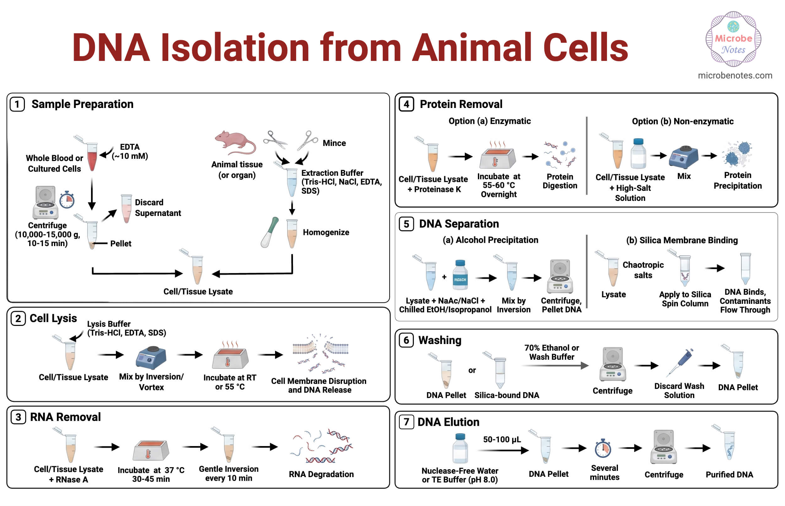
Step 1: Sample Preparation
For blood or cultured animal cells
- Transfer the required volume of whole blood or harvested cultured cells into a sterile centrifuge tube.
- Add EDTA to a final concentration of approximately 10 mM to inhibit DNase activity.
- Centrifuge at 10,000-15,000 x g for 10–15 min.
- Carefully discard the supernatant without disturbing the pellet.
- Repeat centrifugation if a visible cell pellet is not obtained (Lahiri & Nurnberger, 1991).
For animal tissue samples
- Excise a small piece of animal tissue (e.g., mouse tail or organ tissue).
- Mince the tissue finely using sterile scissors or a scalpel to increase surface area.
- Transfer minced tissue into a tube containing extraction buffer (Tris-HCl, NaCl, EDTA, SDS).
- Gently homogenize using a pestle or homogenizer, avoiding excessive foaming to prevent DNA shearing (STEMCELL Technologies, 2019; Green & Sambrook, 2023).
Step 2: Cell Lysis
- Add lysis buffer containing Tris-HCl, EDTA, and SDS to the cell pellet or tissue homogenate.
- Mix gently by inversion or brief vortexing until the sample is uniformly suspended.
- Incubate at room temperature or 55 °C to ensure complete disruption of plasma and nuclear membranes.
- EDTA chelates divalent cations, protecting DNA from nuclease-mediated degradation (Green & Sambrook, 2023).
- If immediate processing is not possible, resuspend samples in PBS and store at 4 °C temporarily (STEMCELL Technologies, 2019).
Step 3: RNA Removal
- Add RNase A to the lysate to a final concentration of approximately 100 µg mL⁻¹.
- Incubate at 37 °C for 30–45 min.
- Gently invert the tube every 10 min to ensure uniform RNA digestion.
- This step minimizes RNA contamination in the final DNA preparation (QIAGEN, 2023).
Step 4: Protein Removal
Enzymatic digestion approach
- Add Proteinase K to a final concentration of 100–200 µg mL⁻¹.
- Incubate at 55–60 °C for several hours or overnight.
- Proteinase K digests histones and other DNA-associated proteins, improving DNA purity and yield (Sigma-Aldrich, n.d.; Green & Sambrook, 2023).
Non-enzymatic approach
- Add a high-salt solution to the lysate.
- Mix gently to promote protein aggregation and precipitation.
- This method enables rapid isolation of high-molecular-weight DNA without enzymatic digestion (Lahiri & Nurnberger, 1991).
Step 5: DNA Separation
Choose one of the following DNA separation methods based on experimental needs:
Alcohol precipitation
- Add sodium acetate or NaCl to the lysate.
- Add chilled ethanol or isopropanol and mix gently by inversion.
- Incubate briefly and centrifuge to pellet the DNA.
Silica membrane binding
- Apply the lysate to a silica spin column in the presence of chaotropic salts.
- Allow DNA to bind selectively to the membrane while contaminants pass through (QIAGEN, 2023).
Step 6: Washing
- Wash the DNA pellet or silica-bound DNA using 70 % ethanol or manufacturer-provided wash buffers.
- Centrifuge and discard the wash solution carefully.
- Repeat washing if necessary to remove residual salts, detergents, and proteins.
- Ensure complete removal of ethanol to avoid inhibition of downstream enzymatic reactions (QIAGEN, 2023).
Step 7: DNA Elution
- Elute DNA using nuclease-free water or a low-salt buffer such as TE buffer (pH 8.0).
- Typical elution volumes range from 50–100 µL.
- Allow the elution buffer to remain in contact with DNA for several minutes before centrifugation to maximize recovery (Green & Sambrook, 2023).
Observations and Results
Typically, successful DNA isolation yields a clear or slightly viscous solution. Agarose gel electrophoresis reveals a prominent high-molecular-weight band with minimal smearing. Spectrophotometric analysis shows absorbance ratios consistent with high-purity DNA, confirming effective removal of proteins and other contaminants.
Modifications of DNA Isolation from Animal Cells
- Non-enzymatic extraction for rapid screening: This modification eliminates the use of proteinase K, thereby significantly reducing reagent cost and overall processing time. It is particularly useful for rapid screening studies where high-molecular-weight DNA is required for RFLP analysis rather than ultra-pure DNA (Lahiri & Nurnberger, 1991).
- Extended proteinase K digestion: Prolonged proteinase K digestion enhances lysis efficiency and protein removal, especially in fibrous, connective, or collagen-rich tissues. This modification increases DNA yield and purity by ensuring complete degradation of histones and structural proteins that tightly bind genomic DNA (STEMCELL Technologies, 2019).
- Silica column purification: The use of silica membrane–based spin columns improves consistency and reproducibility across samples. This modification allows selective binding of DNA while efficiently removing contaminants, making the isolated DNA highly suitable for sensitive downstream applications such as PCR, sequencing, and epigenetic analyses (QIAGEN, 2023).
- Adaptation for fixed or FFPE tissues: For fixed or formalin-fixed paraffin-embedded (FFPE) tissues, additional steps are incorporated to reverse formaldehyde-induced cross-linking. These modifications improve DNA recovery and fragment quality, enabling reliable molecular analysis from archived or preserved animal tissue samples (Green & Sambrook, 2023).
Troubleshooting of DNA Isolation from Animal Cells
| Problem | Likely Cause | Solution |
| Low DNA yield | Incomplete lysis or digestion | Increase incubation time or proteinase K concentration |
| DNA degradation | DNase activity | Ensure sufficient EDTA and gentle handling |
| Protein contamination | Inadequate protein removal | Extend digestion or add additional wash steps |
| RNA contamination | RNase omitted | Treat with RNase A |
| Poor downstream performance | Residual salts or ethanol | Ensure complete washing and drying |
(STEMCELL Technologies, 2019; QIAGEN, 2023; Green & Sambrook, 2023)
Quality Assessment of the Isolated DNA
- Spectrophotometric purity (A260/A280 ratio): Ratios ~1.8 indicate minimal protein contamination (QIAGEN, 2023).
- Agarose gel electrophoresis: High-molecular-weight DNA appears as a distinct band with minimal smearing (Lahiri & Nurnberger, 1991).
- Amplifiability by PCR: Confirms absence of inhibitors (Green & Sambrook, 2023).
Safety Tips and Precautions of DNA Isolation from Animal Cells
- Use personal protective equipment (PPE): Laboratory personnel should consistently wear gloves, lab coats, and protective eyewear during DNA extraction procedures. Proper use of PPE minimizes the risk of exposure to biological samples, chemical reagents, and accidental splashes or spills during sample processing.
- Handle detergents and chaotropic salts carefully: Detergents such as SDS and chaotropic salts used in lysis and binding buffers must be handled with caution, as they can cause skin or eye irritation. Proper labeling, careful pipetting, and appropriate waste disposal are essential for laboratory safety (QIAGEN, 2023).
- Avoid vigorous mixing: Excessive vortexing or harsh mechanical agitation should be avoided during DNA extraction, particularly after cell lysis. Vigorous mixing can cause mechanical shearing of high-molecular-weight genomic DNA, reducing fragment size and compromising downstream analytical performance.
Storage and Long‑Term Stability of Isolated DNA
- Short-term storage at 4 °C: Isolated DNA may be stored at 4 °C for short periods when immediate downstream applications such as PCR or restriction digestion are planned. This temperature slows enzymatic degradation while allowing convenient access without repeated freeze-thaw cycles.
- Long-term storage at −20 °C or −80 °C: For prolonged storage, DNA should be kept at −20 °C or −80 °C to minimize hydrolytic and enzymatic damage. These conditions preserve DNA integrity and concentration for months to years, ensuring reliability for future molecular analyses (Green & Sambrook, 2023).
- Use of TE buffer: Storage of DNA in TE buffer improves long-term stability by maintaining an optimal pH and chelating divalent cations through EDTA. This inhibits residual nuclease activity and reduces degradation during extended storage or repeated handling.
Applications of DNA Isolation from Animal Cells
- Genotyping and RFLP analysis: Genotyping and restriction fragment length polymorphism (RFLP) analyses require intact high-molecular-weight genomic DNA to ensure accurate restriction enzyme digestion and reliable interpretation of fragment patterns. Degraded DNA can lead to incomplete digestion or ambiguous banding profiles, affecting genetic analysis outcomes (Lahiri & Nurnberger, 1991).
- PCR and sequencing: Polymerase chain reaction and DNA sequencing require highly purified DNA free from protein, salt, or detergent contaminants. Such impurities can inhibit polymerase activity or interfere with signal detection, making high-quality DNA essential for reliable amplification, base calling, and downstream bioinformatic analysis (QIAGEN, 2023).
- Epigenetic and methylation studies: Epigenetic and DNA methylation analyses depend on minimally degraded genomic DNA to preserve native methylation patterns. Fragmentation or chemical damage can compromise bisulfite conversion efficiency and lead to inaccurate methylation profiling results (Green & Sambrook, 2023).
- Whole-genome amplification: Whole-genome amplification techniques require high-quality, intact DNA templates to ensure uniform amplification across the genome. Poor-quality or degraded DNA can introduce amplification bias, resulting in uneven genomic coverage and reduced reliability of downstream genomic analyses (Sigma-Aldrich, n.d.).
Advantages of DNA Isolation from Animal Cells
- High yield of genomic DNA: Suitable for multiple analyses.
- Flexibility across sample types: Applicable to blood, tissues, and cultured cells.
- Compatibility with modern molecular techniques: Particularly when kit-based purification is used.
Limitations of DNA Isolation from Animal Cells
- Time-consuming for tissue samples, especially with overnight digestion steps.
- Cost of commercial kits: May limit use in resource-restricted settings.
- Risk of DNA shearing: If handling is not gentle.
Conclusion
DNA isolation from animal cells is a critical prerequisite for molecular and genomic analyses. The methods discussed-ranging from rapid non-enzymatic protocols to enzymatic and silica-based approaches-offer flexibility depending on research needs. By understanding the underlying principles, reagent functions, and potential pitfalls, researchers can reliably obtain high-quality genomic DNA suitable for a wide array of applications.
References
- Green, M. R., & Sambrook, J. (2023). Isolation of genomic DNA from mammalian cells and fixed tissue. Current Protocols, 3(7), e818. https://doi.org/10.1002/cpz1.818
- Lahiri, D. K., & Nurnberger, J. I., Jr. (1991). A rapid non-enzymatic method for the preparation of HMW DNA from blood for RFLP studies. Nucleic Acids Research, 19(15), 4043. https://doi.org/10.1093/nar/19.19.5444
- QIAGEN. (2023). DNeasy blood & tissue handbook. QIAGEN. https://www.qiagen.com/us/resources/resourcedetail?id=68f29296-5a9f-40fa-8b3d-1c148d0b3030&lang=en
- Sigma-Aldrich. (n.d.). Animal tissue DNA extraction & whole genome amplification protocol. Merck KGaA. https://www.sigmaaldrich.com/US/en/technical-documents/protocol/genomics/dna-and-rna-purification/extraction-protocol-animal-tissue
- STEMCELL Technologies. (2019). Protocol for genomic DNA isolation from mouse tail, animal tissue, or cultured cells. STEMCELL Technologies. https://www.stemcell.com/protocol-for-genomic-dna-isolation-from-mouse-tail-animal-tissue-or-cultured-cells.html
- Brown, T. A. (2020). Gene cloning and DNA analysis: An introduction (8th ed.). Wiley-Blackwell.
- Kathmandu University. (2025). Phenol–chloroform extraction manual for human urine samples. Unpublished laboratory protocol.
