
Campylobacter jejuni is a bacterium that causes campylobacteriosis, typically residing in the intestinal tracts of animals- especially poultry.
Transmission to humans mainly occurs through undercooked meat, contaminated water, and unpasteurized milk. Infection often results in symptoms such as fever, diarrhea, abdominal pain, cramps, and nausea. In some cases, complications like Guillain-Barré syndrome may develop.
Taxonomy and Classification of Campylobacter jejuni
Domain: Bacteria
Kingdom: Pseudomonadati
Phylum: Campylobacterota
Class: Campylobacteria
Order: Campylobacterales
Family: Campylobacteraceae
Genus: Campylobacter
Species: C. jejuni
Morphology and Microscopy of Campylobacter jejuni
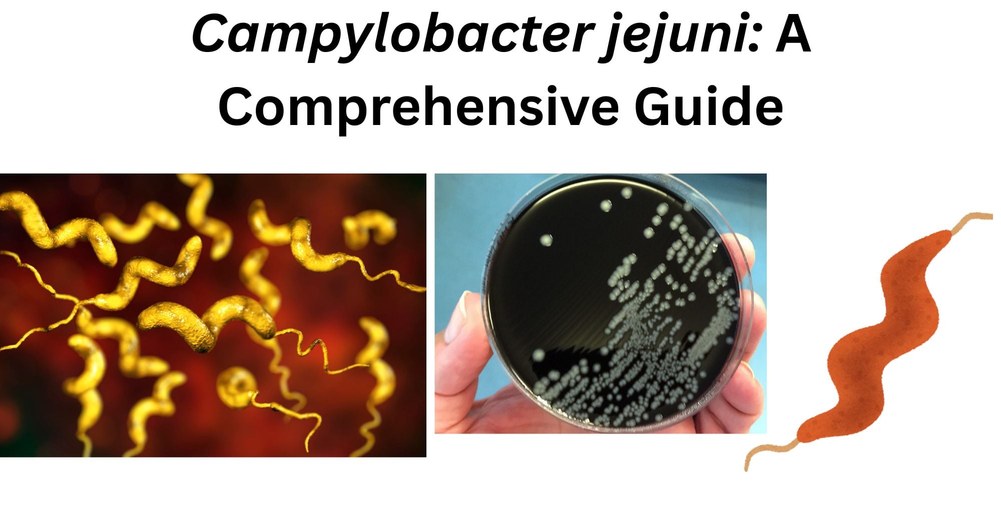
- Gram-negative, curved, rod or spiral-shaped.
- It shows a typical gull-winged shape.
- Approximately 0.2-0.8 µm width and 0.5-5.0 µm length.
- Motile with amphitrichous flagella.
- Non-spore-forming
Under the microscope, after Gram staining, Campylobacter appears as slender, spiral, or curved Gram-negative rods, often displaying a distinctive gull-wing or S-shaped appearance.
Cultural and Growth Characteristics of Campylobacter jejuni
- Microaerophilic (requires 3-10% O2) and capnophilic (requires 5-10% Co2).
- Optimum temperature: 30˚C to 47˚C
- Optimum pH: 5.5 to 7
- It requires selective media to grow.
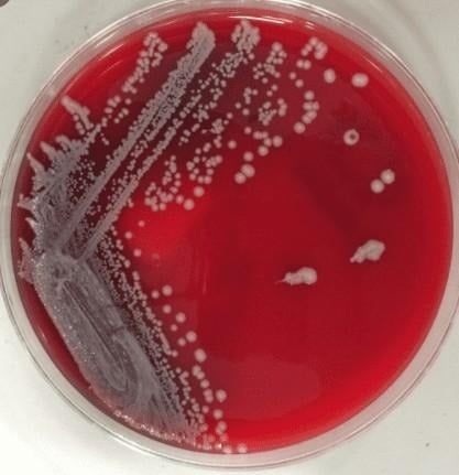
- Skirrow’s Media (Horse Blood Agar): small, gray, smooth surface, flat to low-convex, moist and slightly translucent, and may show slight spreading on the medium.
- This selective medium contains antibiotics including vancomycin, polymyxin B, cephalothin, and trimethoprim, which inhibit the growth of most non-Campylobacter bacteria, thereby facilitating the isolation of Campylobacter species from clinical specimens.
- Sheep Blood Agar: small, gray, flat, smooth, moist, non-hemolytic colonies are formed.
- This selective medium contains antibiotics including bacitracin, colistin, cephalothin, and actidione, which inhibit the growth of most non-Campylobacter bacteria and fungi, thereby enabling the isolation of Campylobacter species from clinical specimens.
- Butzler Medium: small, round or irregular in shape, smooth, moist, translucent to slightly opaque colonies are formed.
Biochemical Tests of Campylobacter jejuni
| Tests | Results |
| Gram Staining | Negative |
| Catalse | Positive |
| Oxidase | Positive |
| H2S | Negative |
| KOH | Positive |
| Nitrate Reduction | Positive |
| Nitrite Reduction | Negative |
| OF (Oxidative-Fermentative) | Non-fermentative |
| Urease | Negative |
| Fermentation of | |
| Glucose | Negative |
| Dnase | Negative |
| Enzymatic Reactions | |
| Hippurate | Positive |
| Indoxyl Acetate | Positive |
| Alkaline Phosphatase | Variable |
| Arylsulphatase | Negative |
| Tween 40 | Positive |
| Tween 60 | Positive |
| Tween 80 | Negative |
Pathogenesis of Campylobacter jejuni
- Campylobacter infection is most commonly transmitted by consuming contaminated food, particularly undercooked poultry, pork, unpasteurized milk, or untreated water. Direct contact with infected animals can also serve as a source of infection.
- Bacteria withstand gastric acid and reach the ileum and jejunum of the small intestine.
- In response to unfavorable conditions, Campylobacter produces virulence proteins, including toxins and invasion factors, which damage host cells and cause inflammation of the intestinal epithelium.
- These proteins, referred to as Campylobacter virulence-associated factors, include adhesion molecules, chemotaxis proteins, flagellar components, cytolethal distending toxin, and invasion factors, all of which contribute to the bacterium’s ability to colonize and cause disease in the host.
- The bacteria demonstrate corkscrew-like motility, which facilitates colonization of the host. Their polar flagella and chemoreceptors enable effective movement through the mucus lining of the gastrointestinal tract.
- Cytolethal distending toxin (CDT) consists of three subunits—CdtA, CdtB, and CdtC—each of which contributes to the toxin’s pathogenic effects through distinct mechanisms.
- The CdtA and CdtC subunits function as binding components, helping to deliver the CdtB subunit to enterocytes.
- The CdtB subunit acts as a DNase, causing DNA damage in enterocytes. This disrupts normal cell function, leads to cell cycle arrest, and ultimately results in a reduction in the number of enterocytes.
- Campylobacter displays specific adhesion proteins on its cell surface that enable the bacterium to bind to and colonize epithelial cells of the host.
- Fibronectin domain-containing lipoproteins such as FlpA, CadF, JlpA, and FlaA are proteins that assist in the attachment of the pathogens.
- Likewise, it produces 4 proteins, viz. CiaB, CiaC, CiaD, and Cial, which support the internalization of the pathogen.
- After entering the host cell, the bacterium propagates in a membrane-bound vacuole called a Campylobacter-containing vacuole.
- Then the human immune system activates the production of IL-8, which triggers the T-cells and B-cells.
- The host immune response causes local inflammation, and the Cia protein destroys the tight connection, causing diarrhea.
Virulence Factors of Campylobacter jejuni
- Flagella
-It aids in the bacterial penetration of intestinal mucus.
-It assists in bacterial colonization of the epithelial surface.
- Chemotaxis
-It allows bacteria to shift towards suitable conditions or away from harmful situations, causing colonization of optimal niches by bacteria.
- Adhesions
-It is the surface proteins that help in the attachment of bacteria to intestinal epithelial cells.
-It is crucial for colonization and persistence.
- Invasion Proteins
-It enhances the entry of bacteria into host epithelial cells.
-It promotes intracellular survival and spread.
- Lipooligosaccharide (LOS)
-It functions as an endotoxin and triggers a strong inflammatory response.
- Cytolethal Distending Toxin (CDT)
-It causes DNA damage and leads to cell cycle arrest and apoptosis.
-It contributes to mucosal injury.
- Campylobacter invasion proteins (Cia proteins)
-It helps in the invasion of cells and intracellular survival.
Epidemiology of Campylobacter jejuni
Initially, in 1886, Escherich witnessed bacteria similar to Campylobacter in children’s stool samples with diarrhea.
Similarly, in Belgium, a clinical microbiologist first identified Campylobacter from samples of stool with diarrhea in 1972. C. jejuni is ubiquitous and is commonly found in both developed and developing countries, being the leading cause of bacterial gastroenteritis in developing countries.
C. jejuni infections exhibit seasonal trends, with increased cases during late summer and early fall in developed countries, regardless of the reason. Infection is mainly caused by the handling and consumption of contaminated raw meat. Chicken carcasses have a high number of Campylobacter, which can be easily transmitted during the preparation of food. Most of the outbreaks are sporadic, accounting for only a small number of cases. However, infections are endemic in developing countries, with the most symptomatic cases occurring in young children.
Nearly 1 in 10 people fall sick, and approximately 33 million people die every year from campylobacteriosis caused by C. jejuni. Almost 550 million people suffer from diarrhoeal diseases, with 220 million children under 5 years old. Food-borne diseases can be severe in children under the age of 2 years, sometimes leading to death. Around 2.1 to 2.4 million cases of campylobacteriosis take place every year in the United States.
The prevalence of campylobacteriosis is higher in HIV-infected patients than in normal individuals. Between 1983 and 1987, the campylobacteriosis incidence was reported to be 519 cases per 100,000 in Los Angeles, which is 39 times higher than that found in normal individuals.
Transmission of Campylobacter jejuni
The transmission of C. jejuni takes place primarily through the fecal-oral route, generally through contaminated water, food, or indirect contact with infected animals.
Food-borne Transmission
-It is the most common mode of transmission.
-Bacteria spread through the consumption of undercooked or raw poultry, mainly chicken.
-Cross-contamination can occur from raw meat to ready-to-eat foods while preparing and handling foods.
-The bacterium can also spread through the consumption of unpasteurized milk and dairy products.
Waterborne Transmission
-Bacteria can spread via consumption of contaminated water or recreational water from streams and lakes.
-It is generally associated with poor sanitation and agricultural runoff.
Animal-to-Human Transmission
-Reservoirs involve poultry, sheep, cattle, dogs, and cats.
-Humans get an infection via direct contact with infected feces or animals.
Person-to-Person Transmission
-It is very rare but can occur in settings with poor hygiene, like daycare centers or old-age homes.
-Bacteria can spread through fecal contamination of hands or surfaces.
Clinical Manifestations of Campylobacter jejuni
Symptoms generally start 2-5 days after ingestion of Campylobacter and resolve within 7 days.
Acute Gastroenteritis
Generally, lasts for 5-7 days, but may last longer in some cases, and symptoms are:
- Abdominal Pain: severe, crampy pain similar to appendicitis.
- Fever: ranges from 38-40˚C
- Diarrhea: initially watery but often bloody
- Nausea and Vomiting
- Malaise and Fatigue
Intestinal Infection
- Causes inflammation of the jejunum, ileum, and colon.
- Campylobacter infection can result in ulceration and mucosal damage of the intestinal lining, which may cause a dysentery-like illness characterized by bloody diarrhea and abdominal pain.
Dehydration
Signs in children and adults:
- Not peeing often
- Feeling very thirsty
- Warm skin
- Having very dark pee
- Feeling dizziness
Signs in babies and toddlers:
- Less peeing
- Fewer or no tears while crying
- Lack of interest in playing activities
- Extreme sleepiness
Complications
Complications include:
- Irritable Bowel Syndrome
- Reactive Arthritis
- Guillain-Barré Syndrome (GBS) can cause weakness in muscles and tingling, which appear first in the legs, then in the arms and upper body. Loss of reflexes, pain, problems in breathing, and facial and speech issues are the common symptoms of GBS. In severe cases, it may progress to paralysis.
People with a weakened immune system may develop severe complications such as bloodstream infection, which can lead to a hazardous immune reaction known as sepsis. Signs of sepsis include:
- Fever more than 103˚F
- Confusion or disorientation
- Fast Heart Rate
- Difficulty breathing
- Excessive pain or discomfort
Laboratory Diagnosis of Campylobacter jejuni
Sample Collection and Transportation
Samples for detection of C. jejuni include:
- Fresh stool samples
- Rectal Swab: if the stool sample is unavailable
- Blood: rarely collected, primarily in suspected cases of bacteremia or sepsis, especially in immunocompromised patients
- Intestinal biopsy: in rare cases
The sample should be collected in a clean, sterile container and transported in Cary-Blair medium. The sample should be stored at 4˚C if the processing is delayed, and should be avoided from drying and exposure to oxygen.
Microscopy
- Direct analysis of a stool sample using contrast microscopy or Gram staining.
- After staining, it is observed as a comma-shaped or spiral-shaped gram-negative bacterium under a microscope.
- Although it provides a rapid presumptive diagnosis, it can be confirmed only by stool culture.
Culture
- Skirrow’s Media (Horse Blood Agar): small, gray, smooth surface, flat to low convex, moist and slightly translucent, and may show slight spreading in the medium.
- Sheep Blood Agar: small, gray, flat, smooth, moist, non-hemolytic colonies are formed.
- Butzler Medium: small, round or irregular in shape, smooth, moist, translucent to slightly opaque colonies are formed.
Biochemical Tests of Campylobacter jejuni
After culture, colonies from incubated plates are tested for biochemical tests and identified as C. jejuni based on the following results:
| Tests | Results |
| Gram Staining | Negative |
| Catalse | Positive |
| Oxidase | Positive |
| H2S | Negative |
| KOH | Positive |
| Nitrate Reduction | Positive |
| Nitrite Reduction | Negative |
| OF (Oxidative-Fermentative) | Non-fermentative |
Molecular Methods of Campylobacter jejuni
Polymerase Chain Reaction (PCR)
- Reverse transcriptase polymerase chain reaction (RT-PCR) is used for the identification of bacteria from stool.
- It identifies Campylobacter 20% to 40% more often than the traditional culture-based methods.
- This method is rapid and highly sensitive.
Nucleic Acid Amplification Tests (NAAT)
- It is used for the detection of specific Campylobacter spp.
- It is more rapid and does not require a live organism for detection.
Immunoassays
- It is a rapid method and is used for the detection of Campylobacter jejuni-specific antigen in a stool sample.
Treatments of Campylobacter jejuni
Infection by Campylobacter jejuni is generally mild and self-limiting, and treatment is typically supportive. Most of the infections resolve without the use of antibiotics; however, in some severe cases, antibiotics are used.
Supportive Treatment
- Oral Rehydration Therapy: It is done to prevent dehydration caused by diarrhea.
- Intravenous Fluid: It is used for severe dehydration.
- Antipyretics and analgesics such as ibuprofen are used to manage pain and fever.
Antibiotic Therapy
- Azithromycin, Erythromycin, or Ciprofloxacin are drugs of choice.
Antimotility drugs such as loperamide are used, especially in patients with bloody diarrhea in severe cases.
Patients with Guillain-Barré Syndrome require hospitalization and specialized care.
Patients with reactive arthritis are managed with anti-inflammatory drugs.
Prevention and Control of Campylobacter jejuni
- Properly cook poultry, especially chicken, before consumption.
- Always be cautious while eating seafood or raw fish.
- Avoid consumption of unpasteurized milk or dairy products.
- Wash utensils and surfaces properly and use separate cutting boards for raw meat and ready-to-eat foods to prevent cross-contamination.
- Always drink treated and clean water.
- Avoid consumption of untreated water from rivers, springs, lakes, or wells.
- Always maintain proper sanitation systems to prevent contamination of water sources.
- Properly wash hands with soap and water after handling raw meat, after using the toilet, and before eating or preparing food.
- Always maintain good hygiene while handling animals.
- Avoid direct contact with the feces of animals and use protective measures for people working in farms and slaughterhouses.
- Do not allow a sick person to handle and prepare food until symptoms disappear.
- Properly wash your hands after touching your pets, their food, water, bed, toys, poop, or pee.
Antimicrobial Resistance of Campylobacter jejuni
The increase in antimicrobial-resistant strains of C. jejuni makes the management of campylobacteriosis hard. The resistance can extend illness and can compromise the treatment of patients suffering from bacteremia. The rate of antimicrobial resistance is highest in the developing world, where the use of antibiotics is restricted in humans and animals.
A study conducted in 1994 demonstrated that the C. jejuni isolated from the U.S troops in Thailand showed resistance to ciprofloxacin. Similarly, around one-third of bacteria isolated from U.S troops located in Hat Yai showed resistance towards azithromycin. An experiment conducted in chickens demonstrated that the fluoroquinolone-susceptible C. jejuni showed resistance to these drugs when administered.
In Europe, during the early 1990s, after the approval of the use of fluoroquinolones in poultry, resistant strains developed promptly in humans. Likewise, in 1995, within 2 years of approval of poultry use of fluoroquinolones in the U.S., the number of domestically obtained human cases of ciprofloxacin-resistant infection doubled in Minnesota.
Also, a study conducted in 1997, in Minnesota, showed that out of 60 C. jejuni isolates, 12 were ciprofloxacin-resistant, which were detected in chicken obtained from grocery stores.
Conclusion
C. jejuni is a Gram-negative, microaerophilic, curved bacterium, generally transmitted through contaminated food, especially poultry, and unpasteurized milk or water. It is one of the leading causes of bacterial gastroenteritis worldwide, which colonizes the intestinal tract and causes acute inflammatory diarrhea, abdominal cramps, fever, and sometimes bloody stools.
Even though most infections are self-limiting, sometimes they can lead to serious complications such as Guillain-Barré Syndrome, irritable bowel syndrome, and reactive arthritis. Its increasing resistance towards fluoroquinolones emphasizes the significance of food hygiene, safe cooking practices, and supportive treatment for prevention and control.
References
- Johns Hopkins Medicine. (n.d.). Campylobacter jejuni. Retrieved April 19, 2026, from https://www.hopkinsmedicine.org/health/conditions-and-diseases/campylobacter-jejuni
- Gundogdu, O., & Wren, B. W. (2020). Microbe profile: Campylobacter jejuni – survival instincts. Microbiology, 166(3), 230–232. https://doi.org/10.1099/mic.0.000906
- Aryal, S. (2022). Biochemical test of Campylobacter jejuni. Microbe Notes. https://microbenotes.com/biochemical-test-of-campylobacter-jejuni/
- Tikhomirova, A., McNabb, E. R., Petterlin, L., Bellamy, G. L., Lin, K. H., Santoso, C. A., Daye, E. S., Alhaddad, F. M., Lee, K. P., & Roujeinikova, A. (2024). Campylobacter jejuni virulence factors: Update on emerging issues and trends. Journal of Biomedical Science, 31, 45. https://doi.org/10.1186/s12929-024-01033-6
- Tikhomirova, A., McNabb, E. R., Petterlin, L., Bellamy, G. L., Lin, K. H., Santoso, C. A., Daye, E. S., Alhaddad, F. M., Lee, K. P., & Roujeinikova, A. (2024). Campylobacter jejuni virulence factors: Update on emerging issues and trends. Journal of Biomedical Science, 31, 45. https://doi.org/10.1186/s12929-024-01033-6
- World Health Organization. (2020, May 1). Campylobacter. https://www.who.int/news-room/fact-sheets/detail/campylobacter
- Altekruse, S. F., Stern, N. J., Fields, P. I., & Swerdlow, D. L. (1999). Campylobacter jejuni—An emerging foodborne pathogen. Emerging Infectious Diseases, 5(1), 28–35. https://doi.org/10.3201/eid0501.990104
https://wwwnc.cdc.gov/eid/article/5/1/99-0104_article - Centers for Disease Control and Prevention. (2024, May 13). Symptoms of Campylobacter infection. https://www.cdc.gov/campylobacter/signs-symptoms/index.html
- Cleveland Clinic. (n.d.). Campylobacter infection. Cleveland Clinic. https://my.clevelandclinic.org/health/diseases/15251-campylobacter-infection
- Association of Public Health Laboratories. (2023). Campylobacter isolation and characterization: Diagnosis recommendations. https://www.aphl.org/docs/default-source/technical/FS-Campylobacter-Diagnosis-Recommendations.pdf
- Marler Clark. (n.d.). Diagnosis of a Campylobacter infection. Marler Clark. https://marlerclark.com/foodborne-illnesses/campylobacter/campylobacter-diagnosis
- Altekruse, S. F., Stern, N. J., Fields, P. I., & Swerdlow, D. L. (1999). Campylobacter jejuni—An Emerging Foodborne Pathogen. Emerging Infectious Diseases, 5(1), 28-35. https://doi.org/10.3201/eid0501.990104.

Microbiology 1st chapter notes