
Habitat of Streptococcus pneumoniae
- Streptococcus pneumoniae are found worldwide.
- Found in primates, livestock and felines.
- They are the part of normal flora of upper respiratory tract infection in humans.
- Mostly found in throat and nasal passage.
- The infection mostly affects children in winter seasons.
- They are Mesophilic, 30-35°C.
Morphology of Streptococcus pneumoniae
- They are Gram-positive bacteria.
- May lose gram +ve character in older cultures and appear gram –ve.
- They are non-sporing and non-motile bacteria.
- May have pili for adherence.
- They are 0.5 x 1.25 µm in diameter.
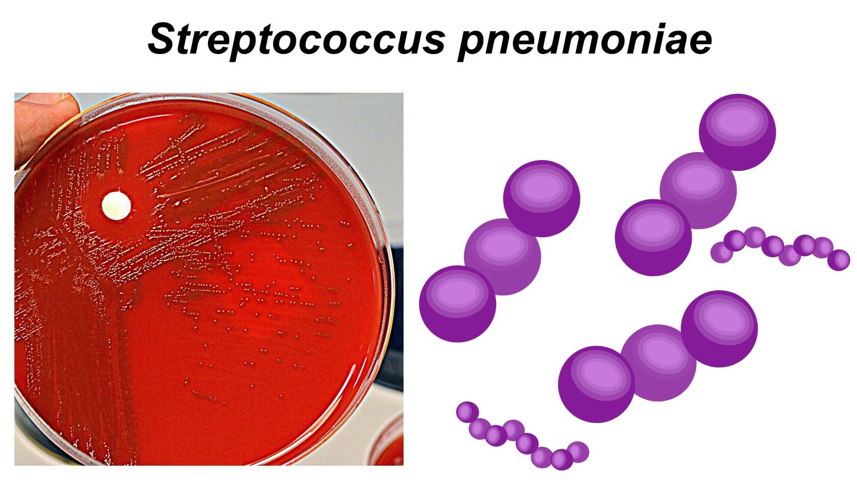
- They are mostly found in pairs (diplococci).
- Also found singly and in short chains isolated from body liquids.
- They are lancet-shaped or bullet-shaped.
- They are capsulated.
- Capsules has 90+ serotypes.
- They are alpha (α) hemolytic.
- They contains more than 500 different surface proteins.
Genomes of Streptococcus pneumoniae
- Single circular chromosome.
- 2 million base pair.
- GC Content: 39.7%.
- Codes for 1914 proteins and 73 RNA’s.
Cultural characteristics of Streptococcus pneumoniae
- The growth of most Streptococcus species on simple nutrient agar is poor in contrast to other Gram-positive bacteria.
- For the efficient growth of S. pneumoniae, the media is supplemented with blood, serum or fermentable carbohydrate source. This is required for primary isolation of the bacteria.
- On artificial medium, S. pneumoniae requires a supplement of carbon dioxide during incubation. Continued growth on the medium results in chain formation in the bacteria.
- The species are susceptible to vancomycin that can exhibit growth in the presence of 6.5% NaCl-containing agar medium.
- The complex nutritional requirements of S. pneumoniae include amino acids, peptides, purines, pyrimidine and vitamins.
- In order to avoid competition and inhibition of Gram-positive bacteria, Selective Strep Agar is used as a selective media. Besides, Crystal Violet-Nalidixic acid-Gentamycin agar is also used as a selective medium for the isolation of S. pneumoniae.
- Some strains of S. pneumoniae can grow at 40% bile which enables the production of pigments in the presence of 5% carbon dioxide.
- The ideal growth of S. pneumoniae occurs between 30-35°C, but no growth is observed below 10°C or above 45°C.
- S. pneumoniae mostly favour acidic and neutral pH, but no growth occurs at alkaline pH of 9.
- Common tests used for the differentiation of S. pneumoniae include esculin hydrolysis and the ability to survive at high concentration of bile.
- The following are some cultural characteristics of S. pneumoniae on different culture media:
- Nutrient Agar
- The colonies of S. pneumoniae are small, circular and dome-shaped or flattened. Glistening or mucoid colonies are observed due to the production of the capsule.
- The colour of the colonies is clear or whitish or light, depending on the strains. The structure of the colonies is transparent or opaque.
- On prolonged incubation, the colonies become flat with raised edges and a central combination of concentric rings.
- Blood Agar
- On blood agar, the colonies of S. pneumoniae are typically smooth and non-pigmented with an entire margin.
- The growth of S. pneumoniae occurs readily on blood agar and exhibits α-hemolysis in the form of opaque colonies surrounded by an area of green colouration.
Biochemical characteristics of Streptococcus pneumoniae
Virulence factors of Streptococcus pneumoniae
Polysaccharide capsule
- The capsule is antiphagocytic, inhibiting complement deposition and phagocytosis.
Cell wall associated polymers and proteins
- Teichoic acid – binds to epithelial cells and activates alternative complement pathway
- Protein adhesion – binds to epithelial cells
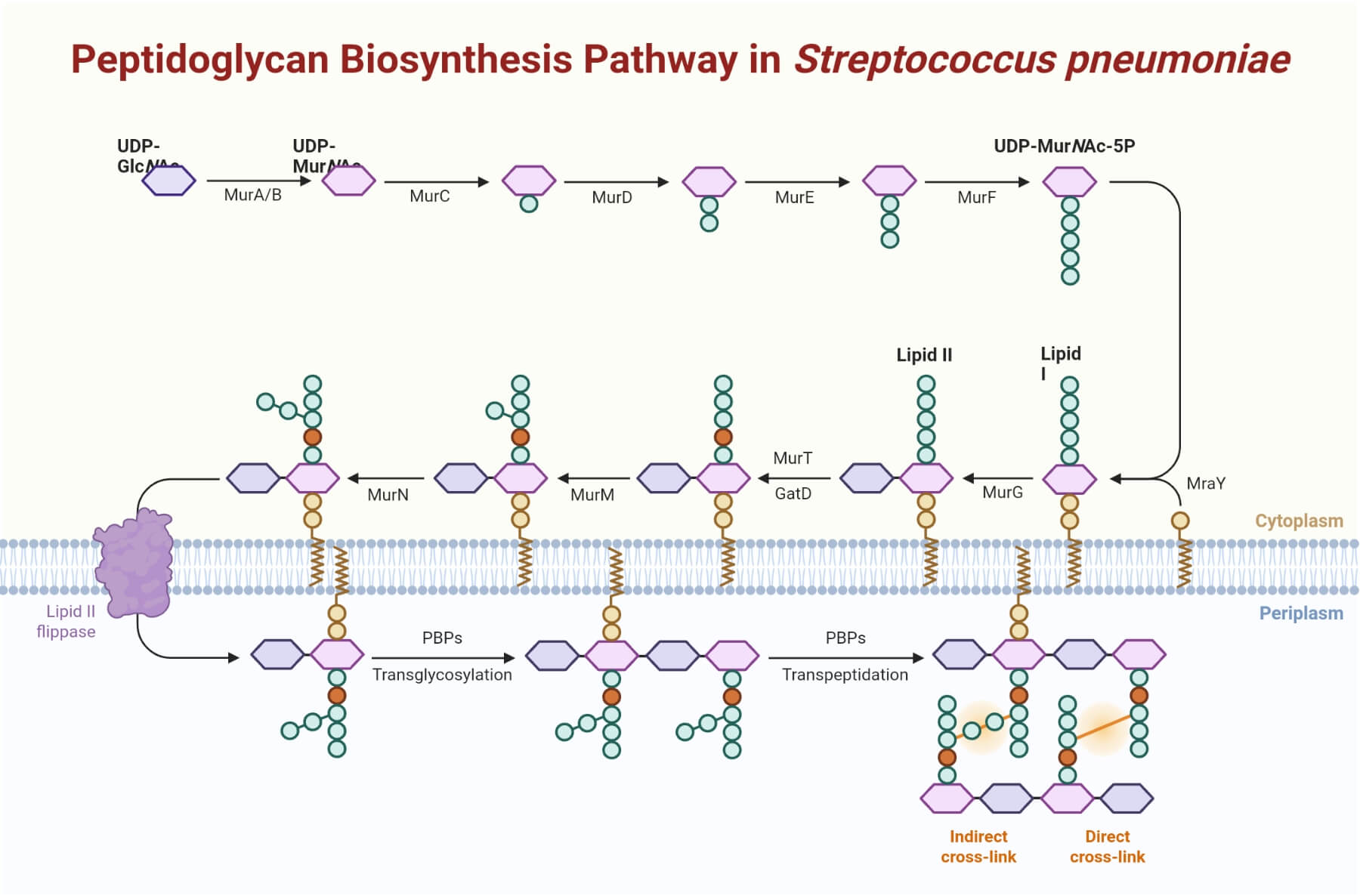
- Peptidogylcan – activates alternative complement pathway
- Phosphorylcholine – mediates invasion of host cell
- F protein – mediates attachment to epithelial cells
Pneumolysin
- It is intracellular membrane-damaging toxin produced by autolysis.
- Pneumolysin binds to cholesterol and therefore interacts indiscriminately with all cell types.
- This toxin stimulates production of proinflammatory cytokines.
- Pneumolysin inhibits:
- neutrophil chemotaxis
- phagocytosis and the respiratory burst
- lymphocyte proliferation and immunoglobulin synthesis.
Pili
- Pili enable the attachment of encapsulated pneumococci to the epithelial cells of the upper respiratory tract.
- However it is not present in all strains.
IgA1 protease
- It is extracellular protease that specifically cleaves human IgA1 in the hinge region.
- This protease enables these pathogens to evade the protective functions of the principal immunoglobulin isotype of the upper respiratory tract.
Autolysins
- Autolysins are enzymes that hydrolyze the components of a biological cell in which it is produced.
- When activated, the pneumococcal breaks the peptide cross-linking of the cell wall peptidoglycan, leading to lysis of the bacteria.
- Autolysis enables the release of pneumolysin and, in addition, large amounts of cell wall fragments.
Pathogenesis of Streptococcus pneumoniae
- Transmission from a sick person, or more commonly from an asymptomatic carrier, occurs through droplets of respiratory secretions that remain airborne over distances of a few feet.
- Infecting organisms can also be carried on hands contaminated with secretions.
- S. pneumoniae colonizes the oropharynx by adhering to the epithelial cells of pharynx.
- This adhesion is mediated by pneumococcal neuraminidase or by pneumococcal cell-surface ligands of the cocci called adhesins.
- Subsequent migration of the organism to the lower respiratory tract can be prevented if the bacteria are enveloped in mucus and removed from the airways by the action of ciliated epithelial cells.
- The bacteria counteract this envelopment by producing secretory IgA protease and
- The secretory IgA protease destroys the secretory IgA and thereby enhances the ability of the cocci to colonize mucosa of the upper respiratory tract.
- Pneumolysin, on the other hand, destroys the ciliated epithelial cells and phagocytic cells by binding to cholesterol in the epithelial cell membrane.
- This activity can destroy the ciliated epithelial cells and phagocytic cells.
- The process of tissue destruction is mediated by factors, such as cell wall teichoic acid, peptidoglycan, and phosphorylcholine.
- The destructive action is further supplemented by hydrogen peroxide produced by the bacteria caused by reactive oxygen intermediates.
- Teichoic acid and the peptidoglycan fragments activate the alternative complement pathway, producing C5a, which being chemotaxic attracts neutrophils to the site of inflammation and mediates the inflammatory process.
- Migration of inflammatory cells to the site of infection is the key feature of pneumococcal infection.
- In turn, cytokines, such as interleukin-1 (IL-1) and tumor necrosis factor-α (TNF-α), are produced by the activated leukocytes, leading to the further migration of inflammatory cells to the site of infection, fever, tissue damage, and other signs characteristic of pneumococcal infection.
- Finally, phosphorylcholine present in the bacterial cell wall can bind to receptors for platelet-activating factor that are expressed on the surface of endothelial cells, leukocytes, platelets, and tissue cells, such as those in the lungs and meninges.
- By binding these receptors, the bacteria can enter the cells, where they are protected from opsonization and phagocytosis, and pass into sequestered areas, such as blood and the central nervous system.
- This activity facilitates the spread of disease.
- S. pneumoniae survives phagocytosis because of the antiphagocytic protection afforded by its capsule and the pneumolysin-mediated suppression of the phagocytic cell oxidative burst, which is required for intracellular killing.
- Antibodies directed against the type-specific capsular polysaccharides protect against disease caused by immunologically related strains.
- These antibodies appearing in serum 5–8 days after the onset of infection are protective against the pneumococcal serotype causing the infection.
- Humoral factors, including antibodies, complement, and perhaps C reactive protein (CRP), assist macrophages in the spleen, liver, and lymph nodes in carrying out their filtering function.
Clinical manifestations of Streptococcus pneumoniae
A. Pneumonia
- Pneumonia is a leading cause of death, especially in older adults and those whose resistance is impaired and is caused most frequently by S. pneumonia.
- Since the disease is associated with aspiration and is localized in the lower lobes of the lungs, it is called lobar pneumonia.
- However, children and the elderly can have a more generalized bronchopneumonia.
- Pneumococcal pneumonia develops when the bacteria multiply in the alveolar spaces.
- Erythrocytes, leaking from congested capillaries, accumulate in the alveoli, followed by the neutrophils, then the alveolar macrophages.
- The onset of the clinical manifestations of pneumococcal pneumonia is abrupt, consisting of a severe shaking chill and sustained fever of 39° C to 41° C.
- Most patients have a productive cough with blood-tinged sputum, and they commonly have chest pain (pleurisy).
- Occasionally, lung necrosis and intrapulmonary abscess formation occur with the more virulent pneumococcal serotypes.
- Pleural effusions are seen in approximately 25% of patients with pneumococcal pneumonia and empyema (purulent effusion) is a rare complication.
B. Sinusitis and otitis media
- The disease is usually preceded by a viral infection of the upper respiratory tract, after which polymorphonuclear neutrophils (leukocytes) (PMNs) infiltrate and obstruct the sinuses and ear canal.
- The viral infection lowers the mucosal immunity, facilitating the invasion by S. pneumonia.
- Sinusitis caused by the pneumococci occurs in patients of all ages, but middle ear infections (otitis media) caused by the bacteria is seen only in young children.
C. Bacteremia
- In the absence of a focus of infection bacteremia/sepsis is commonly caused by pneumococcus, especially in individuals who are functionally or anatomically asplenic.
- In contrast, bacteria are generally not present in the blood of patients with sinusitis or otitis media.
- Bacteremia from pneumonia has a triad of severe complications: meningitis, endocarditis, and septic arthritis.
D. Meningitis
- S. pneumoniae is among the three leading causes of bacterial meningitis.
- Meningitis is always secondary to other pneumococcal infections, such as pneumonia, bacteremia, infections of the ear or sinuses.
- Pneumococcus is the most common cause of pyogenic meningitis in children, although the condition can occur in all age groups.
- The bacteria reach the brain through blood stream or from nasopharynx (following head trauma or dural tear particularly with cerebrospinal fluid leak).
- Meningitis caused by S. pneumoniae is associated with a higher mortality and more neurological complications than the meningitis caused by any other bacteria.
Laboratory diagnosis of Streptococcus pneumoniae
Specimen: Sputum, blood, endotracheal aspirate, bronchoalveolar lavage, cerebrospinal fluid (CSF), pleural fluid, joint fluid, abscess fluid, bones, and other biopsy material.
Microscopy
- Gram staining of sputum shows lancet shaped Gram-positive cocci in pairs.
- Fresh emulsified sputum mixed with antiserum causes capsule swelling (the quellung reaction) for identification of pneumococci.
- In acute pneumococcal otitis media, Gram stain of an aspirated fluid smear from middle ear is useful to demonstrate the bacteria.
Culture
- Sputum or blood is plated on blood agar and incubated at 37° C in the presence of 5–10% carbon dioxide.
- Gray colonies with alpha-hemolysis are observed after overnight incubation.
- Diagnosis of pneumococcal meningitis is confirmed by CSF culture.
Identification of bacteria
- Optochin sensitivity test
- S. pneumoniae is identified by its sensitivity to optochin (ethylhydrocupreine dihydrochloride).
- The isolate is streaked onto a blood agar plate and a disk saturated with optochin is placed in the middle of the inoculum.
- A zone of inhibited bacterial growth is seen around the disk after overnight incubation.
- Bile solubility test
- It detects an autolytic enzyme, amidase, present in pneumococci, which breaks the bond between alanine and muramic acid of the peptidoglycan of the pneumococcal cell wall.
- Isolates of S. pneumoniae are lysed rapidly when the autolysins are activated after exposure to bile.
- Thus the organism can be identified by placing a drop of bile on an isolated colony.
- Inulin fermentation test
- It ferments inulin and hence differentiate it from other streptococci.
Animal inoculation
- S. pneumoniae can be isolated from clinical specimens containing few pneumococci by intraperitoneal inoculation in mice.
- Pneumococci are demonstrated in the peritoneal exudate and heart blood of the mice, which die 1–3 days after inoculation.
Antigen detection
- Pneumococcal C polysaccharide is excreted in urine and can be detected using a commercially prepared immunoassay.
- The CIEP is a useful test to detect pneumococcal capsular polysaccharide antigen in the CSF for diagnosis of meningitis, and in the blood or urine for diagnosis of bacteremia and pneumonia.
- Latex agglutination test using the latex particles coated with anti-CRP antibody is employed to detect C reactive protein.
- The CRP is used as a prognostic marker in acute cases of acute pneumococcal pneumonia, acute rheumatic fever, and other infectious diseases.
Antibody detection
- The indirect hemagglutination, indirect fluorescent antibody test, and ELISA are used to demonstrate specific pneumococcal antibodies in invasive pneumococcal diseases.
Nucleic Acid–Based Tests
- Nucleic acid probes and PCR assays are used for identification of S. pneumoniae isolates in culture.
Treatment of Streptococcus pneumoniae
- Most pneumococci are susceptible to penicillin.
- Other antibiotics, such as macrolides or selected fluoroquinolones with activity against pneumococci, are available for patients who are allergic to penicillin.
- Penicillin-resistant pneumococci are being increasingly documented.
- The mechanism of this resistance is an alteration of one or more of the bacterium’s penicillin-binding proteins (PBPs) rather than production of beta-lactamase.
- Most resistant strains remain sensitive to third generation cephalosporins (such as cefotaxime or ceftriaxone), and all are still sensitive to vancomycin.
- For serious pneumococcal infections, treatment with a combination of antibiotics is recommended. Vancomycin combined with ceftriazone is used commonly for empiric treatment, followed by monotherapy with an effective cephalosporin, fluoroquinolone or vancomycin.
- Ceftriaxone can be used for meningitis caused by ceftriaxone-susceptible pneumococci.
- Amoxicillin is the drug of choice for treatment of otitis media, sinusitis, and pneumonia caused by penicillin-resistant pneumococci with intermediate resistance.
- Ceftriaxone is the drug of choice for non-CNS invasive pneumococcal diseases caused by penicillin- and ceftriaxone resistant pneumococci.
- Vancomycin is used if the pneumococcus is resistant to ceftriaxone.
Prevention and control of Streptococcus pneumoniae
- Pneumococcal vaccines: 23 valent pneumococcal polysaccharide vaccine (PPSV23) and polyvalent pneumococcal conjugate vaccine (PCV13) play an important role in prevention of pneumococcal diseases.
- Personal hygiene is another important factor.
References
- Green, S. P., Chuntharapai, A., & Curnutte, J. T. (1996). Interleukin-8 (IL-8), Melanoma Growth-stimulatory Activity, and Neutrophil-activating Peptide Selectively Mediate Priming of the Neutrophil NADPH Oxidase through the Type A or Type B IL-8 Receptor. Journal of Biological Chemistry. https://doi.org/10.1074/jbc.271.41.25400
- Ibrahim, Y. M. (2004). Stress response proteins in Streptococcus pneumoniae. https://core.ac.uk/download/293061334.pdf
- Novembre, E., Mori, F., Pucci, N., Bernardini, R., Vierucci, A., & Martino, M. D. (2007). Systemic treatment of rhinosinusitis in children. Pediatric Allergy and Immunology. https://doi.org/10.1111/j.1399-3038.2007.00636.x
- Computational modelling of dynamics of viral attachment to mucus and epithelial cell surfaces | SMB 2025. http://2025.smb.org/MFBM/PS01-MFBM-16.html
- Pathogenesis and Immunity – Streptococcus pneumonia. https://www.brainkart.com/article/Pathogenesis-and-Immunity—Streptococcus-pneumonia_18031/
- Streptococcus pneumoniae causes. https://hurmanblirrikrftn.web.app/50532/52997.html
