
Neisseria meningitidis (the meningococcus) is a fastidious, encapsulated, aerobic gram-negative diplococcus that transiently colonizes the nasopharynx of healthy subjects and occasionally causes invasive meningococcal disease (IMD).
Despite the availability of therapeutic protocols, IMD, with its most frequent manifestations, sepsis and meningitis, continues to claim victims and to represent a major public health problem.
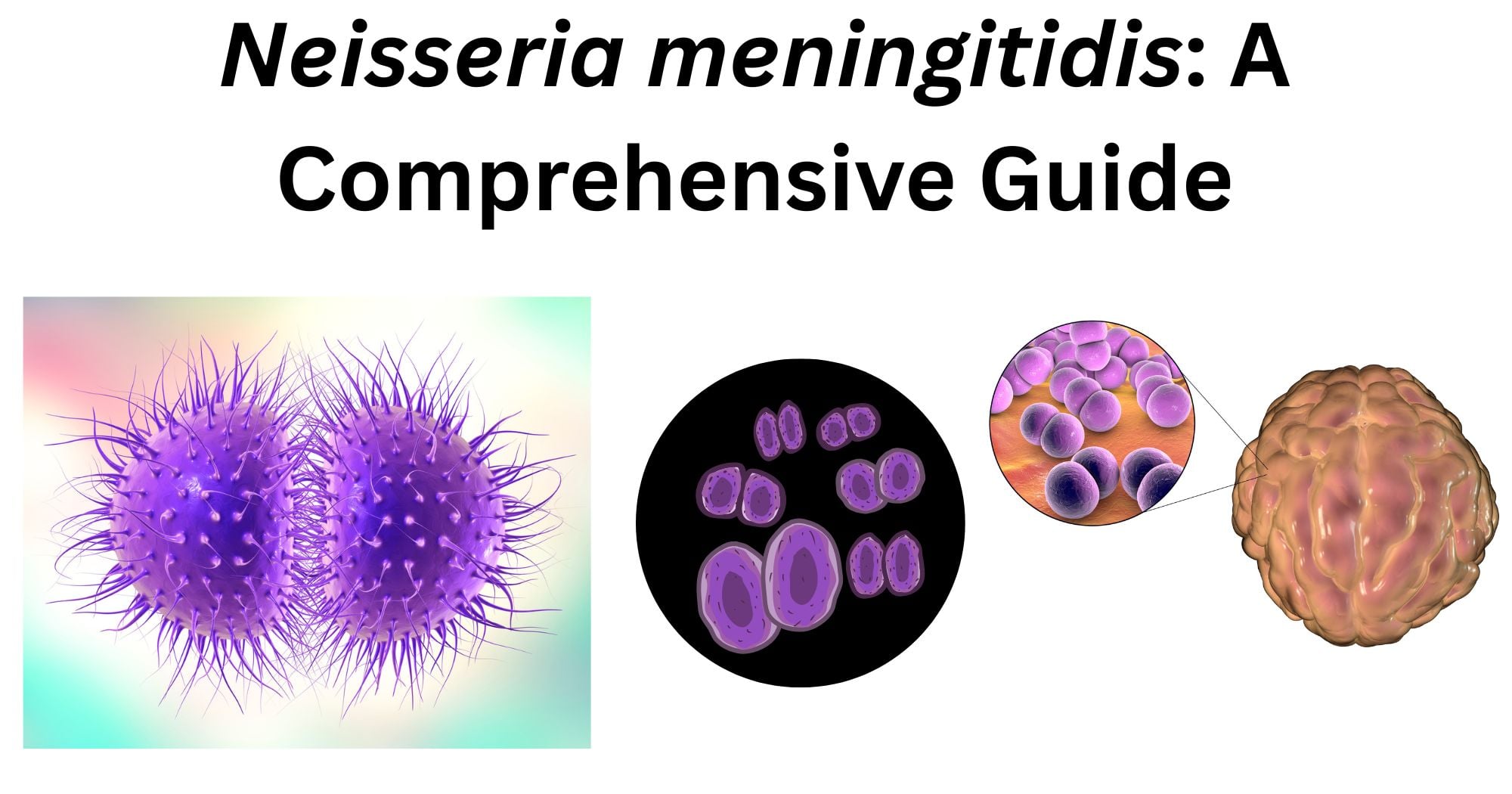
Strains are serogrouped based on their capsular polysaccharides, lipo-oligosaccharide (LOS), and outer membrane proteins. The polysaccharide capsules are necessary for invasive disease and contribute to immune evasion. The first line of antibiotic therapy is penicillin, but resistance can occur. Although new vaccines hold great promise, meningococcal infection continues to be reported in both developed and developing countries, where universal vaccine coverage is absent, and antibiotic resistance is increasingly common.
Taxonomy and Classification of Neisseria meningitidis
Kingdom: Bacteria
Phylum: Pseudomonadota
Class: Betaproteobacteria
Order: Neisseriales
Family: Neisseriaceae
Genus: Neisseria
Species: Neisseria meningitidis
Neisseria meningitidis is classified taxonomically within the family Neisseriaceae, genus Neisseria, which also includes the pathogen N. gonorrhoeae and commensals like N. lactamica. Clinically, strains are divided into serogroups based on their capsular polysaccharide, with groups A, B, C, W, X, and Y responsible for the majority of invasive meningococcal disease worldwide.
Morphology of Neisseria meningitidis
- Neisseria meningitidis is a gram-negative, non-motile, non-spore-forming coccus that appears in pairs known as diplococci. These have a flattened adjacent side to give them a characteristic “coffee bean” or “kidney bean” shape. Singular cells measure approximately 0.6 to 1.0 µm in diameter. They possess polysaccharide capsules external to the cell wall, which is also a major virulence factor that distinguishes it from the non-capsulated Neisseria gonorrhoeae.
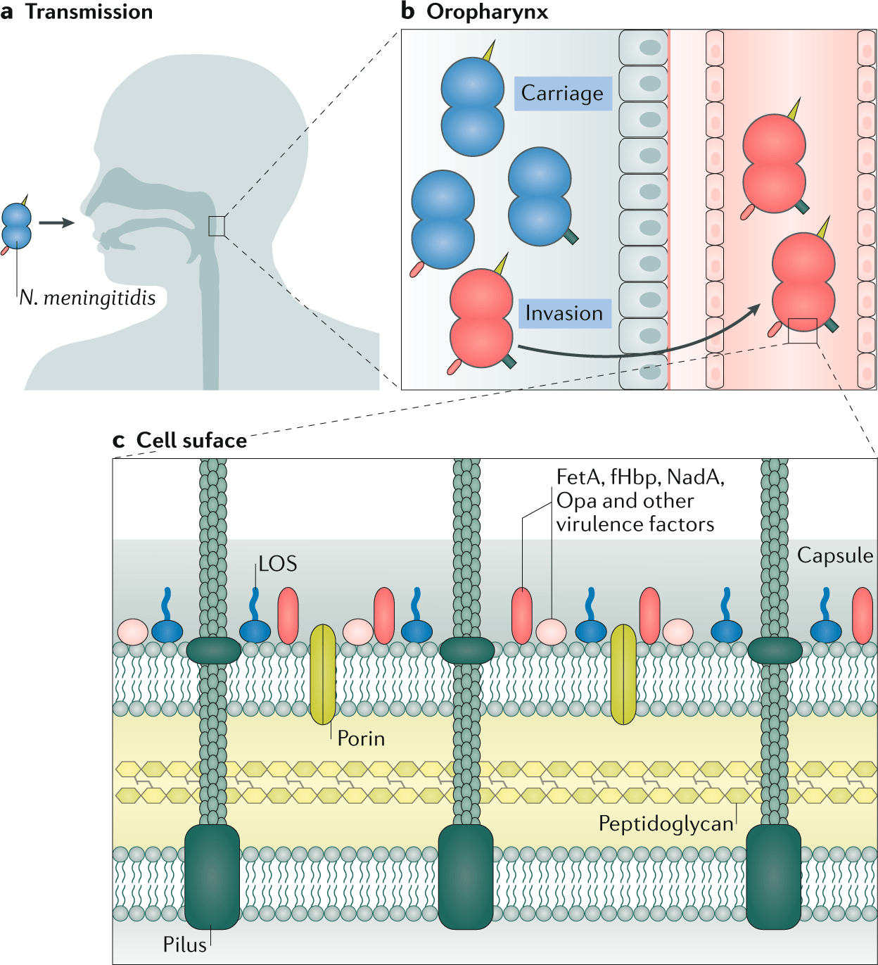
- The cell wall of these bacteria consists of a thin peptidoglycan layer surrounded by an outer membrane containing lipo-oligosaccharide (LOS, formerly called LPS), porin proteins (PorA and PorB), and other surface-exposed proteins that contribute to its antigenic diversity and pathogenicity.
- On solid culture media, like chocolate agar, colonies grown are observed to be greyish-white, non-hemolytic, round, convex, smooth, moist, and glistening with clearly defined entire edges. They typically measure around 1-2 mm in diameter after 24 hours of incubation.
How does Neisseria meningitidis look under a microscope?
- In clinical specimens such as cerebrospinal fluid (CSF) or blood, microscopic examination plays a fundamental role in the initial identification of Neisseria meningitidis.
- Under light microscopy, N. meningitidis is usually observed to be of pink to red-coloured colonies in diplococcus arrangement. This arrangement is often called as “coffee-bean” or “kidney-bean” shaped. The Gram-negative stain result is due to its thin peptidoglycan layer that does not retain the crystal violet-iodine complex and instead takes up the counterstain safranin. Bacteria are frequently observed both extracellularly and inside polymorphonuclear leukocytes in clinical specimens, reflecting the host’s inflammatory response.
- Electron microscopy has revealed detailed ultrastructural features of N. meningitidis not visible by light microscopy. Scanning electron microscopy (SEM) shows the bacterial surface, including the presence of Type IV pili, hair-like structures involved in adhesion to host cells. Transmission electron microscopy (TEM) of ultrathin sections has identified morphological heterogeneity, distinguishing “light” (L) and “dark” (D) type cells. It has demonstrated the trilaminar structure of the outer membrane with its associated capsule.
- The capsule itself can be visualized by using specific stains like Alcian blue-lanthanum nitrate, which preserves and highlights the polysaccharide layer during electron microscopic preparation.
Culture and Growth Characteristics of Neisseria meningitidis
- Neisseria meningitidis is a fastidious and aerobic gram-negative diplococcus exhibiting a specific nutritional requirement for good laboratory cultivation. The growth characteristics of Neisseria meningitidis reflect its ability to adapt to the human nasopharyngeal niche where conditions such as temperature, moisture, and carbon dioxide concentrations are tightly regulated.
- Neisseria meningitidis grows optimally at temperatures between 35-37°C in vitro, given that the atmosphere is enriched with 5-10% CO2, making the environmental conditions closely mimicking the human host. N. meningitidis is strictly aerobic and demonstrates poor survival outside of host-associated environments. They usually undergo rapid autolysis during the stationary phase of growth.
- Since the organism is fastidious, they require enriched media for its primary isolation. They are observed to grow well on blood agar, chocolate agar, and trypticase soy agar. Culturing of N. meningitidis on chocolate agar is often preferred because the heat-lysed blood releases essential growth factors. After 24-48 hours of incubation, colonies typically become visible. The colonies of N. meningitidis grown in chocolate agar are characteristically small to medium-sized, round and smooth, moist and glistening with a non-pigmented appearance. These colonies are generally non-hemolytic and have well-defined edges. The colony phenotype is influenced by the expression of surface structures such as pili and opacity-associated proteins, which in some cases can create variations in colony morphology of Neisseria meningitidis.
- N. meningitidis shows oxidase and catalase positive tests on its routine biochemical tests for identification in the laboratory. Another one of its distinguishing characteristics is its carbohydrate utilization pattern, oxidizing glucose and maltose. However, N. meningitidis does not oxidize sucrose or lactose, which helps to differentiate it from other Neisseria species.
- Timely observation of N. meningitidis culture is very critical for its accurate identification, as it has a high tendency toward autolysis, particularly in older cultures. This autolysis can lead to rapid degradation of the colonies. Hence, timely observation is of importance.
Epidemiology of Neisseria meningitidis
- Neisseria meningitidis remains a major global cause of bacterial meningitis and septicemia. It has been affecting both developed and developing regions. The disease occurs in sporadic cases as well as large-scale epidemics. It has been reported to impact children and young adults.
- The global epidemiology of Neisseria meningitidis is very dynamic as the disease patterns seem to vary dramatically through geography, age, circulating strains, serogroup distribution, environment, and time, resulting in an estimated 1.2 million cases and approximately 135,000 deaths annually worldwide.
- A key determinant of epidemiology is the composition of the capsular polysaccharide that classifies N. meningitidis into multiple serogroups. Among them, most commonly associated with invasive disease are serogroups A, B, C, W-135, X, and Y worldwide. N. meningitidis is an exclusive human pathogen that colonizes the nasopharynx of healthy individuals without causing disease. A reservoir for spread within populations is the asymptomatic carriers that play a crucial role in transmission and epidemiology.
- Certain hypervirulent clonal lineages have a greater capacity to invade the bloodstream, evade host immune responses, and cause systemic infection. These are the same strains that significantly contribute to outbreaks and epidemic patterns observed in different regions.
Pathogenesis of Neisseria meningitidis
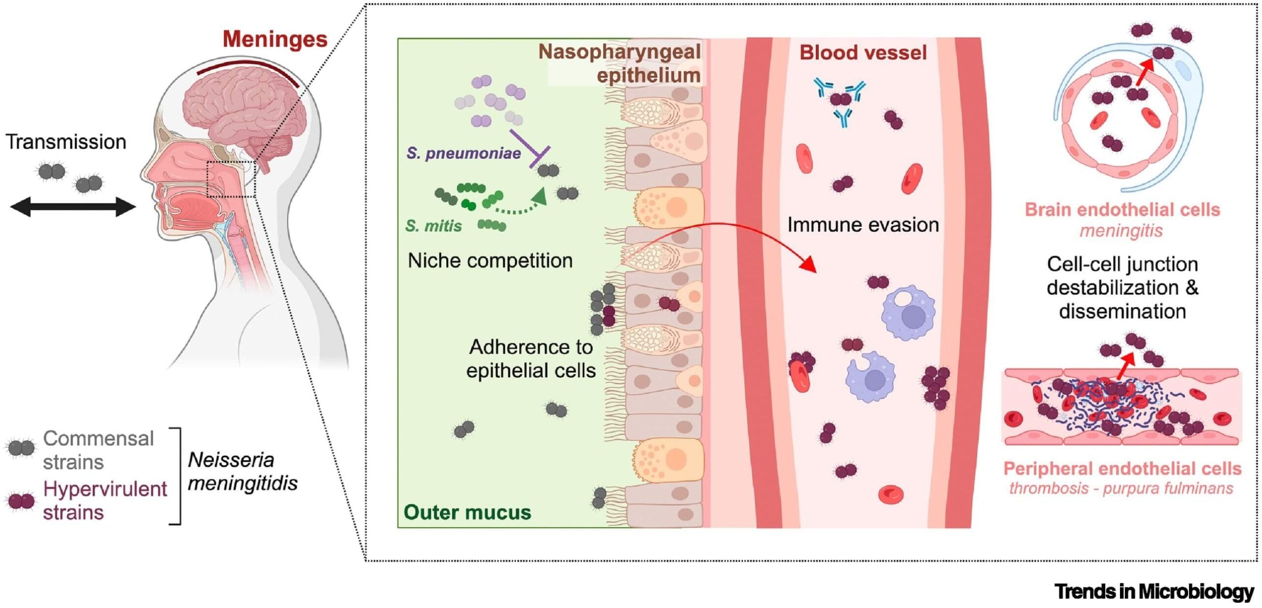
- The pathogenesis of N. meningitidis begins with asymptomatic colonization of the human nasopharynx, a transient and largely benign process that occurs in 8-25% of the population. In a small fraction of individuals, this bacterium transforms into a lethal pathogen unpredictably, causing septicemia and meningitis. To cause invasive disease, N. meningitidis must cross two major barriers- the nasopharyngeal epithelium to enter the bloodstream and subsequently the blood-brain barrier (BBB) or blood-cerebrospinal fluid barrier to reach the meninges. Various virulence factors are employed to accomplish these steps. The polysaccharide capsule provides resistance to antibody/complement-mediated killing and inhibits phagocytosis. This capsule also exhibits molecular mimicry.
- Type IV pili help N. meningitidis in adhesion to host mucosa with the PilC1 and PilC2 proteins. Once they reach the bloodstream, bacteria upregulate iron acquisition systems, detoxifying enzymes (SodC, Kat), and factor H-binding proteins to evade complement. NMB1483, NalP, and LctP are the novel blood survival factors that are essential for the microorganism to resist killing by human blood.
- To cross the blood-brain barrier, Neisseria meningitidis attaches to host cell receptors such as laminin receptor and EGFR. Signaling pathways are also activated, and enzymes that weaken the cell junctions are also produced, which makes the blood vessels more permeable and allows the bacteria to pass between cells. N. meningitidis can survive inside the host cell despite being considered an extracellular pathogen traditionally. During this process, the microorganism interacts with host cell pathways, affects metabolism, and can also influence cell death mechanisms.
- Neisseria meningitidis releases lipooligosaccharide (LOS) once they reach the subarachnoid space, an endotoxin that triggers a strong immune response through TLR4. This leads to the production of inflammatory cytokines such as IL-6 and TNF-α that cause damage to blood vessels, brain swelling, and neuronal injury. More severe disease cases are related to higher levels of LOS.
What are the Virulence Factors of Neisseria meningitidis?
A coordinated set of virulence factors aids in the pathogenicity of Neisseria meningitidis, which enables colonization, immune evasion, bloodstream survival, and invasion of the central nervous system.
Polysaccharide Capsule
The most important virulence determinant of Neisseria meningitidis is its capsule made of polysaccharides. This capsule provides it with protection against phagocytosis and complement-mediated killing and hence allows the bacterium to survive in the bloodstream. Major pathogenic serogroups are defined by the chemical composition of the capsule (serogroups A, B, C, W, X, and Y). The information about the composition of the capsule is essential for both epidemiological tracking and vaccine design.
Type IV Pili
Filamentous appendages on the surface of the Neisseria meningitidis that mediate the initial attachment to epithelial cells in the nasopharynx are the Type IV pili. Apart from adhesion, they also promote the formation of microcolonies, support clustering and sustained colonization, assist in resisting mucociliary clearance, and trigger host cell signaling pathways that encourage bacterial invasion.
Outer Membrane Proteins (OMPs)
The outer membrane proteins are important for adhesion and invasion of host cells. These OMPs include Opa and Opc proteins. These proteins bind to host surface molecules, including integrins, facilitating bacterial uptake by epithelial and endothelial cells.
Lipooligosaccharide (LOS)
LOS is an endotoxin in the outer membrane and is a major mediator of disease severity. It triggers a strong inflammatory response via Toll-like receptor 4 (TLR4). This leads to the release of cytokines such as TNF-α and IL-6. This cascade contributes to vascular damage, septic shock, and meningitis.
Iron Acquisition System
Neisseria meningitidis, like every other pathogen, requires iron for growth and has evolved dedicated systems to scavenge iron from host proteins such as transferrin and lactoferrin. The microorganism thrives in the iron-poor environment of the human body due to this iron-uptake mechanism. It also contributes to the microorganisms’ survival during invasive infection.
Clinical Manifestations of Neisseria meningitidis
When someone encounters Neisseria meningitidis, the outcome can vary widely from carrying the bacterium asymptomatically in the nasopharynx to severe invasive disease, most commonly presenting as meningitis and meningococcemia.
Meningococcal Meningitis
Meningitis ranks among the most common presentations of invasive meningococcal disease. This condition involves swelling of the meninges and usually brings on symptoms such as:
- Fever
- Headache
- Neck stiffness
- Photophobia
- Altered mental status
In infants and young children, symptoms may be less specific and include irritability, poor feeding, vomiting, and a bulging fontanelle. Without prompt treatment, the condition can lead to seizures, coma, and even death.
Meningococcemia (Septicemia)
Meningococcemia refers to a serious infection of the bloodstream that can occur with or without meningitis. It is often associated with:
- High fever and chills
- Fatigue and malaise
- Hypotension
- Disseminated intravascular coagulation (DIC)
One of the distinct features of septicemia is the development of a petechial to purpuric rash, which can rapidly progress to purpura fulminans, a life-threatening condition involving extensive vascular damage and skin necrosis.
Fulminant Disease
In certain individuals, the infection takes an extremely aggressive turn known as fulminant meningococcemia. This is characterized by:
- Septic shock
- Multiorgan failure
- Adrenal hemorrhage
This form has a very high mortality rate and requires immediate intensive care.
Chronic Meningococcemia
A much rarer pattern is chronic meningococcemia, which unfolds over weeks rather than hours or days. Its features include:
- Recurrent fever
- Rash
- Arthralgia (joint pain)
This is generally milder but may still progress if untreated.
The severity of disease is largely driven by the host inflammatory response to bacterial endotoxins, particularly lipooligosaccharide (LOS), which contributes to vascular injury and organ dysfunction.
Laboratory Diagnosis of Neisseria meningitidis
Specimens
Cerebrospinal fluid (CSF), blood, and skin scrapings from petechial rashes from cases and nasopharyngeal swabs from carriers. Specimens should be collected in sterile containers and transported immediately without any delay.
N. meningitidis appears as gram-negative, coffee-bean-shaped diplococci in a Gram-stained smear of the centrifuged deposit of the specimen. They occur intracellularly or extracellularly in PMN leukocytes.
Culture of Neisseria meningitidis
- Isolation of N. meningitidis remains the gold standard as it also provides isolates for strain differentiation and susceptibility testing.
- Samples from sterile body sites are inoculated on either blood or chocolate agar since meningococci are fastidious. The chocolate agar base can be enriched with antibiotics such as vancomycin, colistin, nystatin, and trimethoprim for selective isolation of N. meningitidis. For culture from non-sterile sites like the nasopharynx, a selective medium such as Modified New York City or Modified Thayer Martin medium is required. Culture plates should be incubated for a minimum of 48 hours with a source of 5% CO2.
- Young colonies of N. meningitidis in blood agar are round, smooth, moist, glistening, and convex with a clearly defined edge, whereas actively growing colonies are grey and unpigmented.
- On Modified New York City medium and Thayer Martin Medium, Neisseria meningitidis appears to form large, colourless to bluish-gray mucoid colonies.
Identification of Neisseria meningitidis
- Under the microscope, N. meningitidis appears as gram-negative diplococci, pairs of small pinkish-red spheres with flattened adjacent sides giving them the well-known coffee bean shape.
- For definitive identification, laboratories often turn to matrix-assisted laser desorption ionization time of flight mass spectrometry (MALDI-TOF MS). The results obtained from MALDI-TOF MS should be confirmed, however, by additional identification methods like the carbohydrate utilization test. The CarboFerm Neisseria Kit from Hardy Diagnostics is one example of a system that assesses an isolate’s ability to oxidize various sugars. Neisseria meningitidis produces acid from both glucose and maltose but leaves sucrose and lactose untouched. This pattern distinguishes it from Neisseria gonorrhoeae, which ferments glucose only.
- Other biochemical reactions that help in the identification of N. meningitidis are the catalase test, which results in a positive test. The organism is also negative for the DNase reaction. When it comes to selective media containing VCN inhibitor (vancomycin, colistin, nystatin), meningococcus shows resistance to colistin and therefore grows readily, whereas some other Neisseria do not. The capsular polysaccharide that distinguishes major disease-causing serogroups is also distinguished. Enzyme immunoassays, latex agglutination, and rapid diagnostic kits are some of the serological tests that are most useful for retrospective diagnosis and can detect antibodies against the capsular antigen.
Treatment of Neisseria meningitidis
Meningitidis caused by Neisseria meningitidis are medical emergencies that require immediate antibiotic therapy and supportive care. Due to the rapid progression of the disease, the treatment is often initiated empirically before laboratory confirmation.
Empirical Antibiotic Therapy
Broad-spectrum intravenous antibiotics are to be promptly started upon the suspicion of meningitis. Third-generation cephalosporins like Cefotaxime and Ceftotaxime are commonly used as first-line empirical therapy due to their effectiveness and good penetration into CSF.
Targeted Therapy
Generally, N. meningitidis is susceptible to Penicillin G and Ampicillin. Once Neisseria meningitidis is confirmed and antibiotic susceptibility is known, therapy can be narrowed from the broad-spectrum ones.
Duration of Treatment
Depending upon the clinical response and severity, meningococcal meningitis requires around 5-7 days course of antibiotic therapy, typically.
Supportive Management
In case of severe cases, intensive supportive care is of utmost importance alongside the antibiotic therapy. This may include:
- Fluid resuscitation for hypotension
- Vasopressin in septic shock
- Management of intracranial pressure
- Respiratory support if needed
References
- BioLibreTexts. (n.d.). Neisseria meningitidis. In Microbiology for health sciences. https://bio.libretexts.org/Courses/Mansfield_University_of_Pennsylvania/BSC_3271%3A_Microbiology_for_Health_Sciences_Sp21_%28Kagle%29/15%3A_Selected_Pathogens/15.01%3A_Bacterial_Pathogens/15.1.03%3A_Gram_Negatives/15.1.3.03%3A_Neisseria_meningitidis
- Frontiers in Cellular and Infection Microbiology. (2025). Neisseria meningitidis pathogenesis and host interaction.
https://www.frontiersin.org/journals/cellular-and-infection-microbiology/articles/10.3389/fcimb.2025.1733264/full - IDImages. (n.d.). Neisseria meningitidis atlas entry. https://www.idimages.org/m/atlas/organism/?atlasentryID=23&organism=Neisseria
- Madico, G., Welsch, J. A., Lewis, L. A., McNaughton, A., Perlman, D. H., Costello, C. E., Ngampasutadol, J., Vogel, U., Granoff, D. M., & Ram, S. (2006). The meningococcal vaccine candidate GNA1870 binds the complement regulatory protein factor H and enhances serum resistance. Journal of Immunology, 177(1), 501–510.
https://doi.org/10.4049/jimmunol.177.1.501 - Microbe Online. (n.d.). Neisseria meningitidis: Properties, pathogenesis, and laboratory diagnosis.
https://microbeonline.com/neisseria-meningitidis-properties-pathogenesis-and-laboratory-diagnosis/ - Microbiology Society. (n.d.). Journal of Medical Microbiology article on Neisseria meningitidis. https://www.microbiologyresearch.org/content/journal/jmm/10.1099/jmm.0.45529-0
- PathologyOutlines.com. (n.d.). Neisseria meningitidis. https://www.pathologyoutlines.com/topic/microbiologynmeningitidis.html
- ResearchGate. (n.d.-a). Biochemical properties of Neisseria strains [Figure]. https://www.researchgate.net/figure/Biochemical-properties-of-Neisseria-strains_tbl1_11234112
- ResearchGate. (n.d.-b). Electron microscope image of Neisseria meningitidis showing surface blebbing [Figure].
https://www.researchgate.net/figure/Electron-microscope-image-of-Neisseria-meningitidis-showing-surface-blebbing_fig2_26745654 - Rouphael, N. G., & Stephens, D. S. (2012). Neisseria meningitidis: Biology, microbiology, and epidemiology. Methods in Molecular Biology, 799, 1–20.
https://doi.org/10.1007/978-1-61779-346-2_1 - Vircell. (n.d.). Neisseria meningitidis.
https://www.vircell.com/en/diseases/neisseria-meningitidis - Virji, M. (2009). Pathogenic Neisseria: Surface modulation, pathogenesis and infection control. Nature Reviews Microbiology, 7(4), 274–286.
https://doi.org/10.1038/nrmicro2097
