
A medical ventilator is a core life-support machine in modern medicine that can temporarily take over or assist breathing in situations ranging from routine surgery to severe respiratory failure in intensive care units (ICUs).
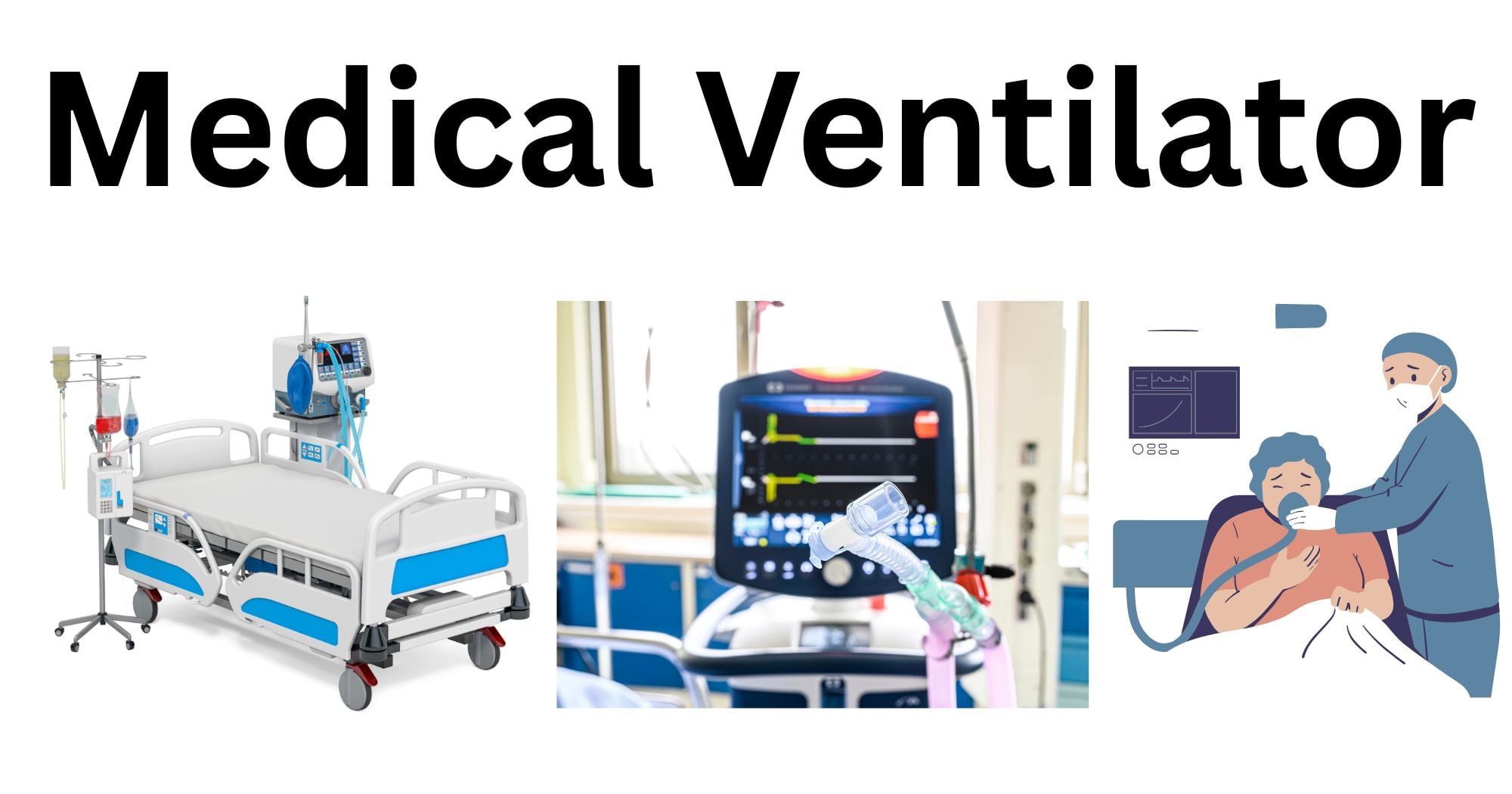
What is a Medical Ventilator?
A medical ventilator, also known as a mechanical ventilator, is a machine designed to move air and oxygen into and out of the lungs, supporting or fully replacing a patient’s spontaneous breathing. It ensures adequate oxygen delivery and carbon dioxide removal while reducing the work of breathing. It is used in anesthesia, critical care, emergency care, and even at home for chronic respiratory failure. It does not cure the underlying disease, but it buys time for the lungs and body to recover while clinicians treat the root cause of respiratory failure.
The main goals of a medical ventilator are to maintain oxygenation, remove carbon dioxide, reduce respiratory muscle workload, prevent complications and lung injury, and buy time to treat the underlying disease.
History and Evolution
The earliest development of mechanical ventilation dates to the 15th-16th centuries, when early anatomists manually inflated animal lungs with bellows, demonstrating that artificial ventilation could sustain life. By the 18th century, experiments were performed with positive-pressure ventilation on drowning victims, although these early attempts often caused complications from excessive pressure. The 19th and early 20th centuries saw the use of negative-pressure ventilators, which cyclically reduced external pressure to draw air into the lungs. The modern mechanical ventilation developed after the mid-20th century polio epidemics, when positive pressure ventilators replaced manual and negative pressure devices.
Early machines were large, cumbersome, and had few safety features. In over 70+ years, ventilators have become smaller, more portable, and technically more sophisticated with multiple modes, alarms, and monitoring. They can deliver lung- and diaphragm-protective strategies and non-invasive ventilation (NIV). Future directions include personalized, AI-guided ventilation to better match each patient’s physiology.
How does a Ventilator Works?
At its core, a ventilator aims to mimic or augment the normal respiratory cycle: inspiration (inflow of air) and expiration (outflow of air). Natural breathing relies on negative pressure: the diaphragm contracts, expanding the chest cavity and drawing air in; exhalation is largely passive. Mechanical ventilators, however, use positive pressure, i.e., they push gas into the lungs at pressures above atmospheric pressure, inflating the lungs, then allow passive exhalation. This process supports oxygenation and ventilation. The key physiological targets are: adequate alveolar ventilation (CO2 removal), adequate oxygenation (PaO2 / SpO2), and acceptable pressures and volumes to limit ventilator-induced lung injury.
Effective support requires that ventilator cycling be aligned with the patient’s own respiratory drive to avoid excess work of breathing.
Key Parts and Components of a Medical Ventilator
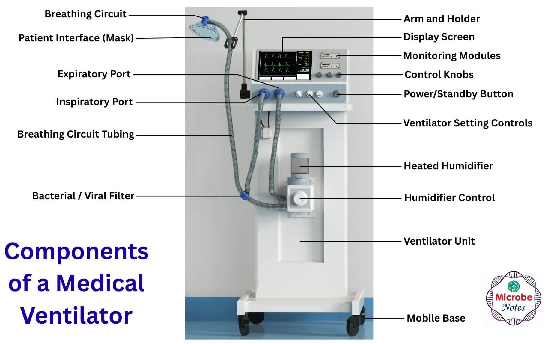
The core components of a medical ventilator are:
- Gas source (compressed air/ oxygen or generator)
- Mixing and delivery system (controls flow, volume, and pressure)
- Patient circuit: Tubing connecting the machine to the patient (inspiratory and expiratory limbs).
- Humidifier: Adds moisture to inspired gas.
- Sensors and monitors: Measure pressure, flow, volume, and oxygen concentration.
- Control panel: For mode and parameter settings.
- Alarms and safety systems: Visual and audible alerts for high pressure, low volume, apnea, or disconnection.
Adjustable settings include tidal volume, respiratory rate, inspiratory time and flow, positive end-expiratory pressure (PEEP), and FiO2 (oxygen concentration).
Applications and Importance of Medical Ventilators
Medical ventilators are indispensable in several clinical settings, such as:
- Respiratory failure, for example, severe pneumonia, acute respiratory distress syndrome (ARDS), COPD exacerbation, etc.
- Post-operative care, especially after major thoracic, abdominal, or neurosurgical procedures.
- Neuromuscular diseases where the respiratory muscles are weak or paralyzed.
- Anesthesia during surgery.
Their importance lies in:
- Stabilizing patients
- Optimizing oxygen delivery
- Resting fatigued respiratory muscles
- In anesthesia, ensuring controlled breathing during surgery.
Limitations
- They cannot treat the underlying disease; they only buy time till therapy works.
- There is a risk of lung and diaphragm injury if settings are inappropriate.
- Prolonged ventilation increases the risk of complications such as ventilator-associated pneumonia (VAP) and ventilator-induced lung injury (VILI).
- Long-term use is associated with muscle atrophy, delirium, and ICU-acquired weakness.
Invasive vs. Non-Invasive Ventilation (NIV)
Invasive ventilation involves an endotracheal tube or tracheostomy, providing direct airway access. Non-invasive ventilation uses masks or helmets, avoiding intubation.
| Aspect | Invasive ventilation | Non-invasive ventilation |
| Airway Access | Endotracheal tube/ Tracheostomy | Mask / Helmet |
| Control Level | High (full support) | Moderate (patient effort required) |
| Risks | Higher infection, Barotrauma | Leakage, skin breakdown, aspiration |
| Patient comfort | Lower (requires sedation) | Higher |
| Typical use cases | Severe ARDS, shock, coma | Mild-moderate COPD, heart failure, and some post-operation monitoring |
Common Ventilation Modes: Control, Assist, and Spontaneous
The core ventilation modes are:
- Control (mandatory) modes: The ventilator delivers a set number of mandatory breaths (each breath fully generated by the ventilator). The patient’s effort may be minimal. E.g., volume or pressure control
- Assist-control: Each breath is either patient-triggered or time-triggered, but every breath is delivered with full preset support.
- Spontaneous/ support modes: The patient initiates breathing, and the ventilator adds pressure to reduce the work of breathing.
Understanding Ventilator Parameters: Tidal Volume, PEEP, and FiO2
Tidal volume (Vt) is the volume of gas delivered per breath. The Vt is typically 6-8 mL/kg of ideal body weight. Overly large volumes cause volutrauma and ventilator-induced lung injury.
PEEP (Positive End-Expiratory Pressure) is the baseline pressure left in the lungs at the end of expiration. It helps keep alveoli open and improves oxygenation, but excessive PEEP can impair circulation or overexpand lungs.
FiO2 is the fraction of inspired oxygen. High levels of FiO2 improve oxygenation, but prolonged 100% oxygen is avoided to reduce oxygen toxicity and atelectotrauma.
Other parameters include respiratory rate, inspiratory time, and peak pressures.
When Does a Patient Need a Ventilator?
The ventilator is typically initiated when these conditions appear:
- Hypoxemic respiratory failure (low oxygen despite supplemental O2).
- Hypercapnic failure (CO2 retention leading to acidosis).
- Inability to protect the airway (e.g., coma, overdose).
- Excessive work of breathing risks fatigue.
- Post-cardiac arrest or major surgery.
The decision is not purely physiological; it is also informed by the patient’s prognosis and the benefit of temporary support.
Clinical Complications: Barotrauma, Volutrauma, and VAP
Mechanical ventilation is life-saving, but complications are inherent and must be minimized. The complications are:
- Barotrauma: Barotrauma is the damage from high airway pressures, leading to pneumothorax and hypotension.
- Volutrauma: Volutrauma is the overdistension from excessive tidal volumes, which may lead to ventilator-induced lung injury.
- Ventilator-Associated Pneumonia (VAP): It is the infection linked to prolonged intubation and invasive support, where microbes colonize the endotracheal tube and airway.
- Others: myopathy, neuromuscular weakness, hemodynamic instability, delirium, organ effects (including kidneys).
The Weaning Process: Testing for Readiness to Extubate
Weaning is the process of liberating a patient from ventilator support and removing the artificial airway. The early consideration of weaning is done as soon as the underlying condition improves. The steps for weaning include:
- Gradual reduction of pressure support or PEEP.
- Spontaneous breathing trials (SBT) using minimal support to assess patient respiratory rate, tidal volume, heart rate, and oxygen saturation.
- Assessment of readiness criteria using clinical and physiological criteria before extubating.
Critical Maintenance: Calibration, Disinfection, and Safety Alarms
Safe ventilator use requires proper maintenance:
- Correct setup and calibration of volumes, pressures, and alarms.
- Regular checks for leaks, disconnections, and circuit problems (calibration).
- Strict disinfection and infection-control practices to reduce VAP and other device-related infections.
- Thoughtful safety alarm settings for pressures, volumes, FiO2, and apnea to detect danger without alarm fatigue.
Conclusion
Medical ventilators are essential tools that can temporarily replace or assist breathing in critically ill or anesthetized patients. Modern ventilators are highly sophisticated with multiple modes and settings for tailoring support, protecting the lungs and diaphragm, and facilitating safe weaning. It is a life-supporting device, but it poses real risks, including barotrauma, volutrauma, infection, and prolonged dependency, so skilled management and careful parameter selection are necessary. Understanding how ventilators work, their indications, and their limitations is crucial to providing safe and effective care for patients who rely on this medical device for survival.
References
- Pham, T., Brochard, L. J., & Slutsky, A. S. (2017). Mechanical Ventilation: State of the Art. Mayo Clinic Proceedings, 92(9), 1382–1400. https://doi.org/10.1016/j.mayocp.2017.05.004
- Staff, D. (2021, June 7). Medical ventilators: A breakdown of the components and sensors needed in essential medical care. KnowHow. https://knowhow.distrelec.com/medical-healthcare/medical-ventilators-a-breakdown-of-the-components-and-sensors-needed-in-essential-medical-care/
- Upadhya, P., H S, S., R, H., Mahesh Babu, V., Balasoupramaniane, K., & Nadaf, Z. (2025). Approach to mechanical ventilation: A simplified approach for a pulmonologist. Monaldi Archives for Chest Disease. https://doi.org/10.4081/monaldi.2025.3476
- Mechanical Ventilation: Background, Classifications of Positive-Pressure Ventilators, Indications for Mechanical Ventilation. (2024). https://emedicine.medscape.com/article/304068-overview#a6
- Ventilator/Ventilator Support – What Is a Ventilator? | NHLBI, NIH. (2022, March 24). https://www.nhlbi.nih.gov/health/ventilator
- Mechanical Ventilation: Purpose, Types & Complications. (n.d.). Retrieved May 27, 2026, from https://my.clevelandclinic.org/health/treatments/15368-mechanical-ventilation
- Tobin, M. J. (2018). Physiologic Basis of Mechanical Ventilation. Annals of the American Thoracic Society, 15(Supplement_1), S49–S52. https://doi.org/10.1513/AnnalsATS.201705-417KV
